


Editado por: Dr. Alberto Calderón Montero
(Doctor Pedro Laín Entralgo Health Center, Alcorcon, Spain)
Dr. José Manuel Fernandez Garcia
(Galicia Health Service, Santiago de Compostela, Spain)
Última actualización: Noviembre 2025
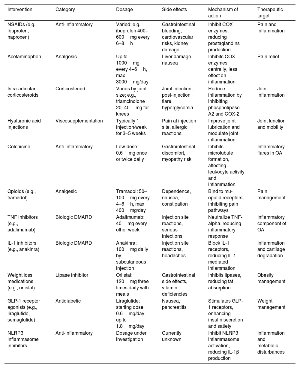
Más datosObesity is a well-established risk factor for osteoarthritis (OA), traditionally attributed to increased mechanical stress on weight-bearing joints. However, recent research suggests a more complex interplay, involving metabolic, biomechanical, and inflammatory pathways. This review delves into these multifaceted connections between obesity and osteoarthritis, extending beyond the conventional understanding of weight-bearing stress. It explores the role of adipokines such as leptin, visfatin, adiponectin, and resistin in OA pathogenesis and progression, highlighting their potential as targets for novel therapeutics. The review also examines how obesity alters the biomechanics of lower extremities, contributing to changes in joint load and movement patterns. Systemic effects, including inflammation and metabolic factors, are discussed to elucidate their roles in exacerbating OA beyond joint loading. Furthermore, the impact of bariatric surgery and weight loss strategies on OA symptoms and progression is evaluated. This comprehensive review aims to provide new insights into obesity-induced OA, paving the way for more targeted and effective treatment strategies.
La obesidad es un factor de riesgo bien establecido para la osteoartritis (OA), tradicionalmente atribuida al aumento del estrés mecánico en las articulaciones de carga. Sin embargo, investigaciones recientes sugieren una interacción más compleja, que involucra vías metabólicas, biomecánicas e inflamatorias. Esta revisión profundiza en estas conexiones multifacéticas entre la obesidad y la osteoartritis, yendo más allá de la comprensión convencional del estrés por carga mecánica. Se explora el papel de las adipocinas, como la leptina, la visfatina, la adiponectina y la resistina, en la patogénesis y la progresión de la OA, destacando su potencial como dianas terapéuticas innovadoras. Además, se analiza cómo la obesidad altera la biomecánica de las extremidades inferiores, contribuyendo a cambios en la carga articular y en los patrones de movimiento. Se abordan los efectos sistémicos, incluyendo la inflamación y los factores metabólicos, para esclarecer su papel en la exacerbación de la OA más allá de la sobrecarga articular. Asimismo, se evalúa el impacto de la cirugía bariátrica y las estrategias de pérdida de peso en los síntomas y la progresión de la OA. Esta revisión integral busca proporcionar nuevas perspectivas sobre la OA inducida por obesidad, allanando el camino para estrategias terapéuticas más específicas y eficaces.
Artículo
Diríjase al área privada de socios de la web de la SEMERGEN, (https://www.semergen.es/index.php?seccion=biblioteca&subSeccion=revistaSEMERGEN ) y autentifíquese.
