


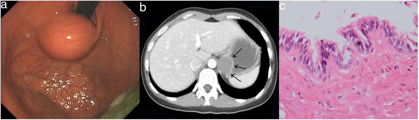
An 18-year-old woman presented with aggravated vomiting for 10 months, without remarkable past history. Both her physical examination and laboratory were normal. Upper endoscopy showed a soft spherical bulge with smooth surface in the gastric cardia and fundus (Fig. 1a). Endoscopic ultrasound demonstrated that the lesion was an extrinsic compression. Enhanced computed tomography scan was performed, revealing a low dense cystic lesion with wall enhancement (Fig. 1b, arrows). The lesion was found to be located in the abdomen, and was completely excised by laparoscopic surgery. Histologic findings reported the presence of ciliated pseudostratified columnar epithelium in the cyst wall (Fig. 1c, H&E 400×), thus an abdominal bronchogenic cyst was finally diagnosed. The patient was discharged without any complications. Her symptom was gone during follow-up.
 Endoscopic image. A soft spherical bulge with smooth surface in the gastric cardia and fundus; (b) computed tomography image. A low dense cystic lesion with wall enhancement; (c) histologic image (H&E, 400×). The presence of ciliated pseudostratified columnar epithelium in the cyst wall.")
(a) Endoscopic image. A soft spherical bulge with smooth surface in the gastric cardia and fundus; (b) computed tomography image. A low dense cystic lesion with wall enhancement; (c) histologic image (H&E, 400×). The presence of ciliated pseudostratified columnar epithelium in the cyst wall.
Bronchogenic cysts below the diaphragm are extremely rare.1 Although most of them are benign and asymptomatic, symptoms can be developed owing to compression of adjacent organs, cyst rupture or secondary infection. Malignancy changes can be also noted.2 As retained cyst wall will cause a recurrence, complete excision of the cyst is of vital importance.
FundingAll authors have no financial relationships relevant to this article to disclose.
Author's contributionFind the patient and get the idea: Liansong Ye, Dan Yang, Xiaobo Qin, Bing Hu.
Collect the information of the patient: Liansong Ye, Dan Yang, Xiaobo Qin.
Discuss the diagnosis and treatment for the patient: Liansong Ye, Dan Yang, Xiaobo Qin, Bing Hu.
Write and revise the article: Liansong Ye, Dan Yang, Xiaobo Qin, Bing Hu.
Conflict of interestAll authors have no conflicts of interest to disclose.
Informed consentInformed consent was obtained from patient for the publication of her information and images.
We acknowledge the support from National Natural Science Foundation of China (81570472).

