Buscar en
Radiología (English Edition)
Toda la web
Inicio Radiología (English Edition) Hyperintense punctiform images in the white matter: A diagnostic approach
Journal Information
Vol. 54. Issue 4.
Pages 321-335 (July - August 2012)
Share
Share
Download PDF
More article options
Visits
32660
Vol. 54. Issue 4.
Pages 321-335 (July - August 2012)
Update in Radiology
Hyperintense punctiform images in the white matter: A diagnostic approach
Imágenes puntiformes hiperintensas en la sustancia blanca: una aproximación diagnóstica
Visits
32660
S. Medrano Martorella,
Corresponding author
santimedra@yahoo.es

Corresponding author.
, M. Cuadrado Blázquezb, D. García Figueredob, S. González Ortiza, J. Capellades Fonta
a Servicio de Radiodiagnóstico, Hospital del Mar/IDIMAR-CRC Mar, Barcelona, Spain
b Servicio de Radiodiagnóstico, Hospital de Granollers, Barcelona, Spain
This item has received
Article information
Abstract
Full Text
Bibliography
Download PDF
Statistics
Figures (20)
Show moreShow less
Tables (1)
Table 1. Semiological characteristics.
Abstract

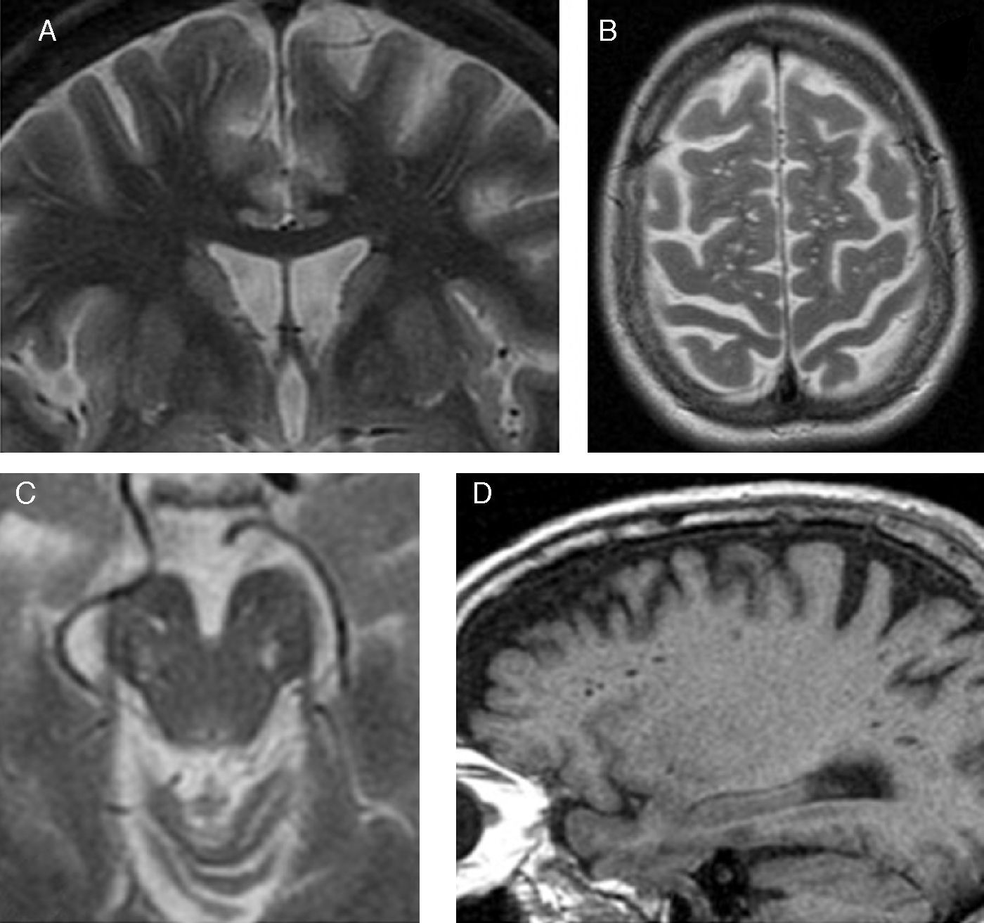
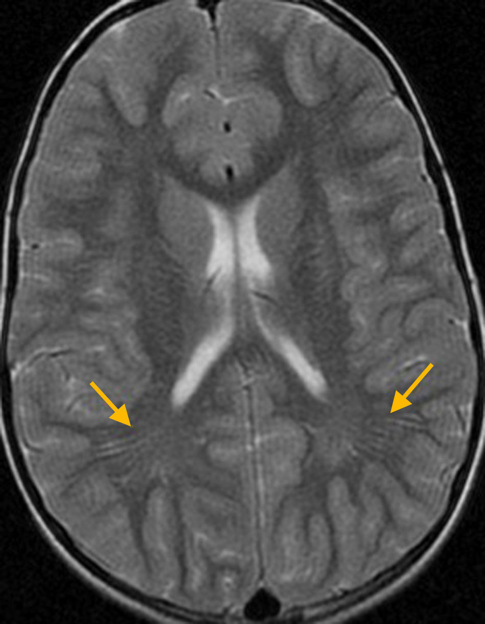
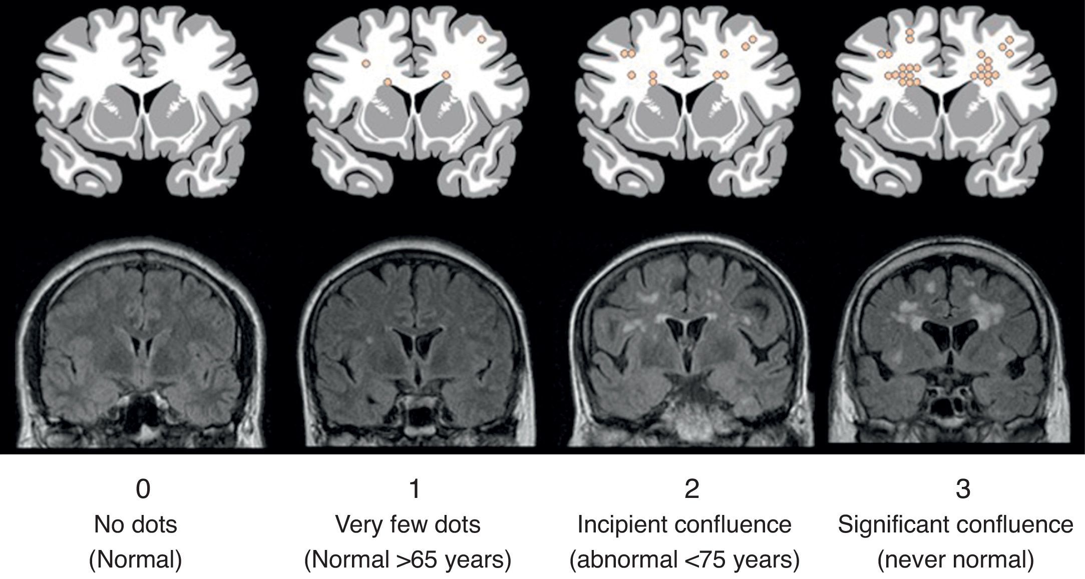
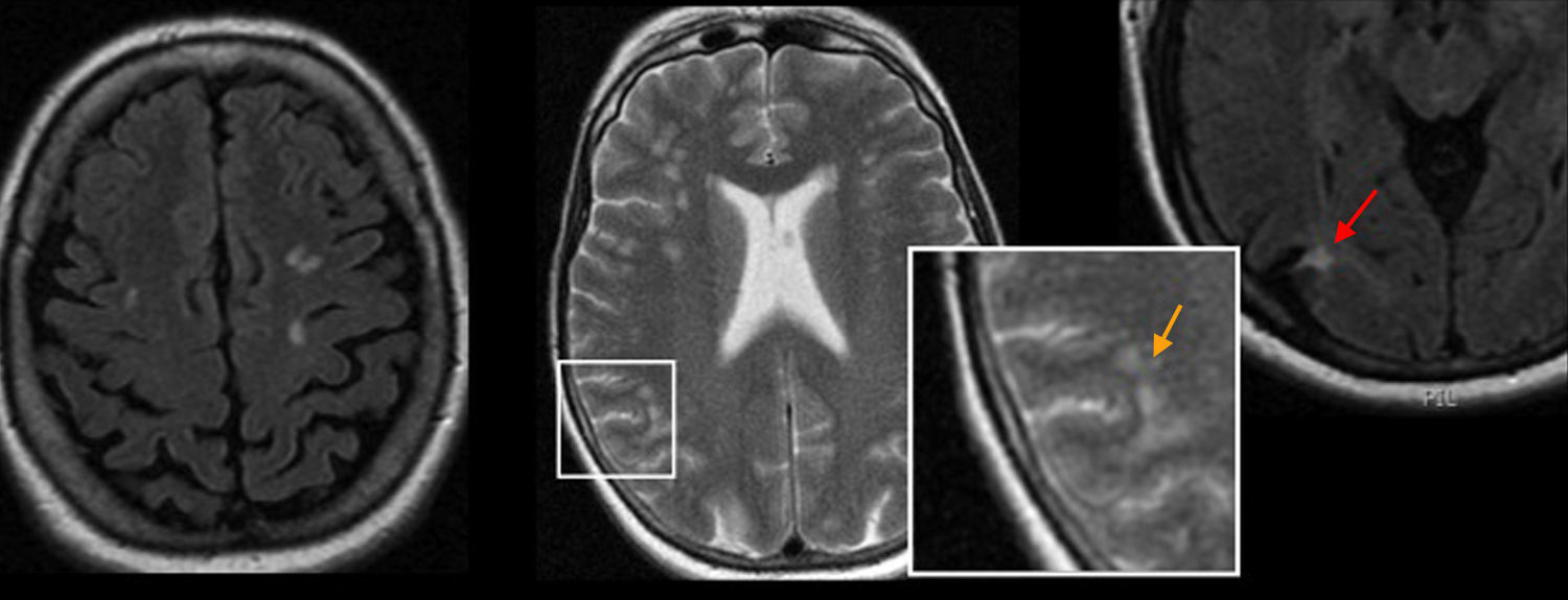
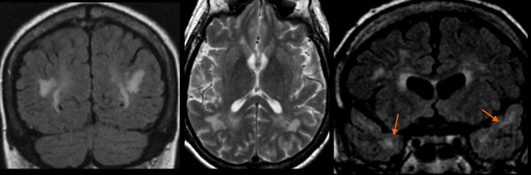
The presence of hyperintense punctiform images in the white matter in T2-weighted magnetic resonance (MR) sequences is a very common finding and is occasionally a diagnostic challenge for the radiologist.

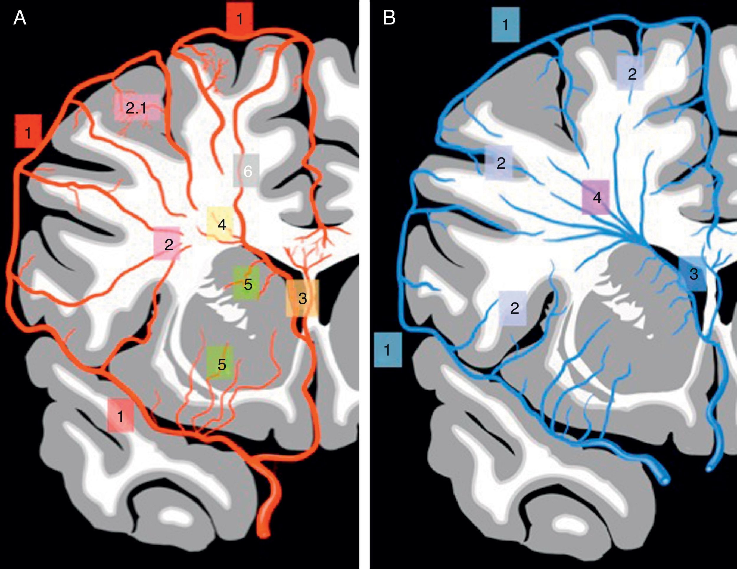
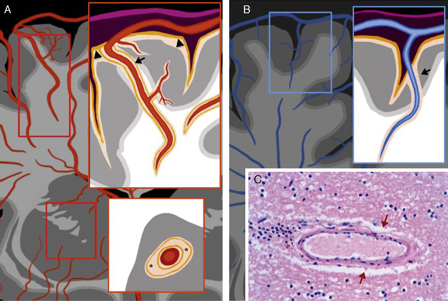
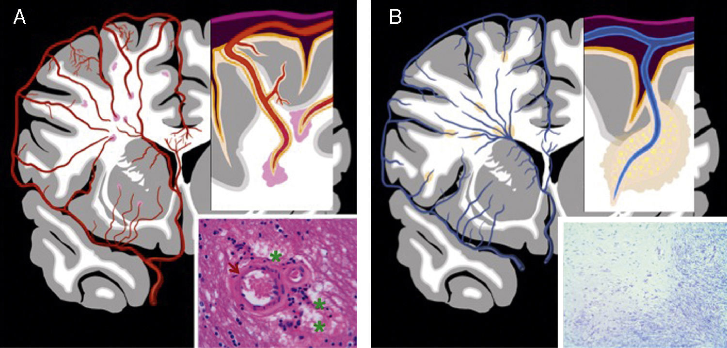
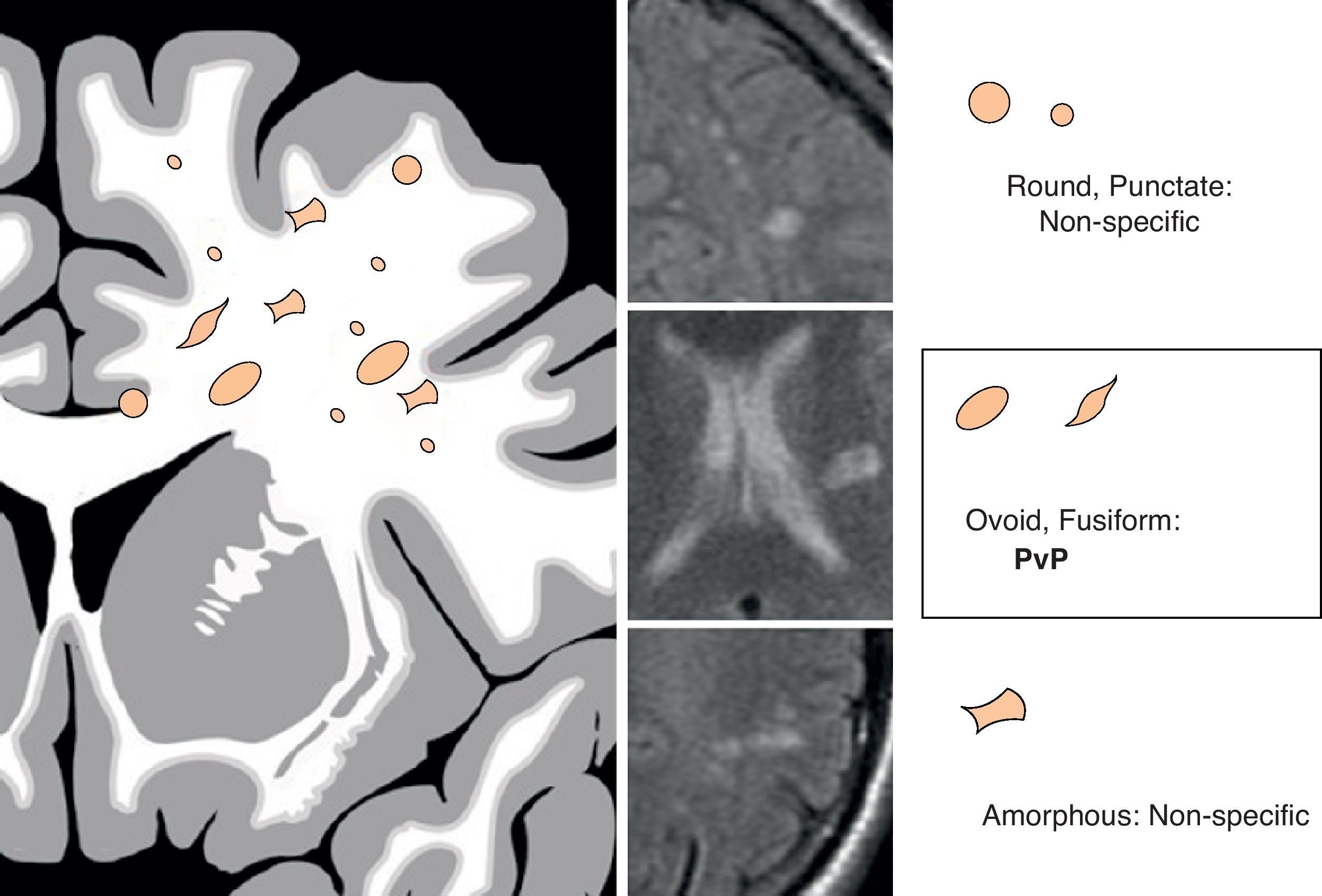
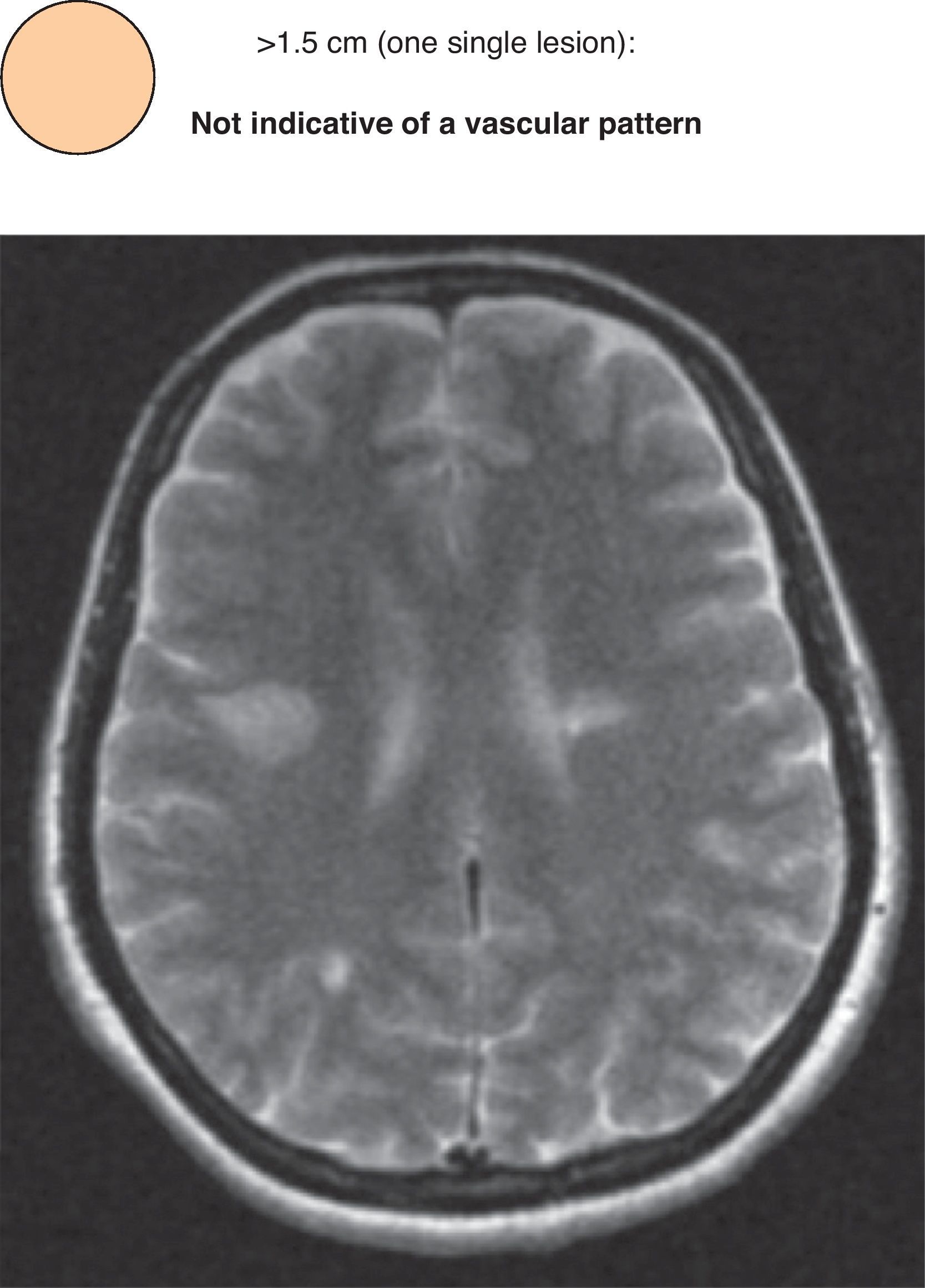
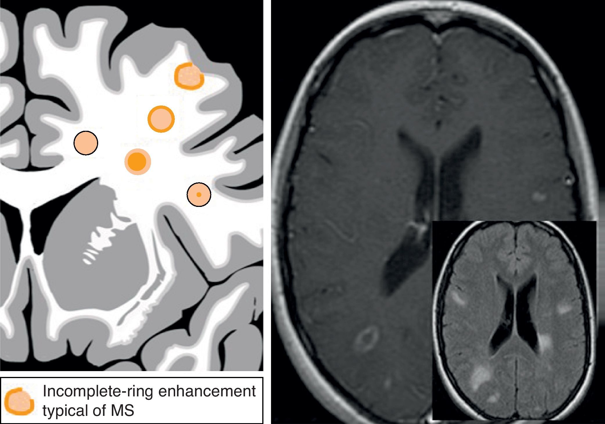
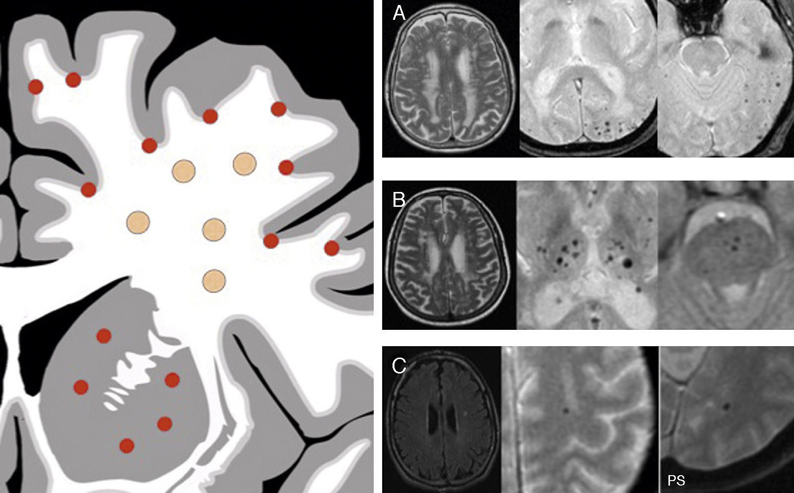
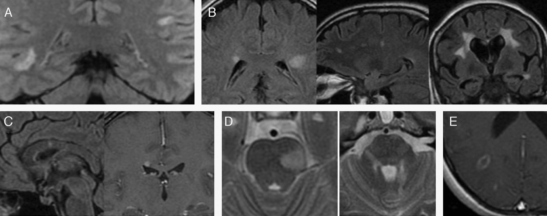
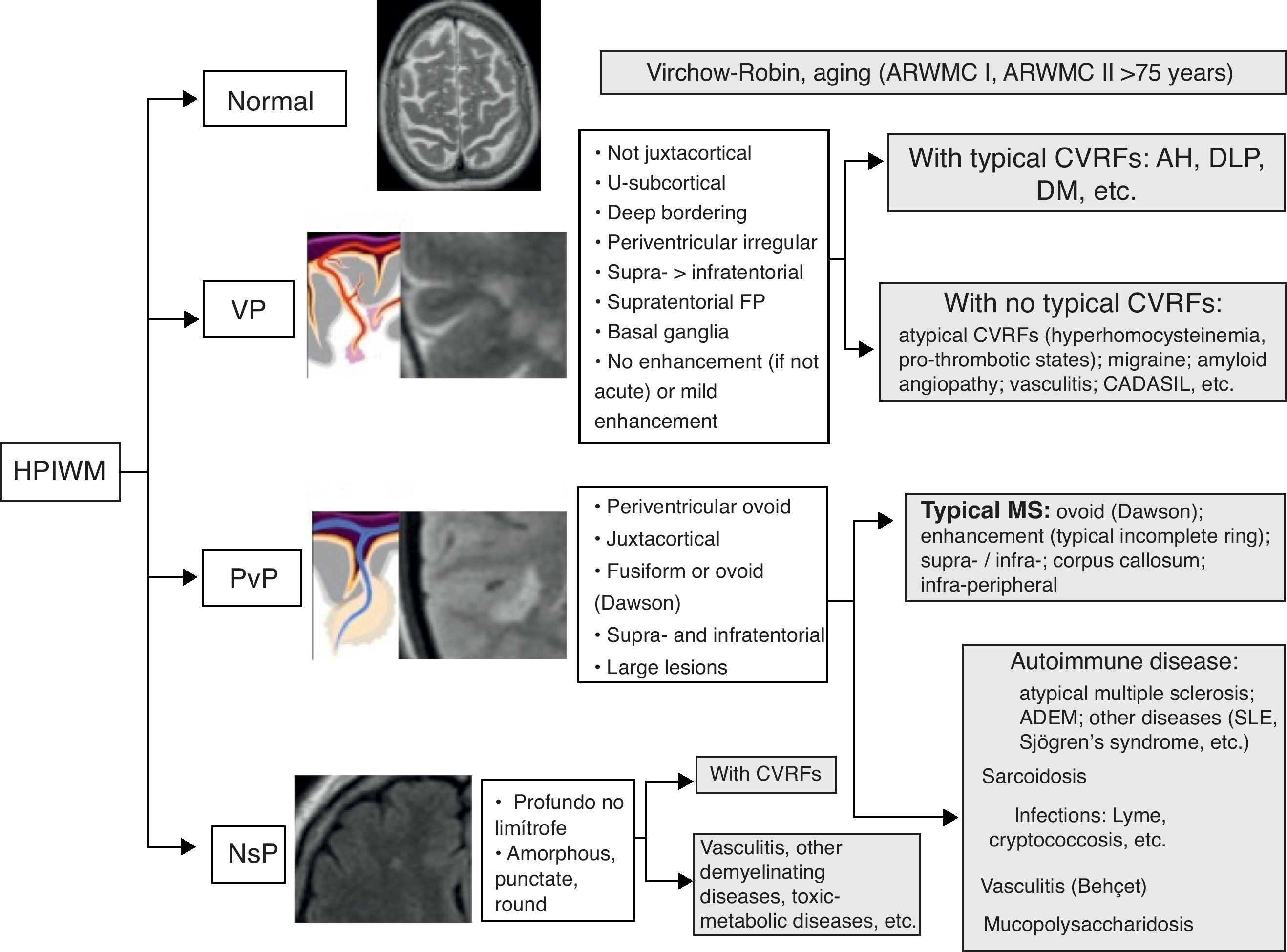
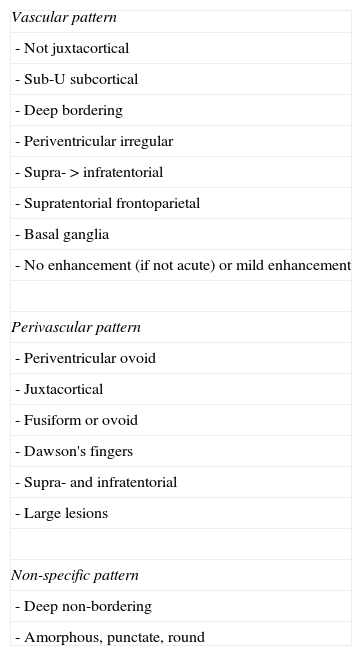
The present article attempts, using an anatomical approach to the brain circulation, as well as from histopathology correlation studies, to simplify the task of interpreting these images from the description of the three main patterns of hyperintense punctiform images in the white matter: vascular pattern, which corresponds to microvascular lesions; perivascular pattern, which represents inflammatory disease of which the paradigm is multiple sclerosis; and a non-specific pattern, which has to be a microvascular disease.

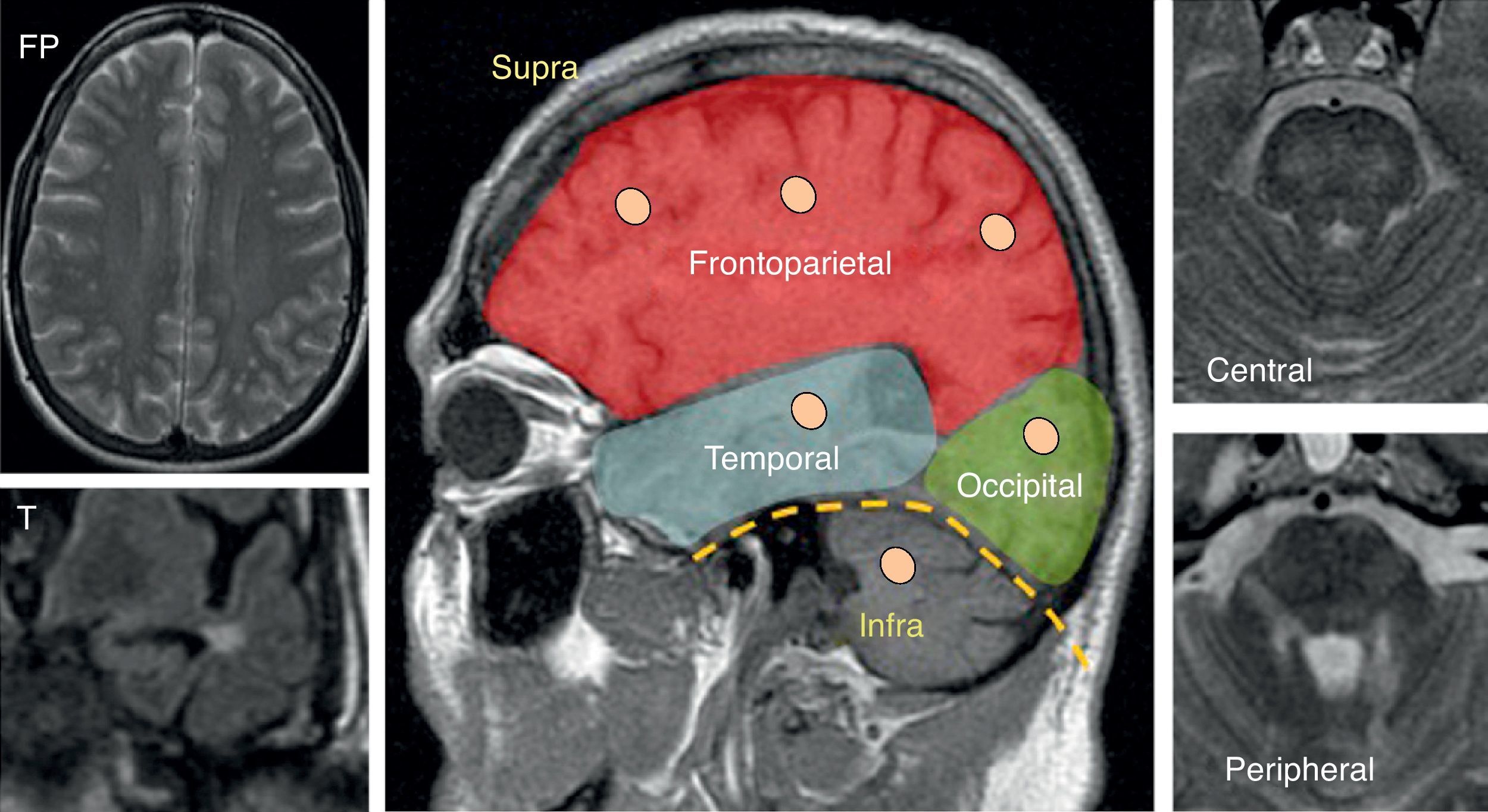
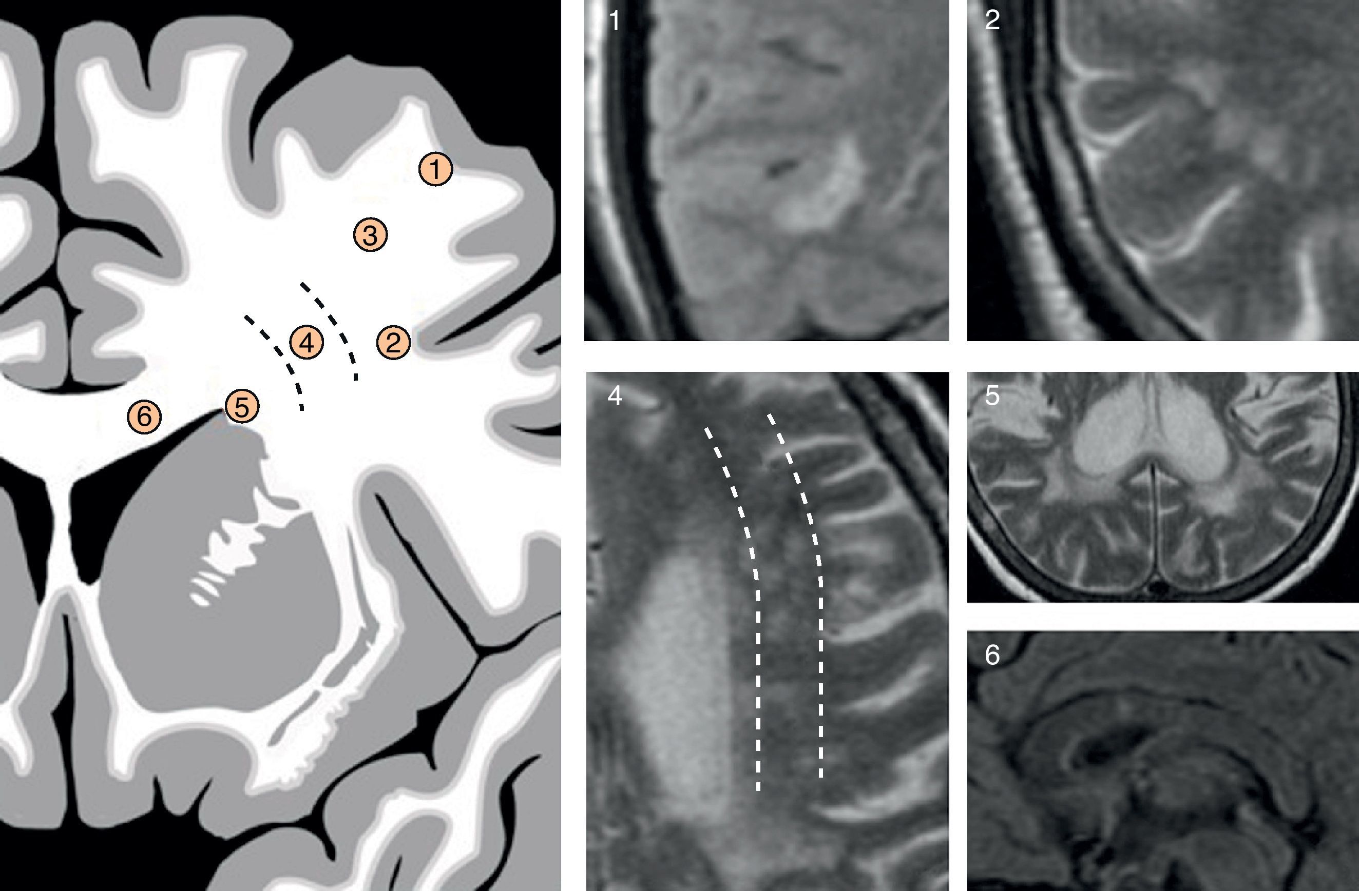
From the various semiological elements in the MR images, a predominant pattern can be determined in each case and, in this way, helps in the differential diagnosis.

Keywords:
White matter
Magnetic resonance
Leukoaraiosis
Multiple sclerosis
CADASIL
Vasculitis
Resumen

La presencia de múltiples imágenes puntiformes hiperintensas en la sustancia blanca (IPHSB) en las secuencias de resonancia magnética (RM) ponderadas en T2 es un hallazgo muy frecuente y, en ocasiones, un reto diagnóstico para el radiólogo.

Este artículo pretende, a través de una aproximación a la anatomía de la microcirculación cerebral, así como a estudios de correlación anatomopatológica, simplificar la tarea de interpretación de estas imágenes a partir de la descripción de tres principales patrones de presentación de IPHSB: patrón vascular (PV), que corresponde a lesiones microvasculares, patrón perivascular (PpV), que representa a la enfermedad inflamatoria cuyo paradigma es la esclerosis múltiple (EM), y patrón inespecífico (PI), que suele deberse a enfermedad microvascular.

A partir de varios elementos semiológicos en las imágenes de RM se puede determinar un patrón predominante en cada caso y, de este modo, acotar el diagnóstico diferencial.

Palabras clave:
Sustancia blanca
Resonancia magnética
Leucoaraiosis
Esclerosis múltiple
CADASIL
Vasculitis

Article

These are the options to access the full texts of the publication Radiología (English Edition)
Subscriber
Subscriber

If you already have your login data, please click here .

If you have forgotten your password you can you can recover it by clicking here and selecting the option “I have forgotten my password”
Purchase
Purchase article

Purchasing article the PDF version will be downloaded

Price 19.34 €

Purchase now
Contact
Phone for subscriptions and reporting of errors
From Monday to Friday from 9 a.m. to 6 p.m. (GMT + 1) except for the months of July and August which will be from 9 a.m. to 3 p.m.
Calls from Spain
932 415 960
Calls from outside Spain
+34 932 415 960
E-mail
Article options
Tools
es en pt

¿Es usted profesional sanitario apto para prescribir o dispensar medicamentos?

Are you a health professional able to prescribe or dispense drugs?

Você é um profissional de saúde habilitado a prescrever ou dispensar medicamentos