


To study the differences in vascular image quality, bone subtraction, and dose of radiation of dual energy CT angiography of the supraaortic trunks using different tube voltages.
Materials and methodsWe reviewed the CT angiograms of the supraaortic trunks in 46 patients acquired with a 128-slice dual source CT scanner using two voltage protocols (80/140kV and 100/140kV). The “head bone removal” tool was used for postprocessing. We divided the arteries into 15 segments. In each segment, we evaluated the image quality of the vessels and the effectiveness of bone removal in multiplanar reconstructions (MPR) and in maximum intensity projections (MIP) with each protocol, analyzing the trabecular and cortical bones separately. We also evaluated the dose of radiation received.
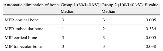
ResultsOf the 46 patients, 13 were studied using 80/140kV and 33 with 100/140kV. There were no significant differences between the two groups in age or sex. Image quality in four segments was better in the group examined with 100/140kV. Cortical bone removal in MPR and MIP and trabecular bone removal in MIP were also better in the group examined with 100/140kV. The dose of radiation received was significantly higher in the group examined with 100/140kV (1.16mSv with 80/140kV vs 1.59mSv with 100/140kV).
ConclusionUsing 100/140kV increases the dose of radiation but improves the quality of the study of arterial segments and bone subtraction.
Estudiar las diferencias en la calidad de imagen vascular, la capacidad de eliminar el hueso y la dosis de radiación de la angio-TC de troncos supraaórticos con la técnica de energía dual utilizando dos diferentes potenciales del tubo.
Material y métodosSe revisaron retrospectivamente los estudios de angio-TC de troncos supraaórticos realizados a 46 pacientes con un equipo de TC de doble fuente de 128 cortes, utilizando dos protocolos de voltaje diferente (80/140kV y 100/140kV). El postproceso se hizo con la herramienta “head bone removal”. Las arterias se dividieron en 15 segmentos. En ellos se evaluó la calidad de los vasos y la capacidad de eliminar el hueso en imágenes multiplanares (MPR) y de proyección de máxima intensidad (MIP) con cada protocolo, analizando de forma separada los huesos trabecular y cortical. También se evaluó la dosis de radiación recibida.
ResultadosSe realizaron 13 estudios con 80/140kV y 33 con 100/140kV, sin diferencias significativas entre los grupos en edad y sexo. Las diferencias fueron significativas en la calidad de los vasos en 4 segmentos, mayor en el grupo de 100/140kV. También en este grupo fue mejor la eliminación automática de hueso cortical en MPR y MIP, y del trabecular en las imágenes MIP. La dosis de radiación (1,16mSv con 80/140kV y 1.59mSv con 100/140kV) fue significativamente mayor en el grupo de 100/140kV.
ConclusiónEl potencial 100/140kV incrementa la dosis de radiación, pero también mejora la calidad del estudio por segmentos arteriales y la eliminación de hueso.

