Buscar en
Archivos de la Sociedad Española de Oftalmología (English Edition)
Toda la web
Inicio Archivos de la Sociedad Española de Oftalmología (English Edition) Eyelid reconstruction in a child with xeroderma pigmentosum
Journal Information
Vol. 91. Issue 9.
Pages 439-441 (September 2016)
Share
Share
Download PDF
More article options
Visits
244
Vol. 91. Issue 9.
Pages 439-441 (September 2016)
Short communication
Eyelid reconstruction in a child with xeroderma pigmentosum
Reconstrucción palpebral en niño con xeroderma pigmentoso
Visits
244
B. Sánchez Cañal, N.E. Monts Cambero
Corresponding author
neleb_pola03@hotmail.com

Corresponding author.
, R. Carrión Campo, J.D. Saldaña Garrido
Unidad de Oculoplastia, Servicio de Oftalmología, Hospital General Universitario de Alicante, Alicante, Spain
This item has received
Article information
Abstract
Full Text
Bibliography
Download PDF
Statistics
Figures (4)
Show moreShow less
Abstract
Clinical case

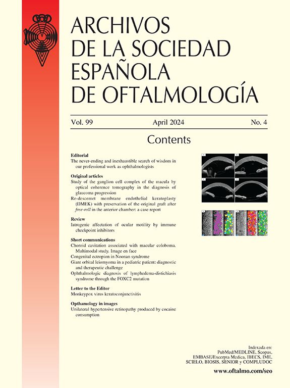
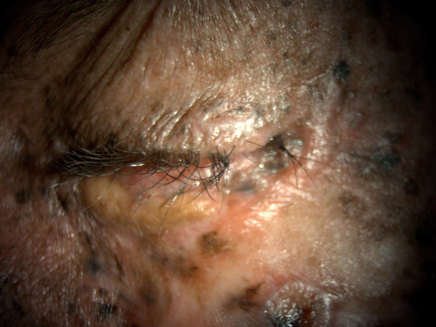
Seven-year-old male patient, affected by xeroderma pigmentosum (XP) who was referred to ophthalmology due to the presence of numerous tumors in his left eye that affected the eyelids and conjunctiva.

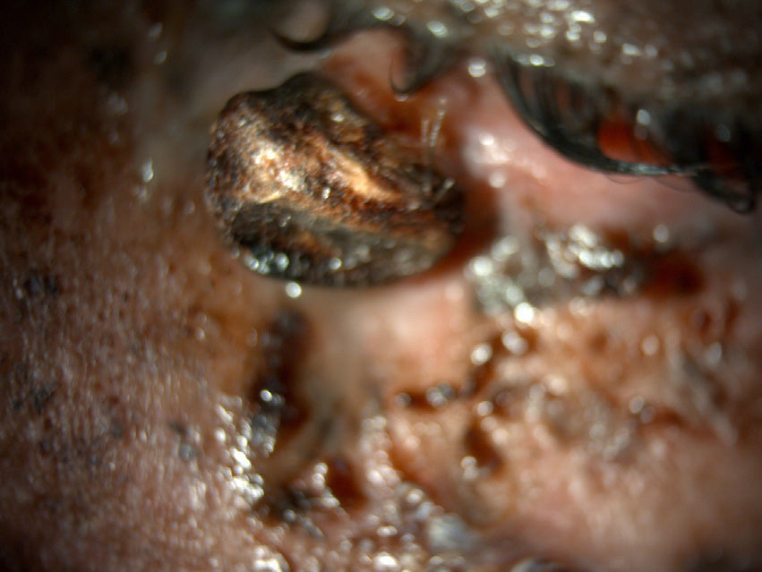
He had a tumor in his lower eyelid that affected the free edge, creating a secondary ectropion, and several lesions with a melanocytic appearance in conjunctiva.

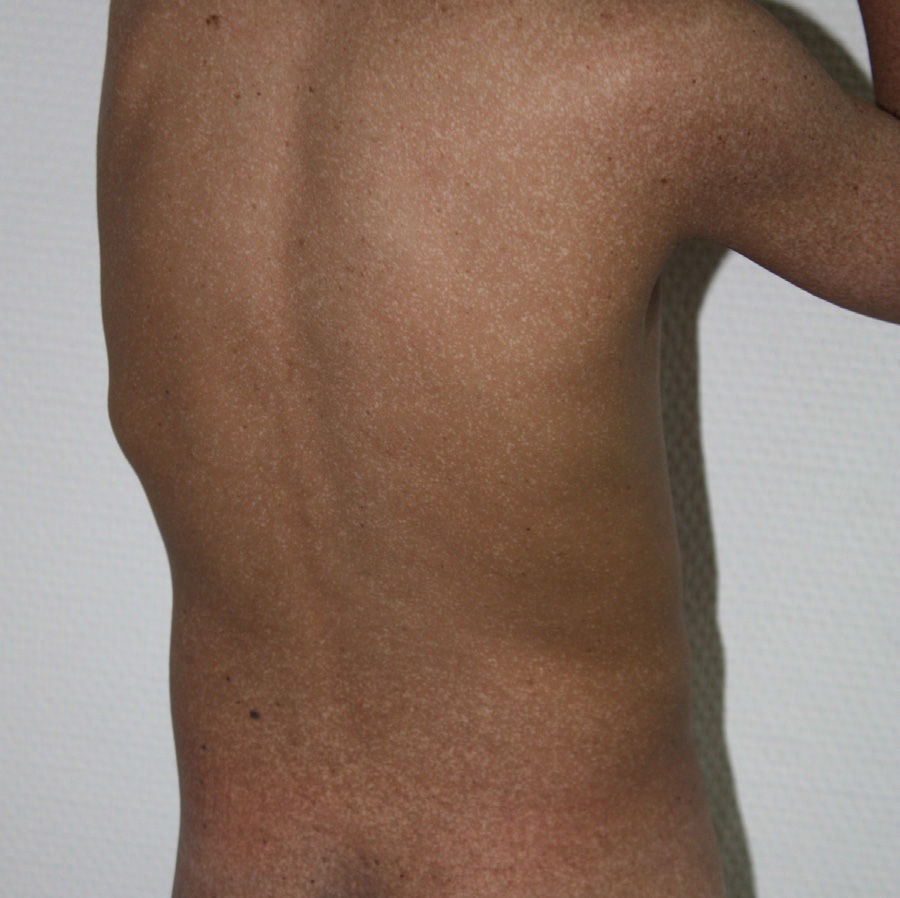
A resection of the eyelid tumor and the conjunctival lesions were performed by placing a skin graft and amniotic membrane, respectively, as covering.

Discussion

XP patients have high probability of developing eye tumors. A thorough ophthalmic examination is necessary to establish an early diagnosis.

Keywords:
Eyelid tumor
Graft
Amniotic membrane
Eyelid
Conjunctiva
Resumen
Caso clínico

Paciente de 7 años, varón, diagnosticado de xeroderma pigmentoso (XP), remitido a oftalmología por presencia de múltiples tumoraciones en el ojo izquierdo, que afectan a párpados y conjuntiva.

Presenta una tumoración en el párpado inferior, que afecta al borde libre, originando un ectropión secundario y varias lesiones de aspecto melanocítico en conjuntiva.

Se realiza resección quirúrgica de la tumoración palpebral y las lesiones conjuntivales colocando un injerto de piel y de membrana amniótica, respectivamente como recubrimiento.

Discusión

Los pacientes con XP tienen una alta probabilidad de desarrollo tumoraciones oculares. Es necesaria una exploración oftalmológica exhaustiva para establecer un diagnóstico precoz.

Palabras clave:
Tumor palpebral
Injerto
Membrana amniótica
Párpado
Conjuntiva

Article

These are the options to access the full texts of the publication Archivos de la Sociedad Española de Oftalmología (English Edition)
Subscriber
Subscriber

If you already have your login data, please click here .

If you have forgotten your password you can you can recover it by clicking here and selecting the option “I have forgotten my password”
Subscribe
Subscribe to

Archivos de la Sociedad Española de Oftalmología (English Edition)

Purchase
Purchase article

Purchasing article the PDF version will be downloaded

Price 19.34 €

Purchase now
Contact
Phone for subscriptions and reporting of errors
From Monday to Friday from 9 a.m. to 6 p.m. (GMT + 1) except for the months of July and August which will be from 9 a.m. to 3 p.m.
Calls from Spain
932 415 960
Calls from outside Spain
+34 932 415 960
E-mail
Article options
Tools
es en pt

¿Es usted profesional sanitario apto para prescribir o dispensar medicamentos?

Are you a health professional able to prescribe or dispense drugs?

Você é um profissional de saúde habilitado a prescrever ou dispensar medicamentos