


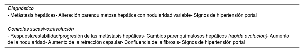
La pseudocirrosis es un diagnóstico radiológico que refleja alteraciones morfológicas hepáticas que simulan la cirrosis en los pacientes oncológicos, sin marcadores histopatológicos de cirrosis verdadera. Su diagnóstico, basado casi exclusivamente en pruebas de imagen, tiene una importante implicación pronóstica, ya que aumenta la morbimortalidad y el riesgo de desarrollar encefalopatía, hemorragia por varices esofágicas/gástricas o insuficiencia/fallo hepático. No obstante, a pesar de su trascendencia, todavía son muchos los interrogantes sobre la fisiopatología, los factores predisponentes y el manejo de esta entidad.
En el contexto de un paciente oncológico con metástasis hepáticas la presencia de alteraciones morfoestructurales hepáticas como la retracción y la nodularidad de la superficie hepática, la atrofia parenquimatosa y/o la hipertrofia del lóbulo caudado deben poner en alerta al radiólogo.
Esta actualización tiene como objetivo presentar el conocimiento actual y la importancia de esta entidad, mostrando sus principales características clínicas y de imagen.
Pseudocirrhosis is a radiological diagnosis that refers to liver morphological changes in oncologic patients that mimic cirrhosis, without histopathological markers of true cirrhosis. Its diagnosis, based almost exclusively on imaging studies, has significant prognostic implications, as it is associated with increased morbidity and mortality, and a higher risk of developing encephalopathy, bleeding from esophageal/gastric varices, or liver insufficiency/failure. However, despite its clinical relevance, many questions remain about the pathophysiology, predisposing factors and management of this entity.
In oncologic patients with liver metastases, the presence of liver morphological alterations, such as surface retraction and nodularity, parenchymal atrophy and/or hypertrophy of the caudate lobe, should alert the radiologist to the possibility of pseudocirrhosis.
This update aims to present the current knowledge and importance of this entity, highlighting its key imaging and clinical features.


