


The aim of this report was to describe the management of periodontitis with hypercementosis.
MethodsA 30-year-old male patient was referred from the Department of Conservation with complaints of pain during root canal filling. The tooth was traumatized 3 years ago. An intraoral assessment showed teeth #11 with grade 2 mobility, probing depth (mesial=6mm, distal: 7mm, buccal: 5mm, palatal: 5mm), extrusion and discolorization. Radiography examination has shown lateral bone destruction and hypercementosis. Regenerative treatment was carried out to resolve the case.
ResultsThe treatment was done using a bone graft combination with Platelet Rich Fibrin (PRF) and has showed improvement after three months evaluation.
ConclusionPeriodontitis with hypercementosis is a rare case. Hence appropriate treatment planning must be done to ensure a good prognosis. Combination of bone graft and platelet-rich fibrin (PRF) in the regenerative periodontal procedure has shown better results after three months evaluation. However, the right diagnosis, appropriate treatment planning, good multidisciplinary collaboration supported by patient cooperation can guarantee the success of treatment.
Hypercementosis (cemental hyperplasia) is a nonneoplastic deposition of excessive cementum which fuses with normal radicular cementum and can involve one tooth, several teeth or the entire tooth. In general, the causes of hypercementosis are idiopathic, but some conditions that are considered to be related to hypercementosis are tooth supra-eruption due to loss of antagonists, chronic periapical lesions, traumatic occlusion and systemic diseases such as Paget's disease, toxic goiter, acromegaly, and gigantism.1,2
Periodontitis is an etiological factor for hypercementosis formation that has never been reported in a review, but periodontal tissues activities such as absorbing, directing and distributing the force received by the tooth can move periodontal fibers and other components of the extracellular matrix, and change the shape of cementoblasts through the cytoskeleton. Cytoskeleton deformation can produce stimuli needed for cellular pressure, resulting in increased mediator release and synthesis of cementum matrix on the root surface.2,3
Periodontitis with hypercementosis reported in this case occur in teeth that were traumatized three years ago, lead to necrotic teeth and over time become supraposition, hence hypercementosis that occurred in this case due to chronic periapical lesions.3
The ultimate goal of periodontal therapy are the elimination of inflammatory process, prevention of periodontal disease progress and regeneration of lost periodontal tissues.4 The treatment success is associated with patient compliance. The patient prognosis is very dependent on patient attitude, esthetic needs, willingness, and ability to maintain oral hygiene.5
Based on clinical examination and radiography it was decided to perform a regenerative periodontal treatment procedure using a bone graft combined with Platelet Rich Fibrin. The objective of this report was to describe the management of periodontitis with hypercementosis using periodontal regenerative procedure through a surgical approach using bone graft combined with Platelet Rich Fibrin.3
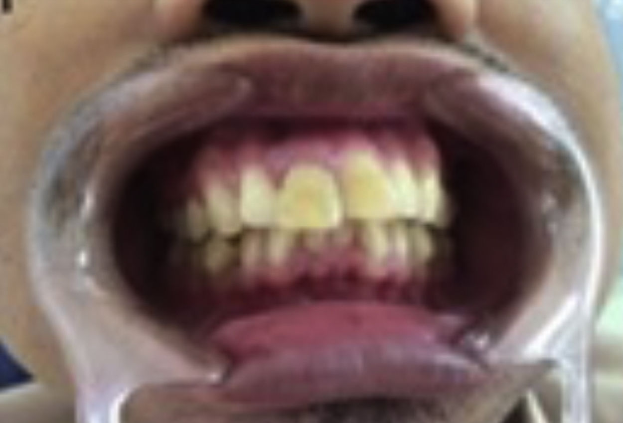
Case reportThe 30-year-old male patient was referred from the Department of Conservation with complaints of mobile tooth and pain during root canal filling. The tooth was traumatized 3 years ago. An intra-oral examination showed #11 with grade 2 mobility, probing depth (mesial: 6mm, distal: 7mm, buccal: 5mm, palatal: 5mm) with extrusion and discolorization of the tooth (Fig. 1).

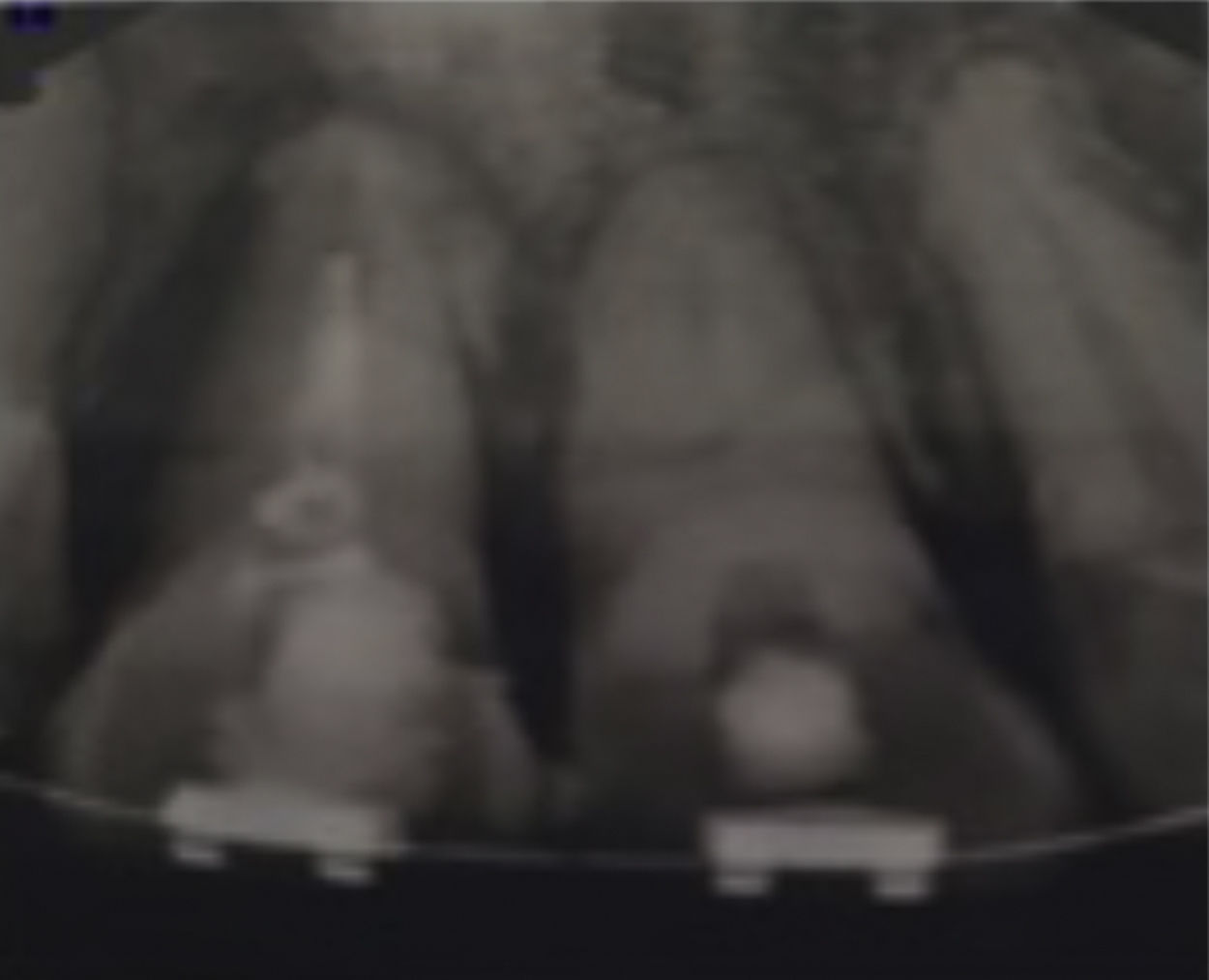
Radiography examination showed lateral bone destruction and hypercementosis in the apical third of the mesial part (Fig. 2a and b). The patient was healthy systematically.

The treatment plan for this case includes non-surgical phase (dental health education, scaling and consult to Department of Orthodontic to overcome extrusion problem), surgical phase (regenerative periodontal), and then restorative phase. Informed consent was obtained before treatment procedure.
Platelet-rich fibrin (PRF) preparation was done based on Choukroun et al. before surgical phase. Intravenous blood was taken then stored in a sterile tube without anticoagulant and immediately centrifuged at a speed of 2700rpm for 12min.6
In the surgical procedure, extraoral and intraoral disinfection were performed with a 2% povidone-iodine solution (Fig. 3) then infiltrated local anesthesia with lidocaine HCL anesthetic and epinephrine 1:80,000 (Fig. 4) was administrated. Sulcular and vertical incisions were performed using no.15 blade (Fig. 5) and the flap was exposed using a rasparatorium (Fig. 6a and b).


Curettage of granulation and necrotic tissue was carried out using Gracey curettage and followed with root planing (Fig. 7a–c).

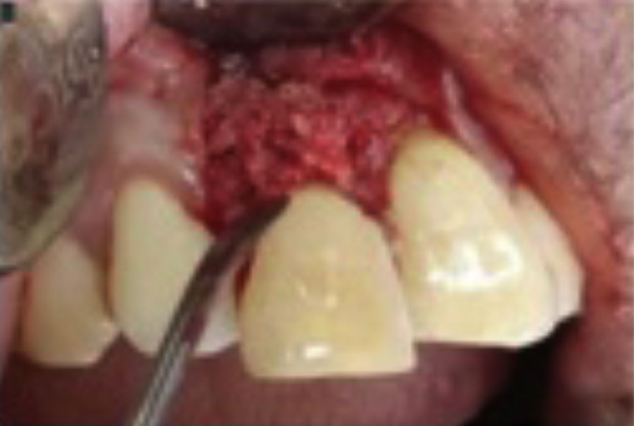
Bone graft was placed on a bone defect and PRF was placed on top of it (Figs. 8 and 9). Flap reposition was done with interrupted suture technique (Fig. 10). The periodontal dressing was placed as the final step (Fig. 11).


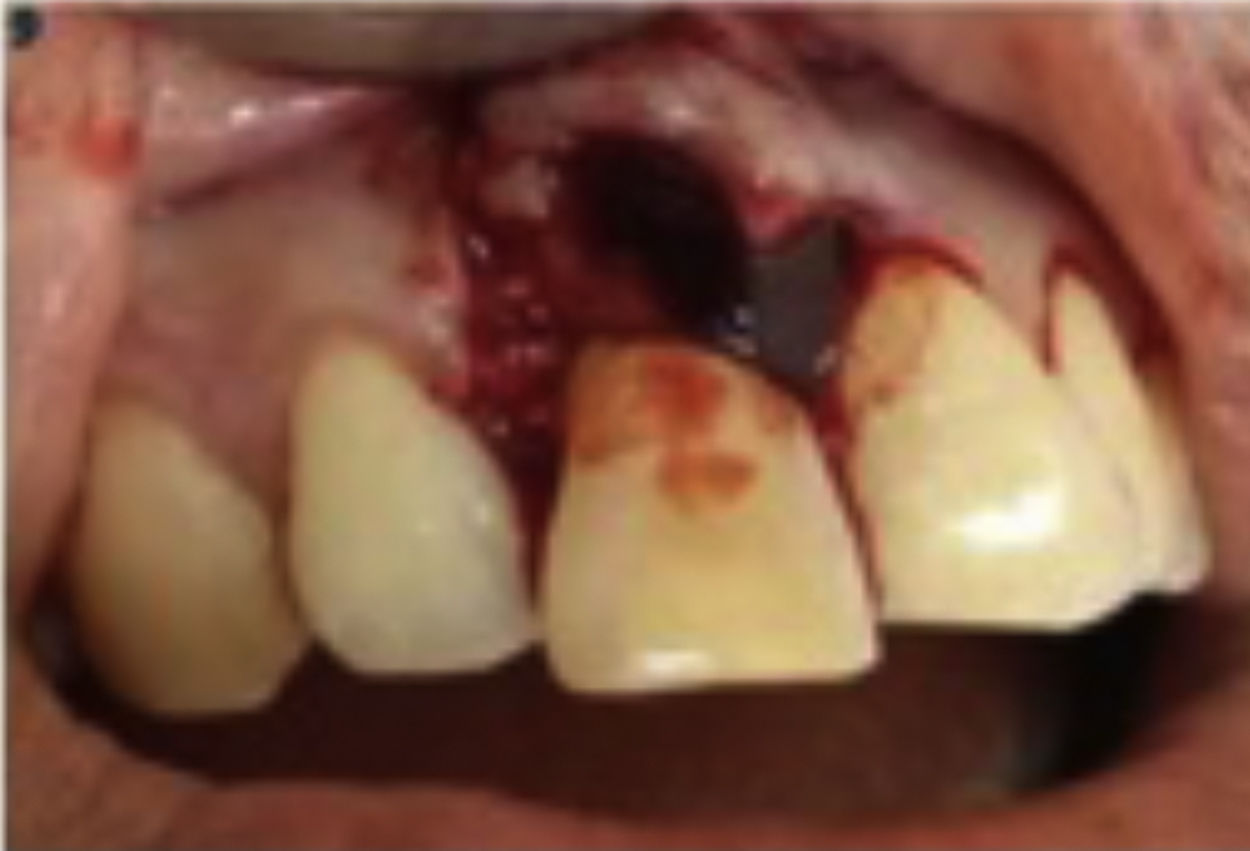
Postoperative instructions, amoxicillin 500mg, mefenamid acid, vitamin C and minocep rinse were prescribed. One week after the procedure, there was little erythema on the sutured area. There was no flap dehiscence or suppuration (Fig. 12a). Three months follow up after procedure showed significant improvement (Fig. 12b). Periapical radiography showed significant bone formation characterized by a decrease in radiolucency and hypercementosis area (Fig. 13).


The patient came back to the Department of Conservation more than one year after the procedure and with complaining tooth mobility and pain during biting food. Radiographic examination showed radiolucency of distal root and root canal filling with improper work length (Fig. 14), lead to disruption of periodontal tissues healing due to infection agent spreading from the pulp chamber.
Discussion
Complications of injuries involving teeth and their supporting structures include pulp necrosis, ankylotic root resorption, inflammatory root resorption, and pulp canal obliteration.7 In this case, the necrotic pulp was caused by blood flow that damage the blood vessels. The necrotic pulp may be infected by bacteria from the gingival margin, thus lead to apical periodontitis. During endodontic treatment, the instrument can be pushed through the apex or side of the root, damaging the periodontal membrane and carrying infected agents from the pulp chamber to lesion.8
Periodontitis with hypercementosis was rarely reported in reviews, but hypercementosis was associated with chronic periapical lesions such as periapical granuloma. On the edge of granuloma, root cementum increases its thickness in response to inflammatory stimuli arising from extensive tissue and cellular mediators. In some cases with clinical periapical lesions extracted, there was hypercementosis found on the lateral part of the tooth.2,3
Management of this case includes endodontic treatment, regenerative treatment of periodontal tissues, and orthodontics to treat extrusion and also function as for splinting. Regenerative treatment of periodontal tissues used bone graft combined with PRF. The result of clinical and radiographs examination showed improvement on 3-month post-treatment evaluation. The combination of bone graft and PRF was expected to provide benefits such as promoting wound healing, bone growth and maturation, graft stabilization, wound sealing and hemostasis, improving properties of graft material Platelet Rich Factor (PRF) membrane to release growth factors for at least one week and up to 28 days, act as anti-infectious agent and aid in immune regulation.6,9,10
PRF can also function as a biological connector between bone particle which releases growth factors such as PDGF, and TGF. These growth factors help in protein synthesis of bone tissue, stimulate angiogenesis, and increase the formation of bone woven.
ConclusionPeriodontitis with hypercementosis is a rare case and in this case report, hypercementosis was occurred due to apical periodontitis from the necrotic tooth. The treatment, in this case, includes endodontics, regenerative periodontal procedures, and orthodontics which can also function as splints. A regenerative periodontal procedure that was carried out, in this case, used a bone graft combination with Platelet Rich Fibrin (PRF). The combination of these two ingredients showed better results after three-month evaluation. The periapical radiograph showed the reduced radiolucent area of bone defect. However, the right diagnosis, appropriate treatment planning, and good multidisciplinary collaboration supported by patient cooperative attitudes can guarantee the success of treatment.
Conflict of interestThe authors declare no conflict of interest.
Peer-review under responsibility of the scientific committee of the International Conference on Women and Societal Perspective on Quality of Life (WOSQUAL-2019). Full-text and the content of it is under responsibility of authors of the article.
