


El tratamiento de los quistes hepáticos requiere del diagnóstico diferencial de quiste simple hepático (QSH) de la neoplasia mucinosa quística (NMQ) hepática. Las características radiológicas no son patognomónicas. Algunos estudios han sugerido la utilidad de los marcadores tumorales (MKT) intraquísticos.
MétodosAnálisis retrospectivo de base de datos prospectiva incluyendo pacientes diagnosticados de QSH sintomático desde el 2003 hasta el 2021. El objetivo del estudio es evaluar los resultados del tratamiento de los QSH sintomáticos y analizar la utilidad de la determinación de «carcinoembryonic antigen» (CEA) y «carbohydrate antigen» CA 19.9 intraquísticos.
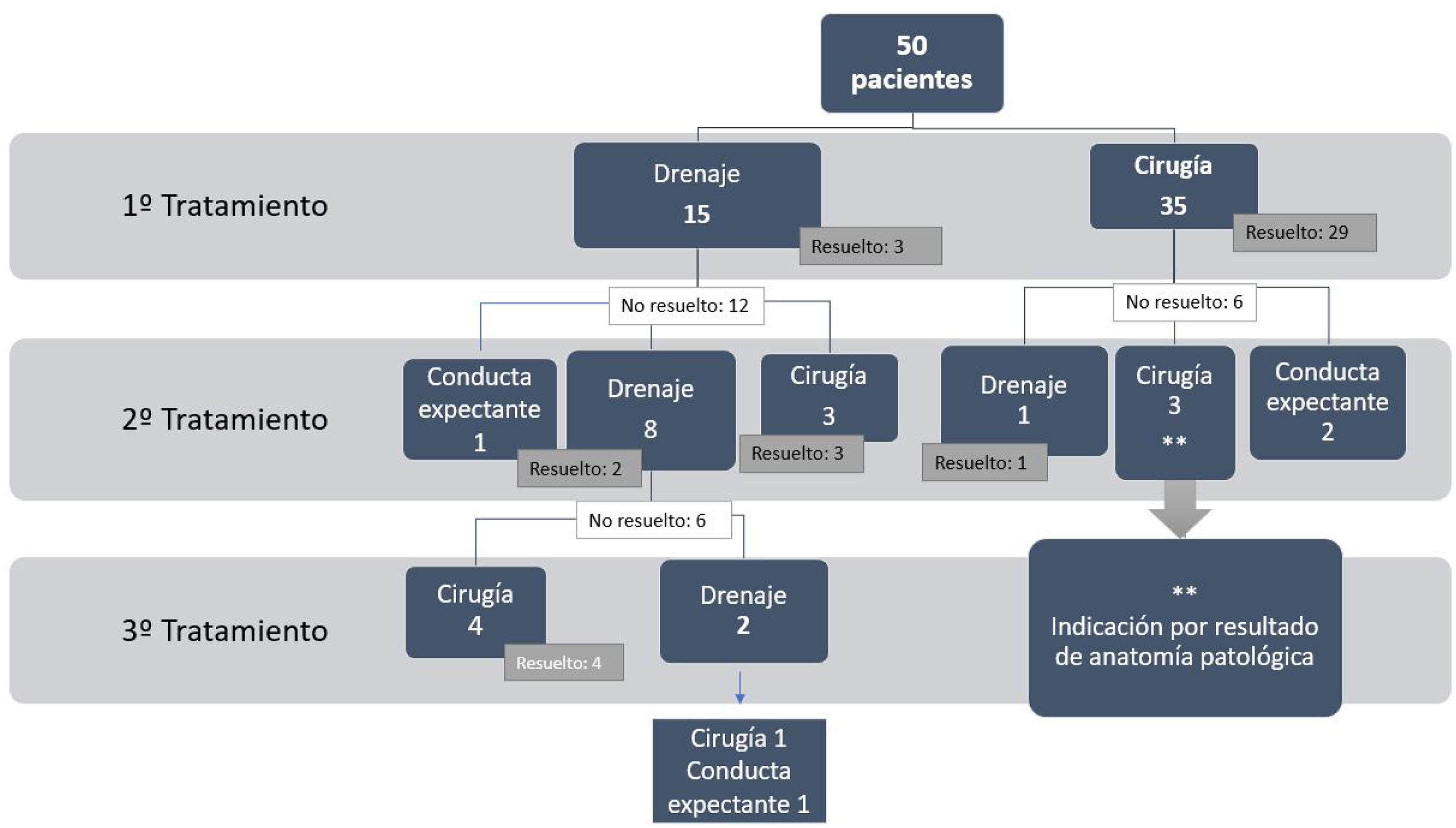
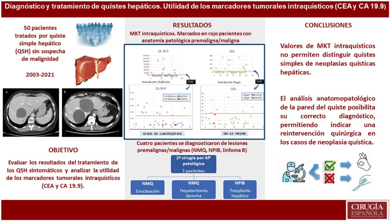
ResultadosSe incluyeron 50 pacientes tratados por quiste sintomático. En 15 pacientes el primer tratamiento fue el drenaje percutáneo. En 35 pacientes se realizó fenestración laparoscópica. Cuatro pacientes se diagnosticaron de lesiones premalignas/malignas (NMQ, NPIB, linfoma B); tres de ellos requirieron una segunda cirugía tras la fenestración y el diagnóstico anatomopatológico.
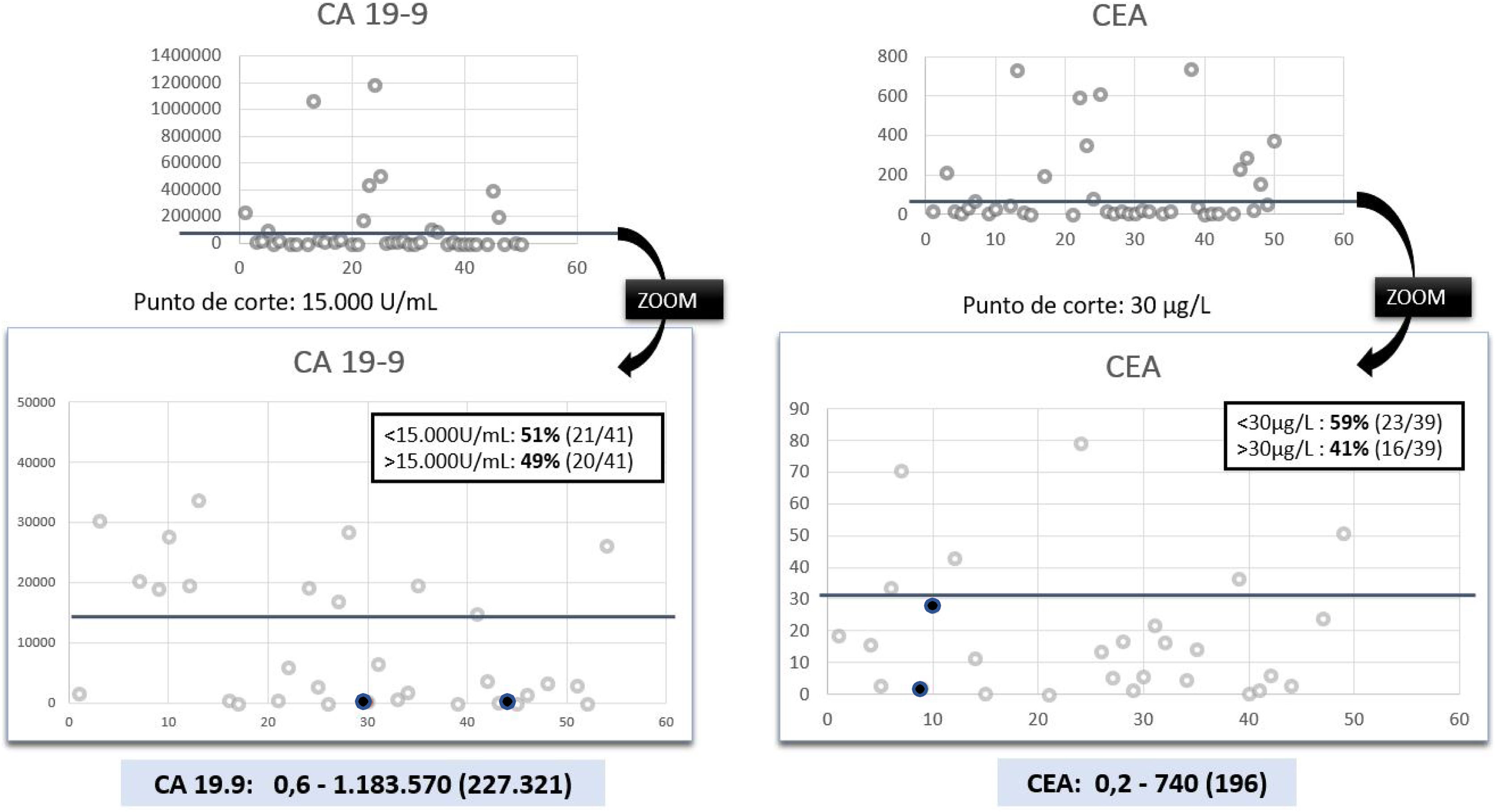
La mediana de los valores de CEA y CA- 19.9 fue de 196μg/L y 227.321U/mL respectivamente. Los pacientes con lesiones premalignas no tuvieron valores elevados de MKT. El valor predictivo positivo fue del 0% en ambos MKT, y el valor predictivo negativo fue de 89% y 91% respectivamente.
ConclusionesLos valores de CEA y CA 19.9 intraquísticos no permiten distinguir los QSH de las NMH. El tratamiento más resolutivo de los QSH sintomáticos es la fenestración quirúrgica. El análisis anatomopatológico de la pared del quiste posibilita su correcto diagnóstico, permitiendo indicar una reintervención quirúrgica en los casos de NMQ.
To decide treatment of hepatic cysts diagnosis between simple hepatic cyst (SHC) and cystic mucinous neoplasm (CMN). Radiological features are not pathognomonic. Some studies have suggested the utility of intracystic tumor markers.
MethodsRetrospective analysis of our prospective database including patients treated due to symptomatic SHC from 2003 to 2021. The aim of the study was to evaluate the results of treatment of symptomatic SHC and the usefulness of the determination of intracystic “carcinoembryonic antigen” (CEA) and “carbohydrate antigen” CA 19.9.
ResultsFifty patients diagnosed and treated for symptomatic SHC were included. In 15 patients the first treatment was percutaneous drainage. In 35 patients the first treatment was laparoscopic fenestration. Four patients were diagnosed of premalignant or malignant liver cystic lesions (MCN, IPMN, and lymphoma B); three of them required surgery after initial fenestration and pathological diagnosis.
Median CEA and CA 19.9 were 196μg/L and 227.321U/mL, respectively. Patients with malignant or premalignant pathology did not have higher levels of intracystic tumor markers. Positive predictive value was 0% for both markers, and negative predictive value was 89% and 91%, respectively.
ConclusionValues of intracystic tumor markers CEA and CA 19.9 do not allow distinguishing simple cysts from cystic liver neoplasms. The most effective treatment for symptomatic simple liver cysts is surgical fenestration. The pathological analysis of the wall of the cysts enables the correct diagnosis, allowing to indicate a surgical reintervention in cases of hepatic cyst neoplasia.

Artículo

Comprando el artículo el PDF del mismo podrá ser descargado
Precio 19,34 €
Comprar ahora

