Buscar en
Archivos de la Sociedad Española de Oftalmología (English Edition)
Toda la web
Inicio Archivos de la Sociedad Española de Oftalmología (English Edition) Assessment of the biometric modifications of the eye in LASIK during suction
Journal Information
Vol. 98. Issue 8.
Pages 434-439 (August 2023)
Share
Share
Download PDF
More article options
Vol. 98. Issue 8.
Pages 434-439 (August 2023)
Original article
Assessment of the biometric modifications of the eye in LASIK during suction
Evaluación de las modificaciones biométricas del ojo durante la succión del LASIK
J.I. Fernández-Vigoa,b,
Corresponding author
, I. Almorín-Fernández-Vigoc, B. Burgos-Blascob, L. De-Pablo-Gómez-de-Liañoa,d, I. Sánchez-Guillénc,e, A. Macarro-Merinoa, J.Á. Fernández-Vigoa,c,f
a Centro Internacional de Oftalmología Avanzada, Madrid, Spain
b Department of Ophthalmology, Hospital Clínico San Carlos, Instituto de Investigación Sanitaria (IdISSC), Madrid, Spain
c Centro Internacional de Oftalmología Avanzada, Badajoz, Spain
d Department of Ophthalmology, Hospital 12 de Octubre, Madrid, Spain
e Department of Ophthalmology, Hospital Perpetuo Socorro, Badajoz, Spain
f Department of Ophthalmology, Universidad de Extremadura, Badajoz, Spain
Article information
Abstract
Full Text
Bibliography
Download PDF
Statistics
Tables (3)
Table 1. Results of the biometric modifications of the eye during the suction maneuvre in LASIK surgery. Mean ± standard deviation, range.
Table 2. Distribution of the eyes by groups according to the modifications suffered by the different intraocular structures during LASIK suction.
Table 3. Comparative analysis of biometric changes during suction by LASIK in patients in whom both eyes were studied.
Show moreShow less
Abstract
Purpose

To study the biometric modifications of the eyeball during suction in Laser assisted in Situ Keratomileusis (LASIK).
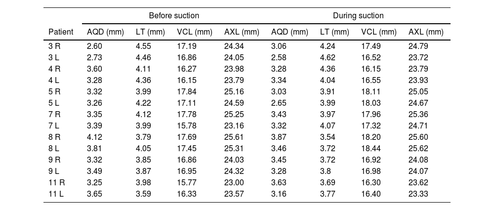
Methods

Observational and cross-sectional study. We studied 43 patients who underwent surgery for myopia and myopic astigmatism. Mean age was 38.3 ± 11.5 years, and 19 were female (44.2%). Conventional LASIK surgery with a manual microkeratome was performed. Before and during the suction maneuvre the following parameters were measured using an 11 Mhz biometric probe: aqueous depth (AQD), lens thickness (LT), vitreous cavity length (VCL) and axial length (AXL). Paired t-test was used to compare the biometric measurements before and during suction.

Results

The mean spherical equivalent refractive error was −4.5 ± 2.3 diopters. During suction, the AQD did not change significantly (p = 0.231). However, AXL and VCL increased by 0.12 mm and 0.22 mm respectively (p = 0.039 and <0.01) and LT decreased by 0.20 mm (p < 0.01). AXL increased in 42% of the eyes and decreased in 16%, VCL increased in 70% of the eyes and decreased in 9%, and the LT was reduced in 67% of the eyes.

Conclusions

Suction maneuvres during LASIK surgery produce changes of little magnitude in the eye globe, mainly a decrease in LT and an increase in VCL and AXL. Therefore, these modifications are expected to produce minimal anatomic alterations.

Keywords:
Biometry
LASIK eye globe suction
Refractive surgery
Myopia
Lens thickness
Vitreous cavity
Resumen
Objetivo

Estudiar las modificaciones biométricas del globo ocular durante la succión en la cirugía de Keratomileusis In Situ asistida por Láser (LASIK).

Métodos

Estudio observacional transversal. Se estudiaron 43 pacientes intervenidos de miopía y astigmatismo miópico. La edad media fue de 38,3 ± 11,5 años, 19 eran mujeres (44,2%). Se realizó una cirugía LASIK convencional con microqueratomo manual. Antes y durante la maniobra de succión se midieron los siguientes parámetros mediante una sonda biométrica de 11 Mhz: profundidad acuosa (AQD), espesor del cristalino (LT), longitud de la cavidad vítrea (VCL) y longitud axial (AXL). Se utilizó la prueba t pareada para comparar las medidas biométricas antes y durante la succión.

Resultados

El equivalente esférico medio fue de -4,5 ± 2,3 dioptrías. Durante la succión, la AQD no aumentó significativamente (p = 0,231). Sin embargo, la AXL y VCL aumentaron en 0,12 mm y 0,2 mm respectivamente (p = 0,039 y <0,01) y LT disminuyó en 0,20 mm (p < 0,01). La AXL aumentó en el 42% de los ojos y disminuyó en el 16%, mientras que la VCL aumentó en el 70% de los ojos y disminuyó en el 9%, y el LT se redujo en el 67% de los ojos.

Conclusiones

Las maniobras de succión durante la cirugía LASIK producen cambios de poca magnitud en el globo ocular, principalmente disminución de LT y aumento de VCL y AXL. Por lo tanto, es esperable que estas modificaciones produzcan alteraciones anatómicas mínimas.

Palabras clave:
Biometría
Succión LASIK
Cirugía refractiva
Miopía
Cristalino
Cavidad vítrea

Article

These are the options to access the full texts of the publication Archivos de la Sociedad Española de Oftalmología (English Edition)
Subscriber
Subscriber

If you already have your login data, please click here .

If you have forgotten your password you can you can recover it by clicking here and selecting the option “I have forgotten my password”
Subscribe
Subscribe to

Archivos de la Sociedad Española de Oftalmología (English Edition)

Purchase
Purchase article

Purchasing article the PDF version will be downloaded

Price 19.34 €

Purchase now
Contact
Phone for subscriptions and reporting of errors
From Monday to Friday from 9 a.m. to 6 p.m. (GMT + 1) except for the months of July and August which will be from 9 a.m. to 3 p.m.
Calls from Spain
932 415 960
Calls from outside Spain
+34 932 415 960
E-mail
Article options
Tools
es en pt

¿Es usted profesional sanitario apto para prescribir o dispensar medicamentos?

Are you a health professional able to prescribe or dispense drugs?

Você é um profissional de saúde habilitado a prescrever ou dispensar medicamentos