
We aimed to compare the detectability of optic disc drusen (ODD), using various non-invasive imaging techniques, including the novel retro-mode imaging (RMI), as well as to analyze the morphological characteristics of ODD on RMI.
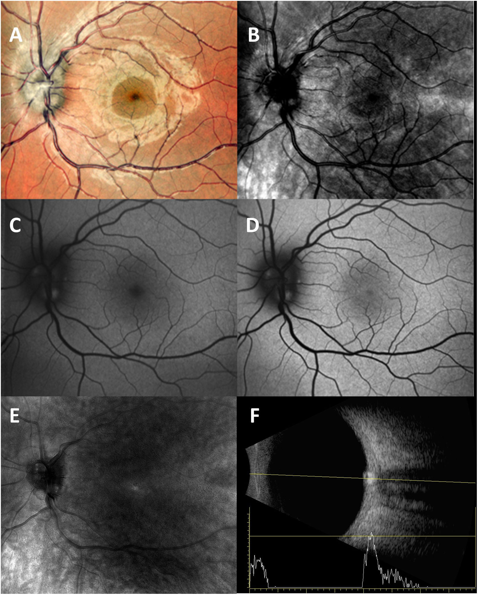
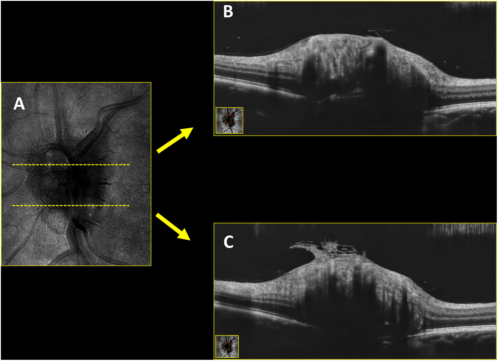
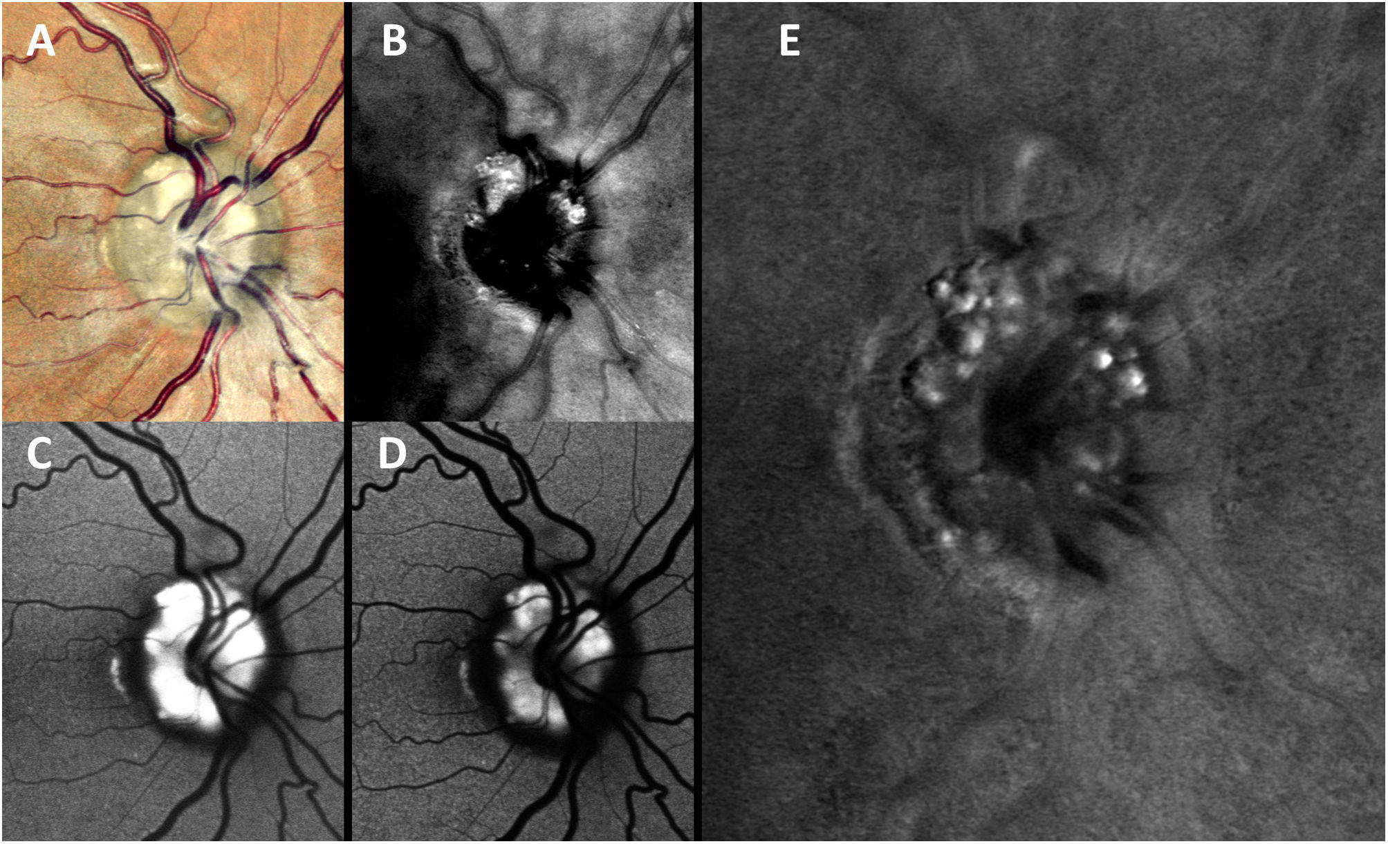
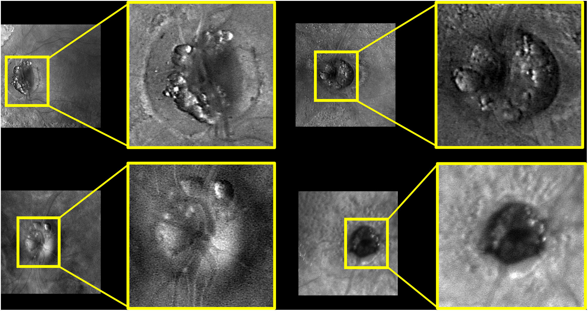
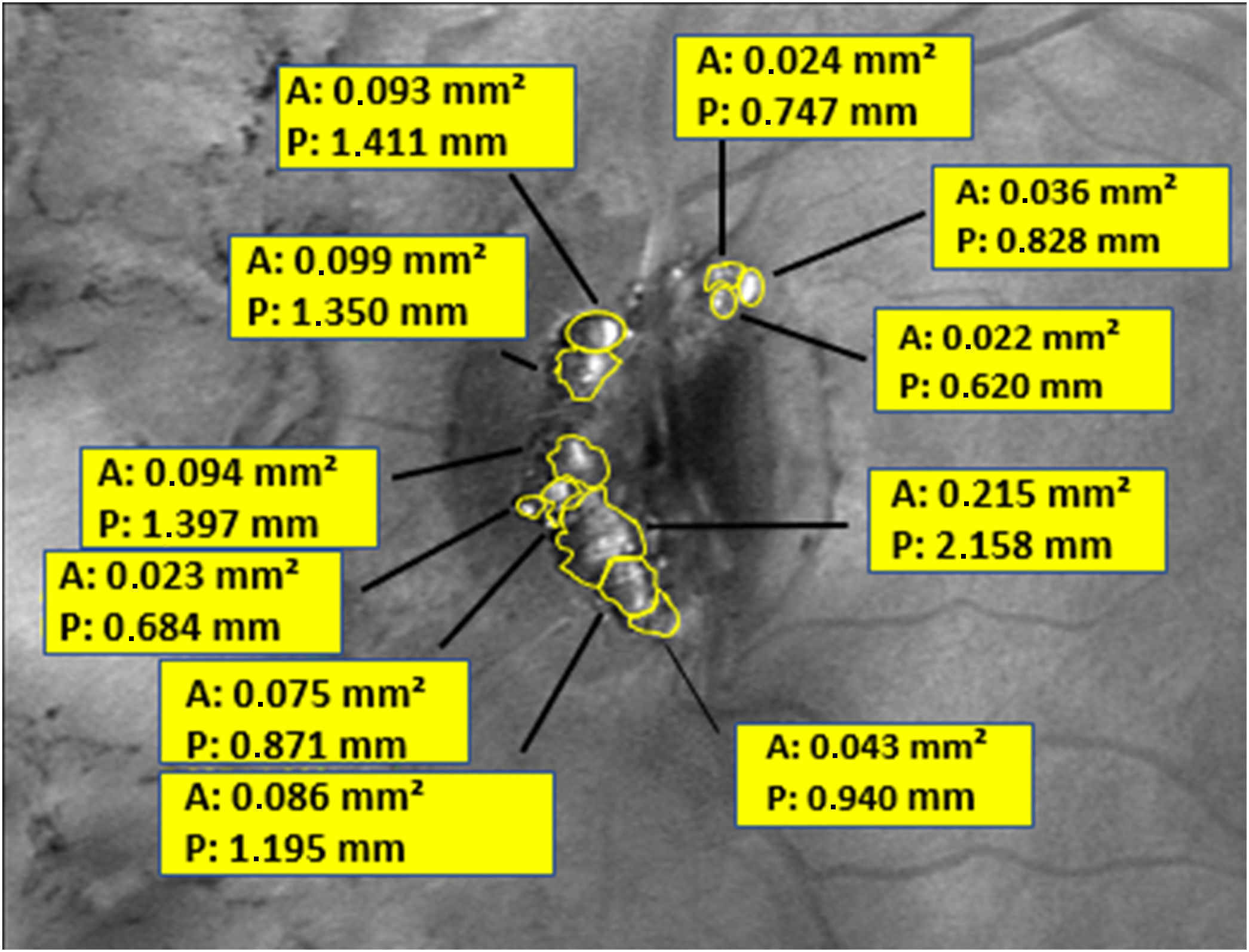
MethodsThis study involved seven patients with bilateral ODD, totaling 14 eyes. Multimodal imaging techniques, including multicolor fundus photography (MC), near-infrared reflectance (NIR), green and blue light fundus autofluorescence (G-FAF and B-FAF, respectively), and RMI were used to examine the eyes. FAF was used as the primary method of identifying ODD, and each method's detection rate was compared by two observers. Quantitative measurements of ODD included the number of ODD visualized by the RMI technique, the perimeter (P) and area (A) of ODD were identified.
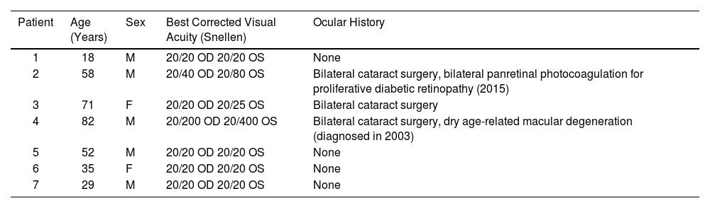
ResultsThe average age of the patients included was 49.28 ± 23.16 years, with five of the seven being men. RMI was able to detect ODD in all cases, with a sensitivity of 100%, compared to MC (sensitivity 60.71%), NIR (sensitivity 60.71%), B-FAF (sensitivity 100%), G-FAF (sensitivity 100%). RMI was the only imaging technique capable of assessing ODD morphology and quantifying ODD.
ConclusionsRMI is a promising imaging modality for diagnosing superficial ODD, providing valuable information on the distribution, location, and size of ODD. We suggest the incorporation of RMI as a complementary tool for diagnosing and monitoring ODD in combination with other multimodal imaging methods.
Nuestro principal objetivo es el de comparar la capacidad para detectar las drusas del disco óptico (ODD) utilizando diversas técnicas de imágenes no-invasivas, incluida la novedosa técnica de imagen de retro-modo (RMI). Como segundo objetivo analizamos las características morfológicas de las ODD bajo esta última técnica.
Materiales y métodosEste estudio incluyó un total de siete pacientes con ODD bilaterales, obteniendo un total de 14 ojos analizados. Se utilizaron técnicas no-invasivas de imágenes multimodales, que incluyeron fotografía multicolor del fondo de ojo (MC), reflectancia en infra-rojo (NIR), autofluorescencia en luz verde y en luz azul (G-FAF y B-FAF, respectivamente), y RMI. La FAF se utilizó como el método principal para el diagnóstico de ODD. Dos observadores realizaron las comparaciones obteniendo las tasas de detección de cada uno de los métodos. Las mediciones cuantitativas de las ODD incluyeron el número, el perímetro (P) y el área (A) de las ODD identificadas mediante la técnica de RMI.
ResultadoLa edad promedio de los pacientes incluidos fue de 49.28 ± 23.16 años, cinco de los siete pacientes fueron de sexo masculino. La técnica de RMI pudo detectar ODD en todos los casos, con una sensibilidad del 100%, en comparación con MC (sensibilidad del 60.71%), NIR (sensibilidad del 60.71%), B-FAF (sensibilidad del 100%), G-FAF (sensibilidad del 100%). RMI fue la única técnica de imagen capaz de evaluar morfológica y cuantitativamente las ODD.
ConclusionesRMI es una prometedora modalidad no-invasiva de imagen para diagnosticar ODD superficiales, proporcionando información valiosa sobre la distribución, ubicación y tamaño de estas, por lo tanto, mediante nuestros resultados sugerimos la incorporación de la novedosa técnica de RMI como una herramienta complementaria para el diagnóstico y seguimiento de ODD en combinación con los otros métodos de imagen multimodales.