To describe the clinical findings and its complications in 2 patients with focal choroidal excavation (FCE).
MethodsA retrospective case-series including 4 eyes of 2 patients with FCE that underwent a comprehensive ophthalmological examination including slit-lamp examination, color fundus photography, spectral-domain optical coherence tomography (SD-OCT), fluorescein angiography (FA), and indocyanine green angiography.
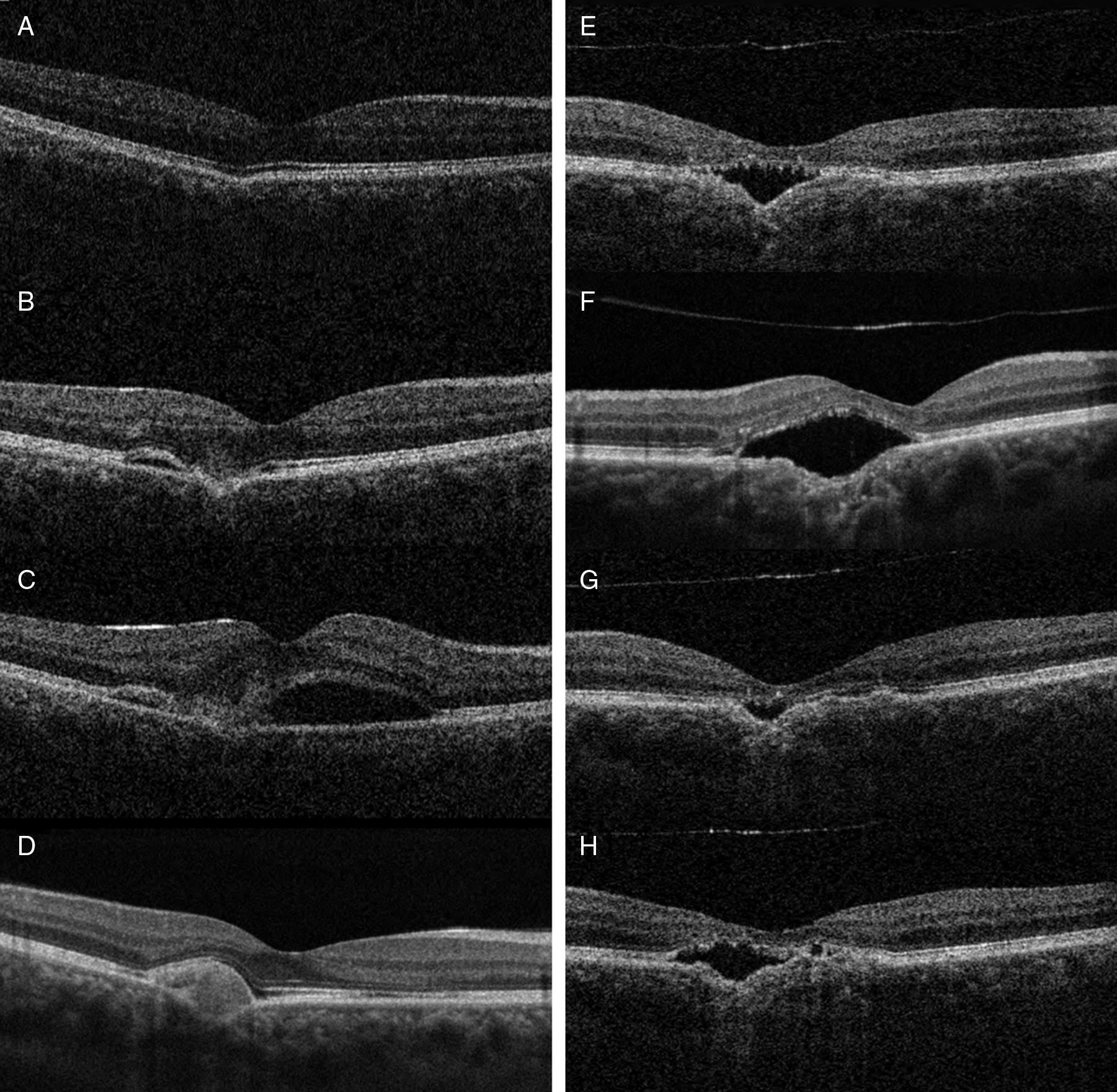
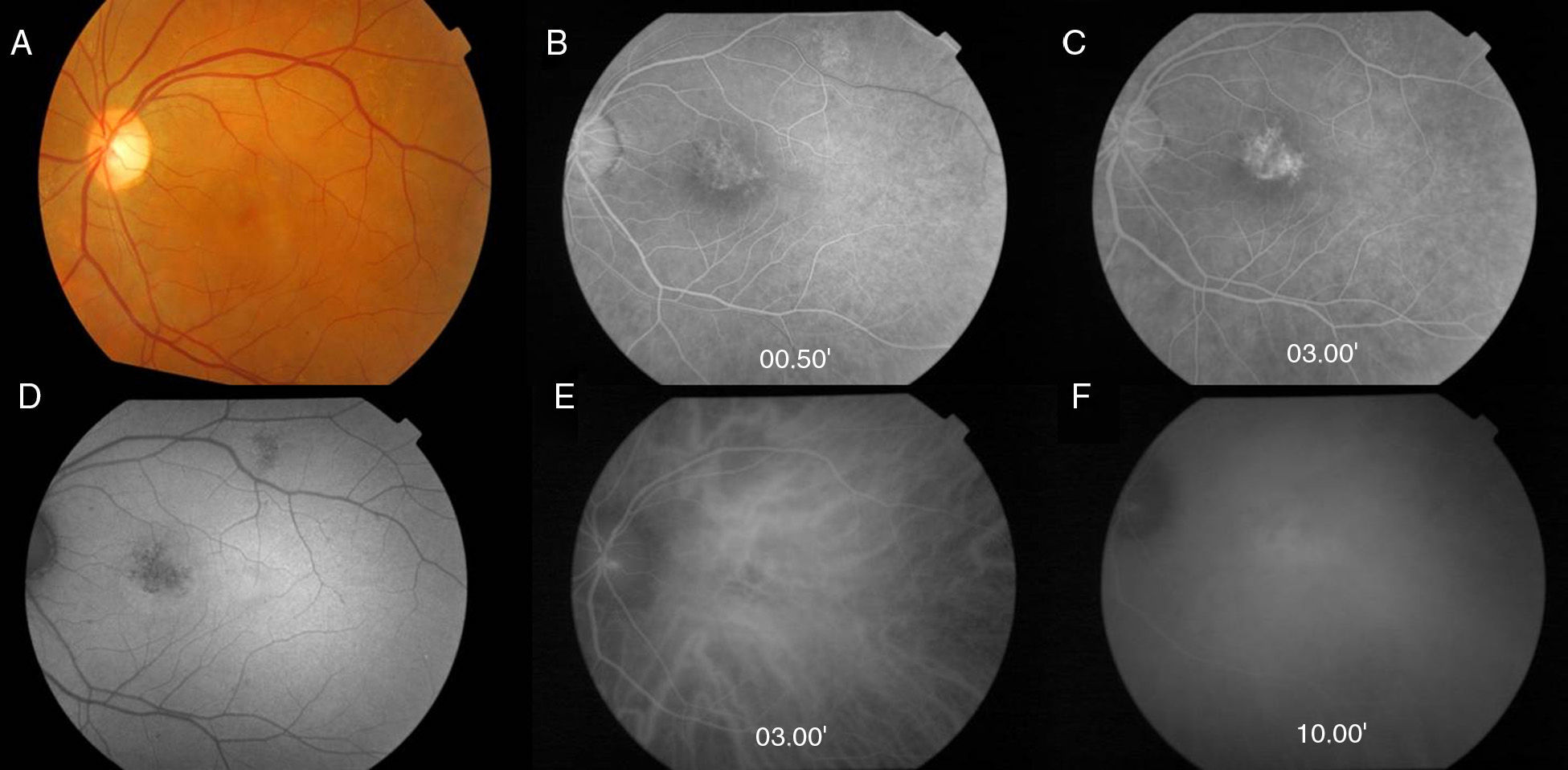
ResultsIn 2 patients, both the anterior and posterior segment evaluations were mostly normal despite the presence of yellowish spots in the macular area of the right eye of patient 1, and of a small yellowish elevated lesion with serous macular detachment in the macular area of the left eye in patient 2. At diagnosis, SD-OCT revealed a conforming FCE in patient 1, and in patient 2, an FCE with perilesional subretinal fluid and a neuroepithelium detachment, suspicious of FCE complicated with central serous retinopathy (CSCR). At one year of follow-up, patient 1 developed choroidal neovascularisation (CNV) over the focal choroidal excavation. FA and indocyanine green angiography examinations revealed areas with hypofluorescence in earlier frames, and a diffuse leakage in late frames. After ranibizumab injections, the SD-OCT of patient 1 revealed no active exudation, while patient 2 showed partial resolution of subretinal fluid.
ConclusionsFCE is a newly described entity of unclear etiology. It is characterized by a choroidal excavation in eyes, with absence of posterior staphyloma, scleral ectasia, trauma, or retinal disease. Although most lesions remain stable, there could be an association with CRSC or CNV.
Describir los hallazgos clínicos y sus complicaciones en dos pacientes con excavación focal coroidea (EFC).
MétodosSerie retrospectiva de casos. Se realizó exploración oftalmológica que incluía examen con lámpara de hendidura, retinografía, tomografía de coherencia óptica de dominio espectral (SD-OCT), angiofluoresceingrafía (AGF) y angiografía con verde de indocianina sobre cuatro ojos de dos pacientes con ECF.
ResultadosEn ambos pacientes la exploración de polo anterior y posterior resulta prácticamente normal a excepción de, en el paciente 1, la presencia de un moteado amarillento sobre al área macular del ojo derecho y, en el paciente 2, de una lesión amarillenta sobreelevada con desprendimiento seroso en el área macular del ojo izquierdo. En el momento del diagnóstico, la SD-OCT mostraba en el paciente 1 una EFC conformadora y en el paciente 2 una EFC con líquido subretiniano perilesional y un desprendimiento del neuroepitelio, compatible con una EFC complicada con una coriorretinopatía serosa central (CRSC). Al año de seguimiento, el paciente 1 desarrolló una neovascularización coroidea (NVC) sobre el área excavada. La AGF y angiografía con verde de indocianina revelaban áreas de hipofluorescencia temprana con hiperfluorescencia difusa tardía. Después del tratamiento con ranibizumab intravítreo, la SD-OCT del paciente 1 mostraba ausencia de exudación mientras que en el paciente 2 se objetivaba una resolución parcial del líquido subretiniano.
ConclusionesLa ECF es una entidad de reciente diagnóstico y etiología desconocida. Se define como un área de excavación coroidea en ausencia de estafiloma posterior, ectasia escleral, trauma o enfermedad retiniana. Aunque la mayoría de las lesiones se mantienen estables, su asociación con una CRSC o NVC puede ocurrir.