Buscar en
Archivos de la Sociedad Española de Oftalmología (English Edition)
Toda la web
Inicio Archivos de la Sociedad Española de Oftalmología (English Edition) Fingerprint microcystic dystrophy: Episodes of irregular astigmatism and their t...
Journal Information
Vol. 86. Issue 12.
Pages 415-418 (December 2011)
Share
Share
Download PDF
More article options
Visits
1791
Vol. 86. Issue 12.
Pages 415-418 (December 2011)
Short communication
Fingerprint microcystic dystrophy: Episodes of irregular astigmatism and their topographical representation
Distrofia en huella dactilar: brotes de astigmatismo irregular y su demostración topográfica
Visits
1791
G. Pérez-Carroa,
Corresponding author
gemurri27@yahoo.es

Corresponding author.
, R. Faua, L. González-Gonzálezb
a Oftalmología, Hospital de la Cruz Roja, Gijón, Asturias, Spain
b Enfermería, Hospital de la Cruz Roja, Gijón, Asturias, Spain
This item has received
Article information
Abstract
Full Text
Bibliography
Download PDF
Statistics
Figures (4)
Show moreShow less
Abstract
Case study

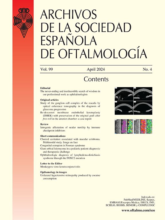
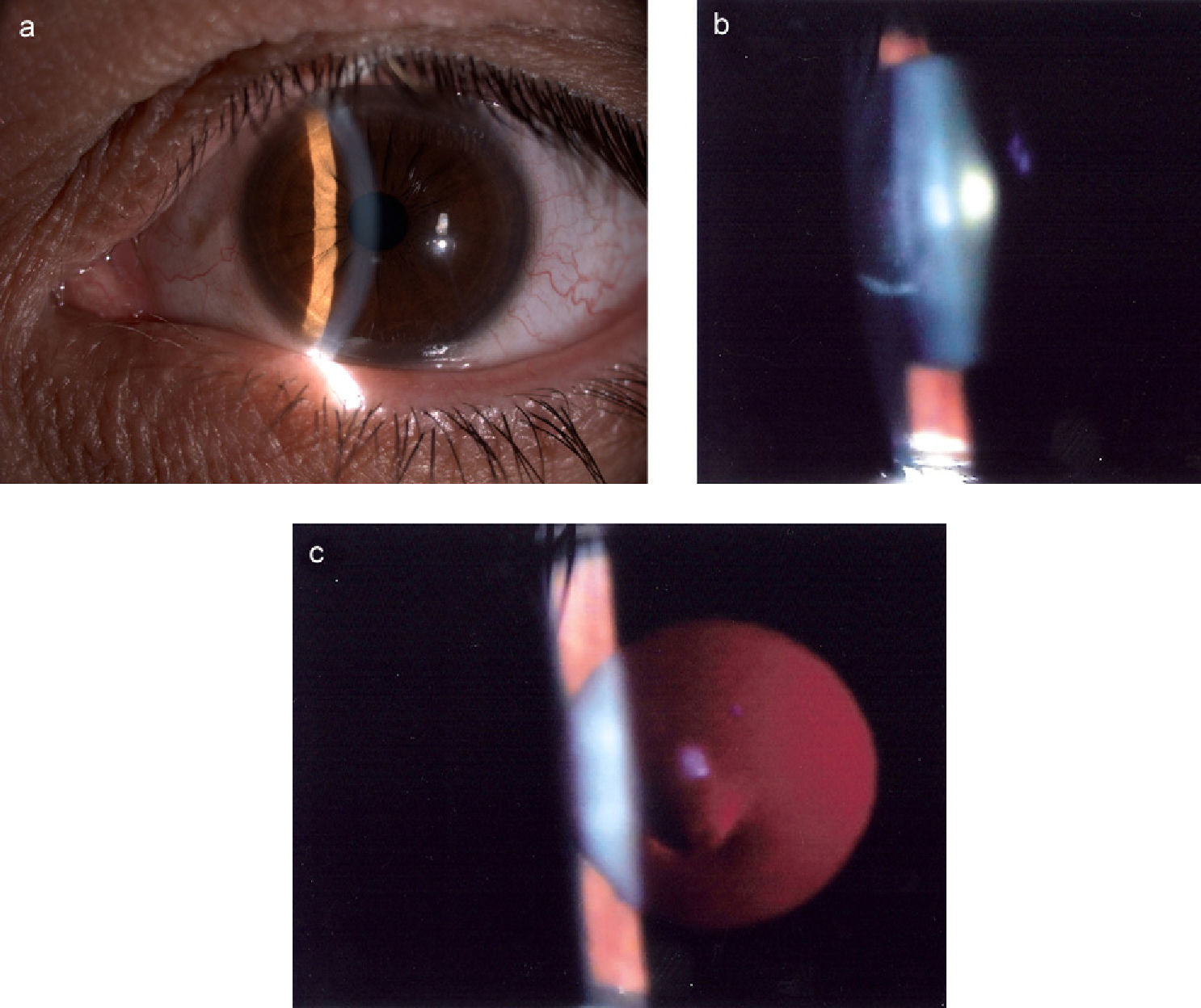
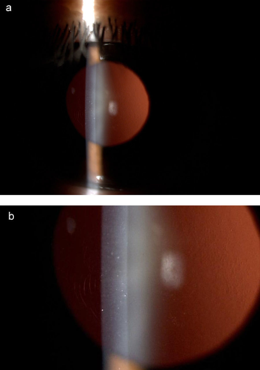
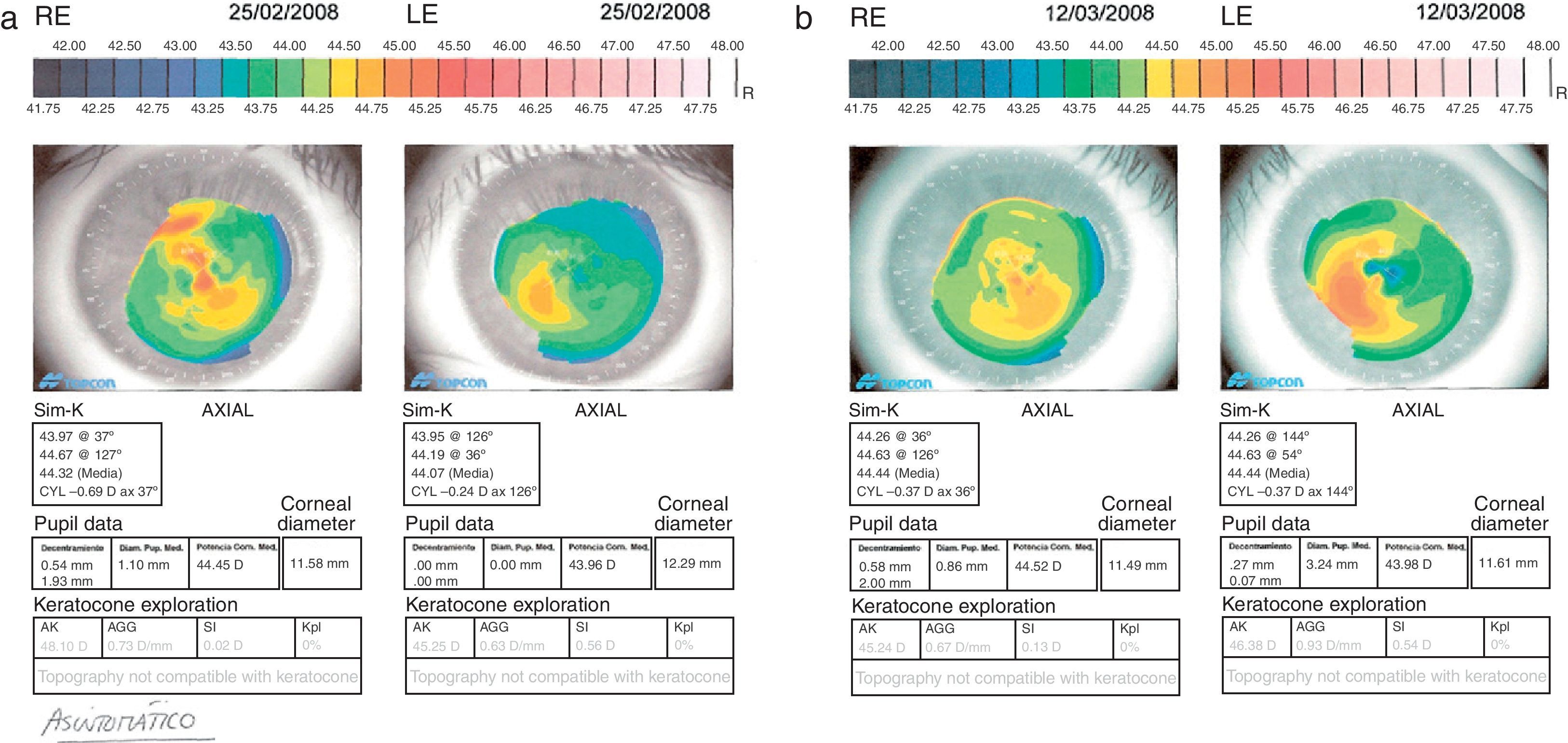
A 44-year-old male patient suffering from painless bouts of blurred vision and with no visible ophthalmological repercussions initially. After one of these clinical episodes we managed to visualise fingerprint images in the anterior pole, as well as corneal, pachymetric and topographical changes, which in turn produce the symptomatic refractive changes.

Discussion

Fingerprint keratopathy is a condition diagnosed through recurring corneal erosion. The pathogenic origin of the condition –an altered epithelial basal membrane –may encourage the separation of the corneal epithelium from its underlying layers. Depending on whether this separation is partial or total, this will lead to spontaneous corneal erosion or, less frequently, episodes of blurred vision caused by oedema and corneal swelling.

Keywords:
Microcystic corneal dystrophy
Cogan keratopathy
Fingerprint lines
Resumen
Caso clínico

Varón de 44 años con brotes indoloros de visión borrosa sin repercusión oftalmológica visible inicialmente. Tras uno de sus episodios, se visualiza en polo anterior imágenes en huella digital así como cambios corneales, paquimétricos y topográficos que originan cambios refractivos sintomáticos.

Discusión

La distrofia en huella dactilar es una entidad diagnosticada por erosiones corneales recurrentes. Su base patogénica, una membrana basal epitelial alterada, favorece la separación del epitelio corneal de las capas subyacentes. En la medida que ésta sea total o parcial ocasionará erosiones corneales recurrentes o, con menor frecuencia, episodios de visión borrosa por edema y engrosamiento corneal.

Palabras clave:
Distrofia microquística corneal
Queratopatía de Cogan
Huella dactilar

Article

These are the options to access the full texts of the publication Archivos de la Sociedad Española de Oftalmología (English Edition)
Subscriber
Subscriber

If you already have your login data, please click here .

If you have forgotten your password you can you can recover it by clicking here and selecting the option “I have forgotten my password”
Subscribe
Subscribe to

Archivos de la Sociedad Española de Oftalmología (English Edition)

Purchase
Purchase article

Purchasing article the PDF version will be downloaded

Price 19.34 €

Purchase now
Contact
Phone for subscriptions and reporting of errors
From Monday to Friday from 9 a.m. to 6 p.m. (GMT + 1) except for the months of July and August which will be from 9 a.m. to 3 p.m.
Calls from Spain
932 415 960
Calls from outside Spain
+34 932 415 960
E-mail
Article options
Tools
es en pt

¿Es usted profesional sanitario apto para prescribir o dispensar medicamentos?

Are you a health professional able to prescribe or dispense drugs?

Você é um profissional de saúde habilitado a prescrever ou dispensar medicamentos