


Dermatophytoses are transmissible infections that affect one billion people worldwide and have a significant impact on public health. Dermatophytes distribution evolves geographically and over time. Consequently, local epidemiology should be periodically assessed to control infection.
AimsTo describe the local distribution of dermatophytes and types of dermatophytosis through the analysis of samples from patients with suspected superficial mycoses, and to identify areas of improvement.
MethodsA retrospective epidemiological analysis of mycological culture results from skin, hair and nail samples referred to the mycology lab in the Hospital Clínico Universitario Lozano Blesa, Zaragoza (Spain) between 2020 and 2023 was performed, and the results statistically analyzed.
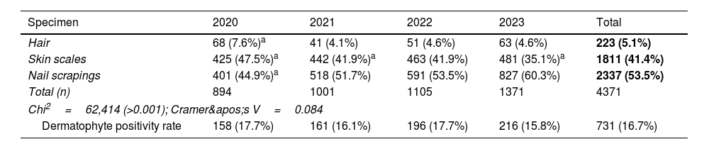
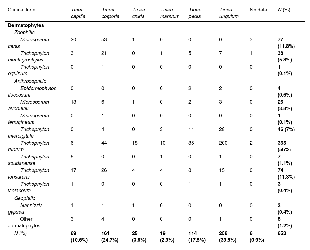
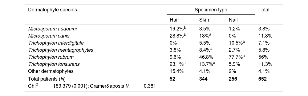
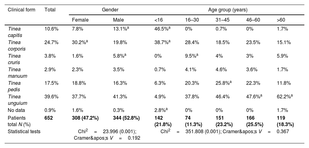
Results4371 specimens (skin: 41.4%; hair: 5.1%; nails: 53.5%) were cultured using standard procedures. The demand for testing increased by 53% over a 4-year time period and a dermatophyte positivity rate of 16.7% (n=731) was found. The species distribution was the following: Trichophyton rubrum (56%), Trichophyton tonsurans (11.3%), Microsporum canis (11.8%), Trichophyton interdigitale (7%), Trichophyton. mentagrophytes (5.8%), Microsporum audouinii (3.8%) and other species (4.9%), with an anthropophilic to zoophilic ratio of 4:1. The dermatophytoses clinical forms found were tinea unguium (39.6%), associated to population over 45 years, tinea corporis (24.7%) and tinea capitis (10.6%), both associated to people with less than 16 years group, tinea pedis (17.5%), mainly observed in people aged 31–45 years; other forms accounted for 7.6%. Finally, the Emergency department requested 11.9% of the mycological tests.
ConclusionsLocal epidemiology of dermatophytoses highlights the predominance of anthropophilic species, whereas 35 years ago zoophilic species represented 80% of the isolates. Interestingly, mild superficial lesions are frequently and inappropriately brought to the Emergency department.
Las dermatofitosis son infecciones transmisibles con impacto en la salud pública al afectar globalmente a más de 1000 millones de personas. Analizar periódicamente la epidermiología local ayuda a controlar la infección, ya que la distribución de los dermatofitos cambia continuamente.
ObjetivosDescribir la distribución local de los dermatofitos y las dermatofitosis mediante el análisis de muestras superficiales con sospecha de micosis e identificar áreas de mejora en su diagnóstico.
MétodosSe realizó, mediante análisis estadístico, un estudio epidemiológico retrospectivo de las muestras de piel, pelo y uñas cultivadas en el laboratorio de micología del Hospital Clínico Universitario Lozano Blesa, Zaragoza, (España) durante el periodo 2020-2023.
ResultadosSe cultivaron 4371 muestras de piel (41.4%), pelo (5,1%) y uñas (53,5%) según procedimientos estandarizados. La demanda de peticiones aumentó un 53% en 4 años. La tasa de positividad para dermatofitos fue del 16,7% (n=731). Las especies aisladas fueron Trichophyton rubrum (56%), Trichophyton tonsurans (11,3%), Microsporum canis (11,8%), Trichophyton interdigitale (7%), Trichophyton mentagrophytes (5,8%), Microsporum audouinii (4%) y otras (4,9%). La proporción de especies antropofílicas/zoofílicas fue de 4:1. Las formas clínicas observadas fueron tinea unguium (39,6%) asociada a mayores de 45 años, tinea corporis (24,7%) y tinea capitis (10,6%), ambas asociadas a menores de 16 años, y tinea pedis (17,5%), asociada al grupo de 31-45 años. Otras formas clínicas representaron el 7,6%. El Servicio de Urgencias solicitó el 11,9% de las peticiones.
ConclusionesLas dermatofitosis causadas por especies antropofílicas predominan en Zaragoza actualmente; en 1997, el 80% eran zoofílicas. Se ha observado un uso inadecuado del servicio de Urgencias para el diagnóstico de lesiones dermatológicas superficiales.

