


This study aimed to determine the functional expression of somatostatin receptors in patients with primary brain tumors including meningiomas and gliomas using 99mTc-HYNIC-octreotide SPECT/CT imaging.
MethodThis cross-sectional study was conducted at Ghaem Hospital, Mashhad, in 2023. Patients with histopathologically confirmed primary brain tumors (gliomas and meningiomas) who had previously received treatment and presented with suspected tumor recurrence based on imaging findings were included. After injection of 99mTc-HYNIC-Octreotide, whole-body SPECT/CT imaging of the head and other suspicious areas was performed. Two nuclear medicine specialists independently reviewed scans. Radiopharmaceutical uptake was assessed qualitatively by comparing absorption in lesions to liver and splenic uptake, with scoring based on the Krenning score. The uptake in lesions (target) and their corresponding mirrored normal brain regions (non-target) was calculated using ROI measurements.
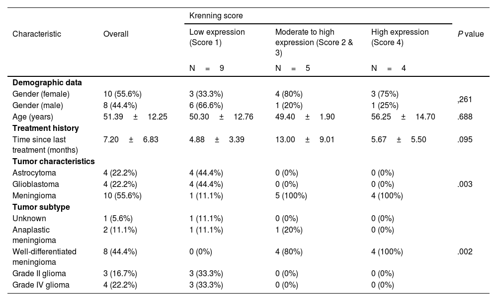
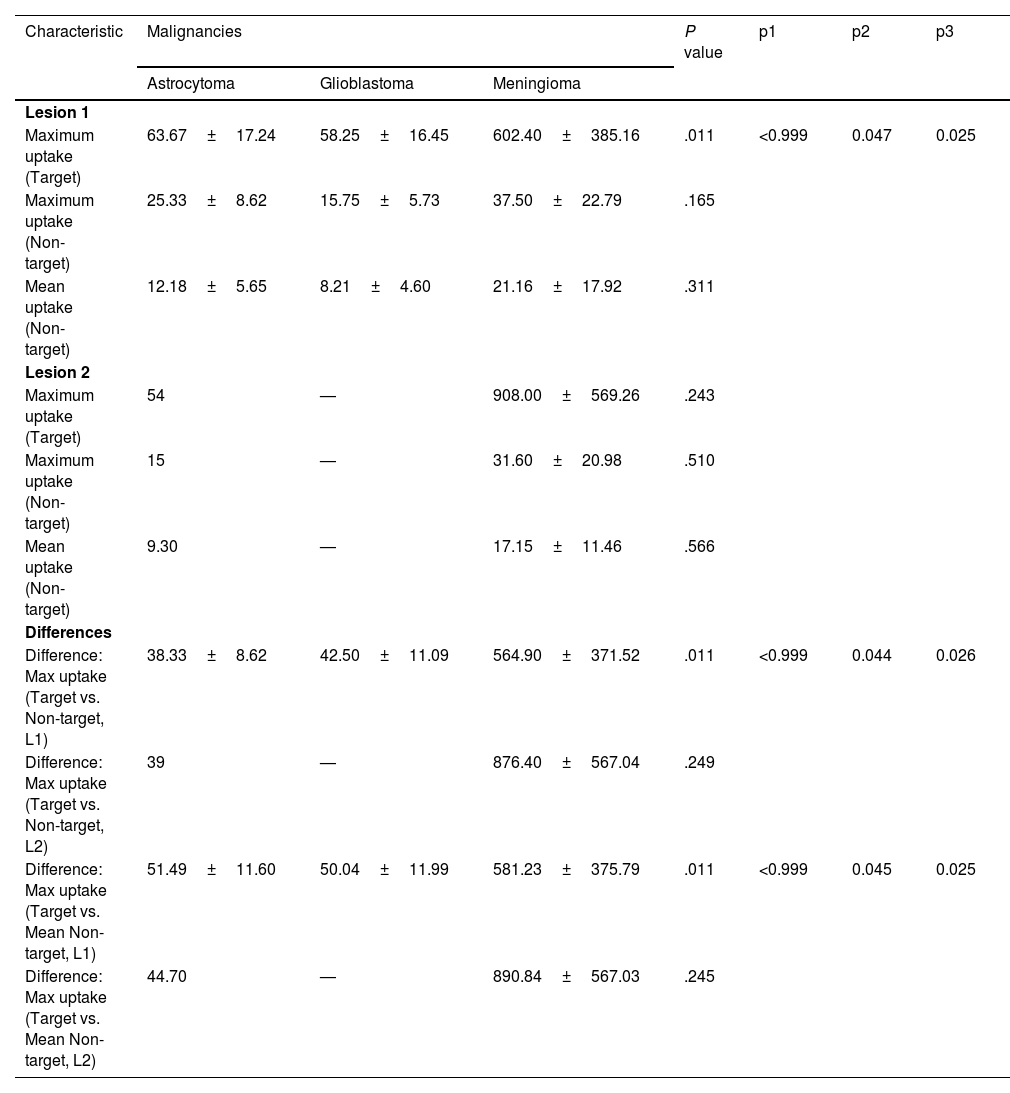
ResultsOut of 24 patients, scans from 18 (mean age: 51.39±12.25 years; range: 35–74) were analyzed due to technical issues and missing images. Of these, 10 (55.6%) were women. Somatostatin receptor expression was observed in all lesions. Meningioma had significantly higher receptor expression compared to gliomas (P=.003). Maximum and target-to-non-target uptake differences were significantly greater in meningioma than in astrocytoma (P=.047, P=.044) and glioblastomas (P=.025, P=.026). Gliomas, including astrocytoma and glioblastomas, consistently showed low receptor expression, and no significant differences in receptor expression were observed between astrocytoma and glioblastomas.
ConclusionThis study demonstrates that 99mTc-HYNIC-octreotide SPECT/CT imaging is an effective method for detecting somatostatin receptor expression in brain tumors, offering a low-cost and accessible alternative to more enhanced imaging techniques, both meningioma and glial tumors express somatostatin receptors, but receptor expression is significantly higher in meningioma.
Este estudio tuvo como objetivo determinar la expresión funcional de los receptores de somatostatina en pacientes con tumores cerebrales primarios, incluidos meningiomas y gliomas, utilizando imágenes de SPECT/CT con 99mTc-HYNICoctreotide.
MétodoEste estudio transversal se llevó a cabo en el Hospital Ghaem, Mashhad, en 2023. Fueron incluidos pacientes con tumores cerebrales primarios (gliomas y meningiomas) confirmados histopatológicamente, que habían recibido tratamiento previamente y presentaban sospecha de recurrencia tumoral basada en hallazgos por imágenes. Después de la inyección de 99mTc-HYNIC-Octreotide, se realizaron imágenes de SPECT/CT de cuerpo completo de la cabeza y otras áreas sospechosas. Dos especialistas en medicina nuclear revisaron las exploraciones de manera independiente. La captación del radiofármaco se evaluó cualitativamente comparando la absorción en las lesiones con la captación en el hígado y el bazo, y la puntuación se basó en el puntaje Krenning. La captación en las lesiones (objetivo) y en las regiones normales correspondientes del cerebro reflejadas (no objetivo) se calculó utilizando mediciones de ROI.
ResultadosDe los 24 pacientes, se analizaron las exploraciones de 18 (edad media: 51,39±12,25 años; rango: 35–74) debido a problemas técnicos y a la falta de imágenes. De estos, 10 (55,6%) eran mujeres. Se observó expresión de receptores de somatostatina en todas las lesiones. Los meningiomas mostraron una expresión significativamente mayor de receptores en comparación con los gliomas (P=0,003). Las diferencias en la captación máxima y en la relación objetivo/no objetivo fueron significativamente mayores en los meningiomas que en los astrocitomas (P=0,047, P=0,044) y los glioblastomas (P=0,025, P=0,026). Los gliomas, incluidos los astrocitomas y los glioblastomas, mostraron consistentemente una baja expresión de receptores, and no se observaron diferencias significativas en la expresión de receptores entre los astrocitomas y los glioblastomas.
ConclusiónEste estudio demuestra que la gammagrafía SPECT/CT con 99mTcHYNIC-octréotido es un método eficaz para detectar la expresión de receptores de somatostatina en tumores cerebrales, ofreciendo una alternativa accesible y de bajo costo frente a técnicas de imagen más avanzadas, tanto los meningiomas como los tumores gliales expresan receptores de somatostatina, pero la expresión de los receptores es significativamente mayor en los meningiomas.
Article
If you experience access problems, you can contact the SEMNIM Technical Secretariat by email at secretaria.tecnica@semnim.es or by phone at +34 619 594 780.

Revista Española de Medicina Nuclear e Imagen Molecular (English Edition)
