


In this study, we aimed to evaluate the diagnostic performance of 18F-PSMA-1007 PET/CT compared to 68Ga-PSMA-11 PET/CT, which is more commonly used in routine practice, for detecting prostate cancer recurrence in prostate cancer patients with biochemical recurrence.
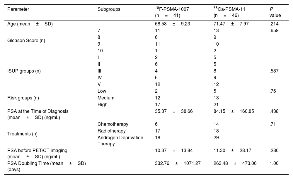
Materials and methodsForty-one prostate cancer patients with biochemical recurrence were prospectively included in the study. Additionally, images from 46 patients in our institution's database, who had undergone 68Ga-PSMA-11 PET/CT imaging for biochemical recurrence, were retrospectively re-evaluated to compare the detection rates with those of 18F-PSMA-1007 PET/CT. SUVmax, total tumor PSMA, PSMA total tumor volume were calculated for local recurrence, lymph node metastasis, and organ metastasis. The diagnostic performances of the two imaging methods were then compared.
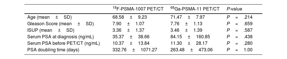
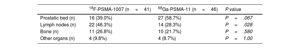
ResultsThe mean age, Gleason scores, ISUP scores, serum PSA levels at diagnosis and at the time of imaging, and PSA doubling times were similar across the 18F-PSMA-1007 and 68Ga-PSMA-11 groups. Pathological uptake was observed in the prostatic bed in 16 patients (39.0%), lymph nodes in 22 patients (46.3%), and bones in 11 patients (26.8%) with 18F-PSMA-1007 PET/CT. 18F-PSMA-1007 PET/CT showed statistically significant superiority over 68Ga-PSMA-11 PET/CT in detecting lymph node metastases (41.6% vs. 25.4%; P=.028). There was no significant difference between the two imaging protocols in the detection rates of local recurrence (P=.067) and bone metastasis (P=.580).
ConclusionAlthough the study included a small sample size, the results revealed that 18F-PSMA-1007 PET/CT had a higher detection rate than 68Ga-PSMA-11 PET/CT in patients with biochemically recurrent prostate carcinoma, particularly for lymph node metastases.
En este estudio, nuestro objetivo fue evaluar el rendimiento diagnóstico del PET/TC con 18F-PSMA-1007 en comparación con el PET/TC con 68Ga-PSMA-11, que se utiliza más comúnmente en la práctica clínica habitual, para la detección de la recurrencia del cáncer de próstata en pacientes con cáncer de próstata que presentan recurrencia bioquímica.
Material y métodosSe incluyeron prospectivamente en el estudio 41 pacientes con cáncer de próstata y recurrencia bioquímica. Además, se reanalizaron retrospectivamente las imágenes de 46 pacientes de la base de datos de nuestra institución que se habían sometido a 68Ga-PSMA-11 PET/TC por recurrencia bioquímica, con el fin de comparar las tasas de detección con las de 18F-PSMA-1007 PET/TC. Se calcularon los valores de SUVmax, PSMA tumoral total y volumen tumoral total de PSMA para recurrencia local, metástasis ganglionares y metástasis orgánicas. Posteriormente, se compararon los rendimientos diagnósticos de ambos métodos de imagen.
ResultadosLa edad media, las puntuaciones de Gleason, las puntuaciones ISUP, los niveles séricos de PSA al momento del diagnóstico y en el momento de la imagen, y los tiempos de duplicación del PSA fueron similares entre los grupos de 18F-PSMA-1007 y 68Ga-PSMA-11. En el grupo 18F-PSMA-1007 PET/TC se observó captación patológica en el lecho prostático en 16 pacientes (39,0%), en ganglios linfáticos en 22 pacientes (46,3%) y en huesos en 11 pacientes (26,8%). 18F-PSMA-1007 PET/TC mostró una superioridad estadísticamente significativa sobre 68Ga-PSMA-11 PET/TC en la detección de metástasis ganglionares (41,6% vs. 25,4%; P=,028). No se observaron diferencias significativas entre ambos protocolos de imagen en las tasas de detección de recurrencia local (P=,067) ni de metástasis óseas (P=,580).
ConclusiónAunque el estudio incluyó una muestra pequeña, los resultados mostraron que 18F-PSMA-1007 PET/TC tiene una mayor tasa de detección que 68Ga-PSMA-11 PET/TC en pacientes con carcinoma de próstata con recurrencia bioquímica, especialmente en la detección de metástasis ganglionares.
Article
If you experience access problems, you can contact the SEMNIM Technical Secretariat by email at secretaria.tecnica@semnim.es or by phone at +34 619 594 780.

Revista Española de Medicina Nuclear e Imagen Molecular (English Edition)
