

metricas

Radiología

Sugerencias
Radiología Guía esencial del arterial spin labelling: revisión de los principales artefac...
Información de la revista
Vol. 67. Núm. 5.
(Septiembre - Octubre 2025)
Cita
Cita
Compartir
Descargar PDF
Más opciones de artículo
Visitas
3257
Vol. 67. Núm. 5.
(Septiembre - Octubre 2025)
Radiología en Imágenes

Guía esencial del arterial spin labelling: revisión de los principales artefactos y aplicaciones

Essential guide to arterial spin labelling: Review of key artifacts and applications
Visitas
3257
R. Sutil Berjóna,b,
Autor para correspondencia
rsutilrad@gmail.com

Autor para correspondencia.
, D. Castanedo Vázqueza,b, D. Herrán de la Galac, M. Drake Péreza,b, E. Marco de Lucasa,b
a Servicio de Radiodiagnóstico, Hospital Universitario Marqués de Valdecilla, Santander, España
b Instituto de Investigación de Valdecilla (Idival), Santander, España
c Neuroradiology Department, APHP, Hospital Pitié-Salpêtrière, París, Francia
Este artículo ha recibido
Información del artículo
Resumen
Texto completo
Bibliografía
Descargar PDF
Estadísticas
Resumen

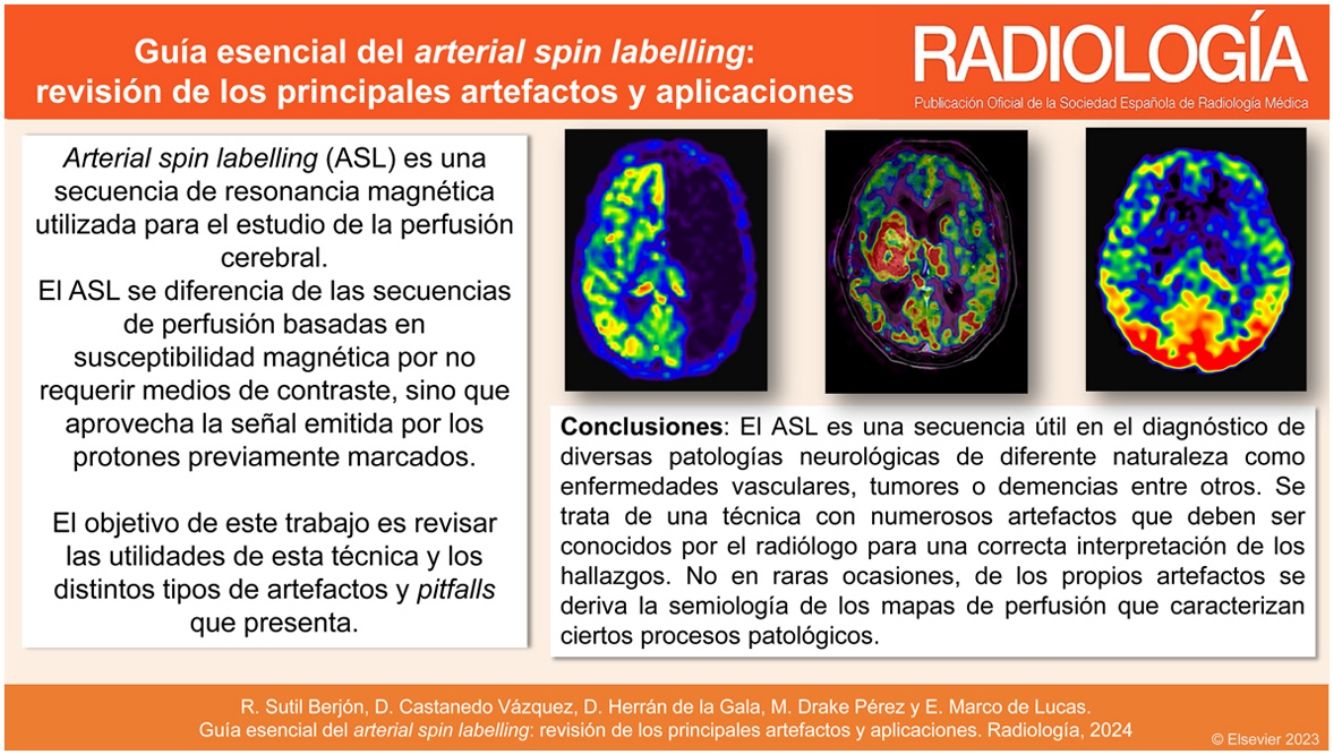
Arterial spin labelling (ASL) es una técnica de perfusión cerebral por de resonancia magnética utilizada para el estudio del flujo sanguíneo cerebral. Se diferencia de las secuencias de perfusión basadas en susceptibilidad magnética por no requerir medios de contraste, sino que aprovecha la señal emitida por los protones previamente marcados. Tiene utilidad en el estudio de diversas patologías neurológicas de diferente naturaleza, como el ictus isquémico, los tumores cerebrales, las demencias o la epilepsia, entre otros. No obstante, presenta numerosos artefactos que deben ser conocidos por el radiólogo para una correcta interpretación de los hallazgos.

Palabras clave:
Resonancia magnética
Artefactos
Estenosis carotídea
Ictus
Tumor cerebral
Demencia
Abstract

Arterial spin labelling (ASL) is a magnetic resonance imaging perfusion technique used to study cerebral blood flow. It differs from magnetic susceptibility-based perfusion sequences in that it does not require contrast media, but rather uses the signals emitted by pre-labelled protons. It is useful in the exploration of several different kinds of neurological pathologies including ischaemic stroke, brain tumours, dementia and epilepsy. However, it presents numerous artefacts that radiologists must be familiar with to ensure a correct interpretation of findings.

Keywords:
Magnetic resonance imaging
Artefacts
Carotid stenosis
Acute stroke
Brain tumour
Dementia
Resumen gráfico

Artículo

Opciones para acceder a los textos completos de la publicación Radiología
Suscriptor
Suscriptor de la revista

Si ya tiene sus datos de acceso, clique aquí.

Si olvidó su clave de acceso puede recuperarla clicando aquí y seleccionando la opción "He olvidado mi contraseña".
Comprar
Comprar acceso al artículo

Comprando el artículo el PDF del mismo podrá ser descargado

Comprar ahora
Contactar
Teléfono para suscripciones e incidencias
De lunes a viernes de 9h a 18h (GMT+1) excepto los meses de julio y agosto que será de 9 a 15h
Llamadas desde España
932 415 960
Llamadas desde fuera de España
+34 932 415 960
E-mail
Bibliografía
[1]
J.A. Guzmán-de-Villoria, P. Fernández-García, J.M. Mateos-Pérez, M. Desco.
Estudio de la perfusión cerebral mediante técnicas de susceptibilidad magnética: técnica y aplicaciones.
Radiología., 54 (2012), pp. 208-220
[2]
A.R. Deibler, J.M. Pollock, R.A. Kraft, H. Tan, J.H. Burdette, J.A. Maldjian.
Arterial spin-labeling in routine clinical practice, Part 1: Technique and artifacts.
AJNR Am J Neuroradiol., 29 (2008), pp. 1228-1234
[3]
T.J. Marini, D. Mistry, S. Jetty, H. Wang, E. Lin, J. Almast, et al.
Arterial spin-labeling perfusion for neuroradiologists: The essentials.
Neurographics., 12 (2022), pp. 135-150
[4]
M. Grade, J.A. Hernandez Tamames, F.B. Pizzini, E. Achten, X. Golay, M. Smits.
A neuroradiologist's guide to arterial spin labeling MRI in clinical practice.
Neuroradiology., 57 (2015), pp. 1181-1202
[5]
T. Iutaka, M.B. de Freitas, S.S. Omar, F.A. Scortegagna, K. Nael, R.H. Nunes, et al.
Arterial spin labeling: Techniques, clinical applications, and interpretation.
RadioGraphics., 43 (2023), pp. e220088
[6]
A. Pfefferbaum, S. Chanraud, A.L. Pitel, A. Shankaranarayanan, D.C. Alsop, T. Rohlfing, et al.
Volumetric cerebral perfusion imaging in healthy adults: regional distribution, laterality, and repeatability of Pulsed Continuous Arterial Spin Labeling (PCASL).
Psychiatry Res., 182 (2010), pp. 266
[7]
H.G. Kim, J.H. Lee, J.W. Choi, M. Han, S.M. Gho, Y. Moon.
Multidelay arterial spin-labeling MRI in neonates and infants: Cerebral perfusion changes during brain maturation.
AJNR Am J Neuroradiol., 39 (2018), pp. 1912-1918
[8]
D. Jaganmohan, S. Pan, C. Kesavadas, B. Thomas.
A pictorial review of brain arterial spin labelling artefacts and their potential remedies in clinical studies.
Neuroradiol J., 34 (2021), pp. 154-168
[9]
D.Y. Chen, Y.S. Kuo, H.L. Hsu, F.X. Yan, H.L. Liu, C.J. Chen, et al.
Loss of labelling efficiency caused by carotid stent in pseudocontinuous arterial spin labelling perfusion study.
Clin Radiol., 71 (2016), pp. e21-e27
[10]
O. Wu, L. Østergaard, A.G. Sorensen.
Technical aspects of perfusion-weighted imaging.
Neuroimaging Clin N Am., 15 (2005), pp. 623-637
[11]
A.R. Deibler, J.M. Pollock, R.A. Kraft, H. Tan, J.H. Burdette, J.A. Maldjian.
Arterial spin-labeling in routine clinical practice, Part 2: Hypoperfusion patterns.
AJNR Am J Neuroradiol., 29 (2008), pp. 1235-1241
[12]
S.A. Amukotuwa, C. Yu, G. Zaharchuk.
3D Pseudocontinuous arterial spin labeling in routine clinical practice: A review of clinically significant artifacts.
J Magn Reson Imaging., 43 (2016), pp. 11-27
[13]
A. Di Napoli, S.F. Cheng, J. Gregson, D. Atkinson, J.E. Markus, T. Richards, et al.
Arterial spin labeling MRI in carotid stenosis: Arterial transit artifacts may predict symptoms.
Radiology., 297 (2020), pp. 652-660
[14]
Y.C. Huang, H.L. Liu, J.D. Lee, J.T. Yang, H.H. Weng, M. Lee, et al.
Comparison of arterial spin labeling and dynamic susceptibility contrast perfusion MRI in patients with acute stroke.
[15]
M. Zhang, Q. Shi, Y. Yue, M. Zhang, L. Zhao, C. Yan.
Evaluation of T2-FLAIR combined with ASL on the collateral circulation of acute ischemic stroke.
Neurol Sci., 43 (2022), pp. 4891-4900
[16]
G. Zaharchuk, I.S. El Mogy, N.J. Fischbein, G.W. Albers.
Comparison of arterial spin labeling and bolus perfusion-weighted imaging for detecting mismatch in acute stroke.
Stroke J Cereb Circ., 43 (2012), pp. 1843-1848
[17]
R.E. Yoo, T.J. Yun, J.H. Rhim, B.W. Yoon, K.M. Kang, S.H. Choi, et al.
Bright vessel appearance on arterial spin labeling MRI for localizing arterial occlusion in acute ischemic stroke.
Stroke., 46 (2015), pp. 564-567
[18]
J. Liu, C. Lin, A. Minuti, M. Lipton.
Arterial spin labeling compared to dynamic susceptibility contrast MR perfusion imaging for assessment of ischemic penumbra: A systematic review.
J Neuroimaging., 31 (2021), pp. 1067-1076
[19]
J.D. Weinstein, O. Hamam, V.C. Urrutia, H. Lu, L.P. Luna, A. Tekes-Brady, et al.
Added value of arterial spin labeling in detecting posterior reversible encephalopathy syndrome as a stroke mimic on baseline neuroimaging: A single center experience.
Front Neurol., 13 (2022), pp. 831218
[20]
V. Adhiyaman, S. Alexander.
Cerebral hyperperfusion syndrome following carotid endarterectomy.
QJM Int J Med., 100 (2007), pp. 239-244
[21]
A. Lavrova, W.H.T. Teunissen, E.A.H. Warnert, M. van den Bent, M. Smits.
Diagnostic accuracy of arterial spin labeling in comparison with dynamic susceptibility contrast-enhanced perfusion for brain tumor surveillance at 3T MRI.
Front Oncol., 12 (2022), pp. 849657
[22]
E.J. Smith, A. Naik, A. Shaffer, M. Goel, D.T. Krist, E. Liang, et al.
Differentiating radiation necrosis from tumor recurrence: A systematic review and diagnostic meta-analysis comparing imaging modalities.
J Neurooncol., 162 (2023), pp. 15-23
[23]
K. Solozhentseva, A. Batalov, N. Zakharova, S. Goryaynov, E. Pogosbekyan, I. Pronin.
The role of 3D-pCASL MRI in the differential diagnosis of glioblastoma and brain metastases.
Front Oncol., 12 (2022), pp. 874924
[24]
R. Ferreira, A.J. Bastos-Leite.
Arterial spin labelling magnetic resonance imaging and perfusion patterns in neurocognitive and other mental disorders: A systematic review.
Neuroradiology., 66 (2024), pp. 1065-1081
[25]
J.M. Pollock, A.R. Deibler, J.H. Burdette, R.A. Kraft, H. Tan, A.B. Evans, et al.
Migraine associated cerebral hyperperfusion with arterial spin-labeled MR imaging.
Am J Neuroradiol., 29 (2008), pp. 1494-1497
[26]
M. Schertz, J. Benzakoun, N. Pyatigorskaya, S. Belkacem, M. Sahli-Amor, V. Navarro, et al.
Specificities of arterial spin labeling (ASL) abnormalities in acute seizure.
J Neuroradiol., 47 (2020), pp. 20-26
[27]
R. Li, S. Jin, Y. Wang, J.F. Li, H.F. Xiao, Y.L. Wang, et al.
Brain perfusion alterations on 3D pseudocontinuous arterial spin-labeling MR imaging in patients with autoimmune encephalitis: A case series and literature review.
AJNR Am J Neuroradiol., 43 (2022), pp. 701-706
[28]
R. Li, P.A. Shi, T.F. Liu, Y. Li, Y. Wang, K. Wu, et al.
Role of 3D pseudocontinuous arterial spin-labeling perfusion in the diagnosis and follow-up in patients with herpes simplex encephalitis.
AJNR Am J Neuroradiol., 40 (2019), pp. 1901-1907
[29]
N. Sollmann, G. Hoffmann, S. Schramm, M. Reichert, M. Hernandez Petzsche, Strobel., et al.
Arterial Spin Labeling (ASL) in neuroradiological diagnostics — Methodological overview and use cases.
Rofo., 196 (2024), pp. 36-51
asdasdasd
Opciones de artículo
Herramientas

