


Chiari malformation type 1 (CM-1) is a structural abnormality characterized by caudal displacement of the cerebellar tonsils (≥5mm) through the foramen magnum, which could compromise the flow of cerebrospinal fluid (CSF) and generate various neurological symptoms.1,2 Although its aetiology is still debated, multifactorial mechanisms have been proposed, including defects in embryological development and alterations in intracranial pressure.3,4
The prevalence of CM-1 has increased in recent decades due to the increased use of neuroimaging techniques, estimated at approximately 0.1–0.5% of the general population, with a female predominance.5,6 Clinical manifestations range from asymptomatic to occipital paroxysmal headache intensified by coughing, laughing or Valsalva manoeuvres; vertigo; dizziness and gait disturbances.1
Despite advances in magnetic resonance imaging (MRI) diagnosis, the management of CM-1 remains challenging, especially in paediatric cases where progression is less predictable. While surgical decompression is the standard treatment for some symptomatic patients, there is no consensus on the approach to mild or asymptomatic cases.7,8
Case presentationA 42-year-old man presented to primary care with a 2-month history of neck pain associated with headache, vertigo, intermittent left upper limb pain and paraesthesia. Current smoker. No relevant past medical history. Neurological examination was unremarkable. A reduced range of neck motion was noted. The vital signs showed slightly elevated blood pressure (131/90mmHg) with a normal heart rate and temperature. He was previously diagnosed with benign paroxysmal positional vertigo (BPPV) as indicated by a positive Dix-Hallpike test with partial response to Epley manoeuvres.
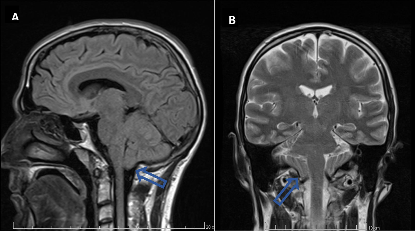
His vertigo was associated with intermittent occipital headaches. MRI brain and cervical spine revealed a herniation of the cerebellar tonsils into the superior cervical spine by 9mm (Fig. 1). Additional imaging reported the absence of hydrocephalus, syrinx, enlarged foramen magnum or other stigmata of Chiari malformation. Conservative management including physiotherapy and analgesia was initiated. The patient was assessed by neurosurgery who agreed with a conservative approach. CM-1 was diagnosed due to cerebellar tonsil herniation ≥5mm below the foramen magnum on neuroimaging. He did not report symptoms before his 40s. A set of blood tests was performed to rule out other possible diagnoses, including C-reactive protein (CRP), electrolytes, kidney function test, liver profile, full blood count, erythrocyte sedimentation rate, calcium and magnesium with all of them within the normal range. CM-1 can present with headache, cervical pain, cerebellar dysfunction, sleep-related breathing disorders and positional vertigo. CM-1 should be considered in cases of BPPV which are refractory to liberatory manoeuvres, especially if other atypical features are present.
Discussion
Headache and vertigo are two common symptoms presented in primary care settings. Most patients with mild or self-limiting symptoms and a normal neurological examination do not require further investigations. However, those patients with persistent symptoms, unclear diagnosis or abnormal findings require further assessment, including neuroimaging (e.g. MRI, computer tomography scan). Although tension-type headache and migraine are the most common causes of headache, less common aetiologies must be considered to guide appropriate investigations and treatment.8 Chiari malformation is characterized by a “cough headache” because it is usually exacerbated by cough, however, it could be triggered by laughing or Valsalva manoeuvres as well.1 Whereas, vertigo is a common condition, the aetiological diagnosis remains challenging and difficult to establish. Acute labyrinthitis, benign paroxysmal positional vertigo, Meniere's disease, migraine associated vertigo, vestibular migraine and vestibular neuritis are some common causes of vertigo.9 However, vertigo could be present in patients with Chiari malformation.1 In fact, the case presented reported vertigo as his main complaint. Because of this, he was diagnosed with benign paroxysmal positional vertigo, however, due to atypical features and persistence of the symptoms an MRI brain was requested. This patient also reported paraesthesia, which has been reported as a common symptom and typical feature of CM-1. Other symptoms described in the literature include nausea, dysphagia, apnoea, clonus, cerebellar symptoms, drop attacks, muscle atrophy and dysphonia.10 With the MRI brain result, clinical features, and other differential diagnoses excluded, we finally reached the diagnosis of CM-1.
The management of CM-1 usually requires multiple approaches. Conservative management remains the most common approach. The indication for surgery is based on clinical symptoms, including their severity and radiological findings. Persistent headache, refractive to conservative management has been reported as the main indication for surgery. Most asymptomatic patients do not need surgery, however, this approach could be advised in the patient with progressive posterior fossa or spinal cord signs, hydrocephalus or syringomyelia.10 Our patient was referred to neurosurgery and neurology and it was agreed to receive conservative management. He was referred for physiotherapy and regular analgesia was prescribed, showing improvement of the symptoms.
CM-1 is typically asymptomatic until late adolescence or adulthood. Our patient remained asymptomatic and he reported his first symptoms in his fourth decade of life. It is also well documented that some patients may remain asymptomatic, with the diagnosis made incidentally.4
In general, the prognosis of CM-1 is good and most patients remain stable or improve over time. Headache is the most common symptom to improve, whilst the paraesthesia tends to persist and remains steady. In our patient, this symptom was persistent in the upper limbs and an MRI cervical spine was requested and reported minimal herniation of the cerebellar tonsils into the superior cervical spine by 9mm, otherwise, unremarkable findings.
ConclusionsIn conclusion, this case report highlights an infrequent but important diagnosis of CM-1 in a patient presenting with headache, neck pain, vertigo, left upper limb pain and paraesthesia on a background of previously diagnosed benign paroxysmal positional vertigo. CM-1 is often underdiagnosed as identification remains challenging in patients presenting with more common symptoms easily attributable to a wide range of diagnoses. In this case the persistence of vertigo and the development of new neurological symptoms warranted further investigation, such as neuroimaging, which lead to the diagnosis of CM-1. Prompt diagnosis facilitates an appropriate treatment plan, which improves patient quality of life. In our case, the patient responded well to conservative management, which was indicated based on the definitive diagnosis. However, some patients require surgery in cases of progressive posterior fossa or spinal cord signs, hydrocephalus or syringomyelia underscoring the need for early diagnosis to achieve optimal patient outcomes.
CRediT authorship contribution statementAll authors have reviewed the final version to be published and agreed to be accountable for all aspects of the work.
Ethical considerationsInformed consent was obtained from patient involved in the case. All data was anonymised.
FundingNone declared.
Conflicts of interestNone declared.


