A 29-year-old woman underwent an abdominal ultrasound due to chronic abdominal pain.
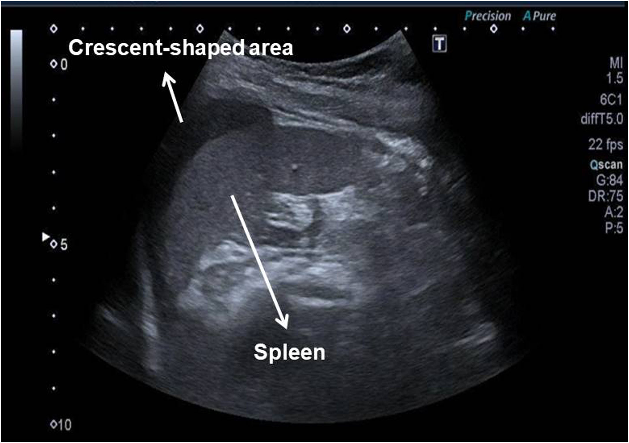
A subdiaphragmatic, hypoechogenic crescent-shaped area with a maximum diameter of 70mm, similar to the characteristic images of splenic hematomas, was observed at the perisplenic level (Fig. 1). The patient had no history of previous abdominal trauma.

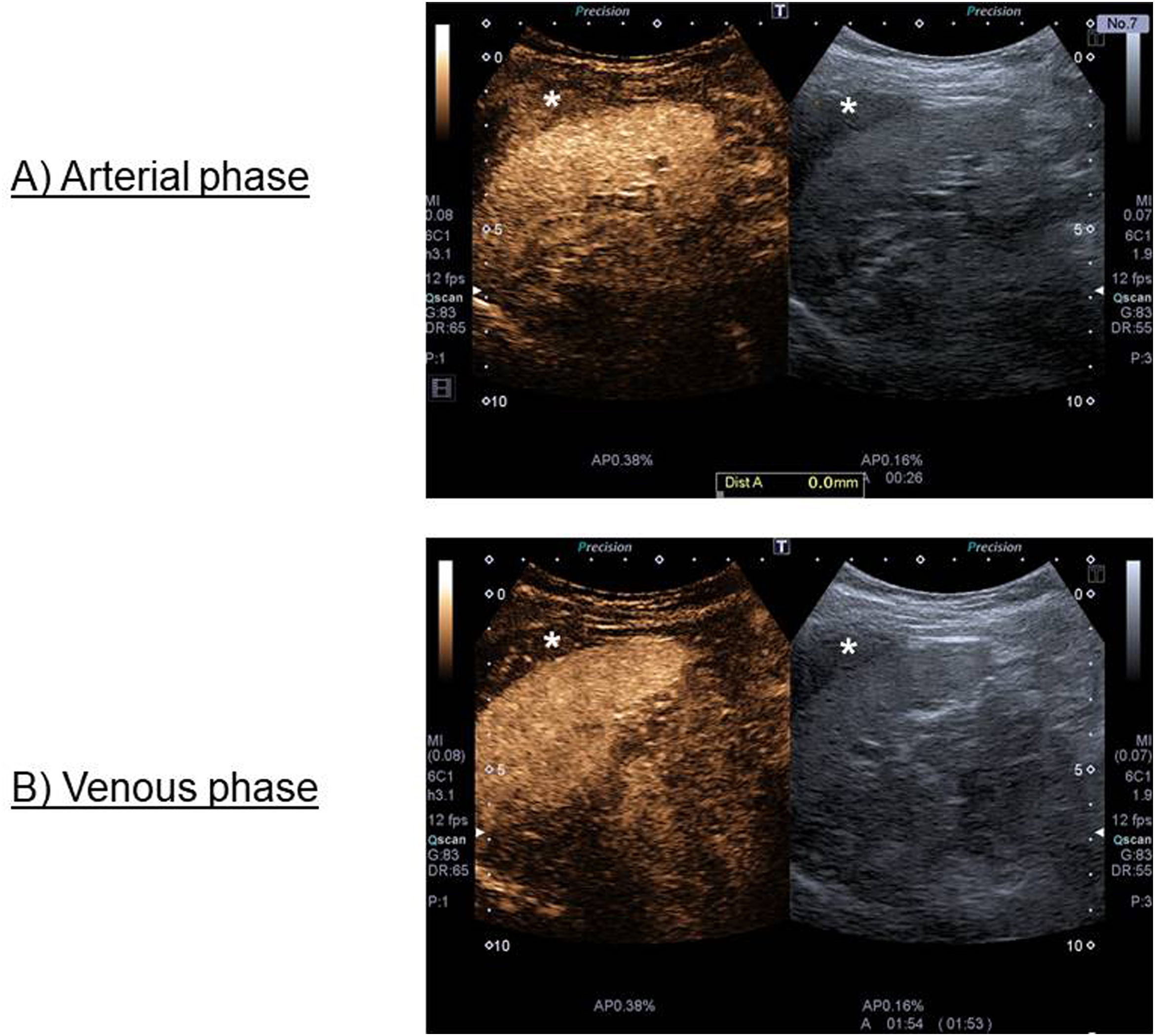
A study with intravenous contrast (SonoVue®) was performed, showing in arterial phase a homogeneous filling of the crescent-shaped formation with less intensity than the splenic parenchyma; in addition, the area showed washout with respect to the splenic surface in very late venous phase (Fig. 2).

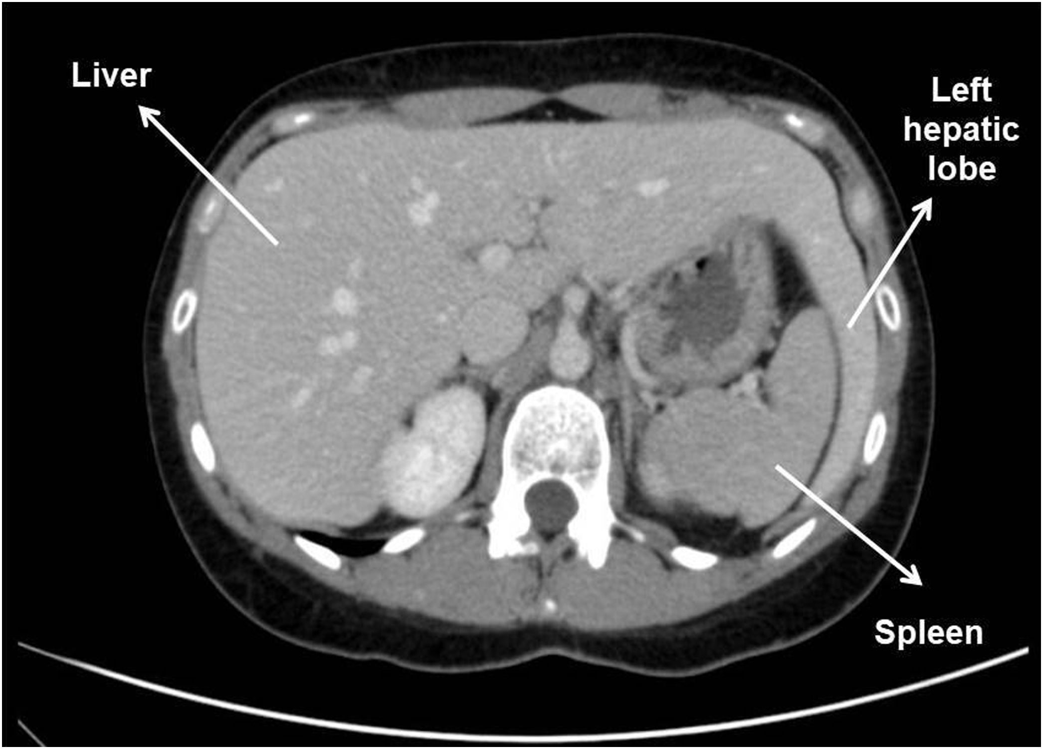
Finally, the dependence of this crescentic image on the hepatic parenchyma was appreciated, being in fact a prolongation of the same (left hepatic lobe) (Fig. 3), confirming by CT scan the variant of normality known as beaver tail liver, a rare hepatic shape with no pathological implications.1,2

It is important to know the possible variants of normality to avoid erroneous interpretations of the imaging tests. Also, the case shows the usefulness of intravenous contrast to increase the characterization capability of B-mode ultrasound.
FundingThere are no financing for this manuscript.
Conflicts of interestThe authors declare they have no conflicts of interest.