


The transtubular potassium gradient (TTKG) is a widely accepted tool for assessing mineralocorticoid activity in hyperkalemia. However, its clinical utility is often limited due to the unavailability of urine osmolality. Identifying alternative, more accessible urinary indices could improve diagnostic performance in suspected hypoaldosteronism.
MethodsWe conducted a retrospective observational study including 106 hyperkalemic patients with hypoaldosteronism referred to Endocrinology Department. Through correlation analyses using Spearman's ρ and Pearson's r, we assessed the relationship between TTKG values and the urinary potassium-to-serum potassium ratio (UK+/SK+), the urinary potassium-to-creatinine ratio (UK+/creatinine), the fractional excretion of potassium (FEK), and the urinary sodium-to-potassium ratio (UNa+/UK).
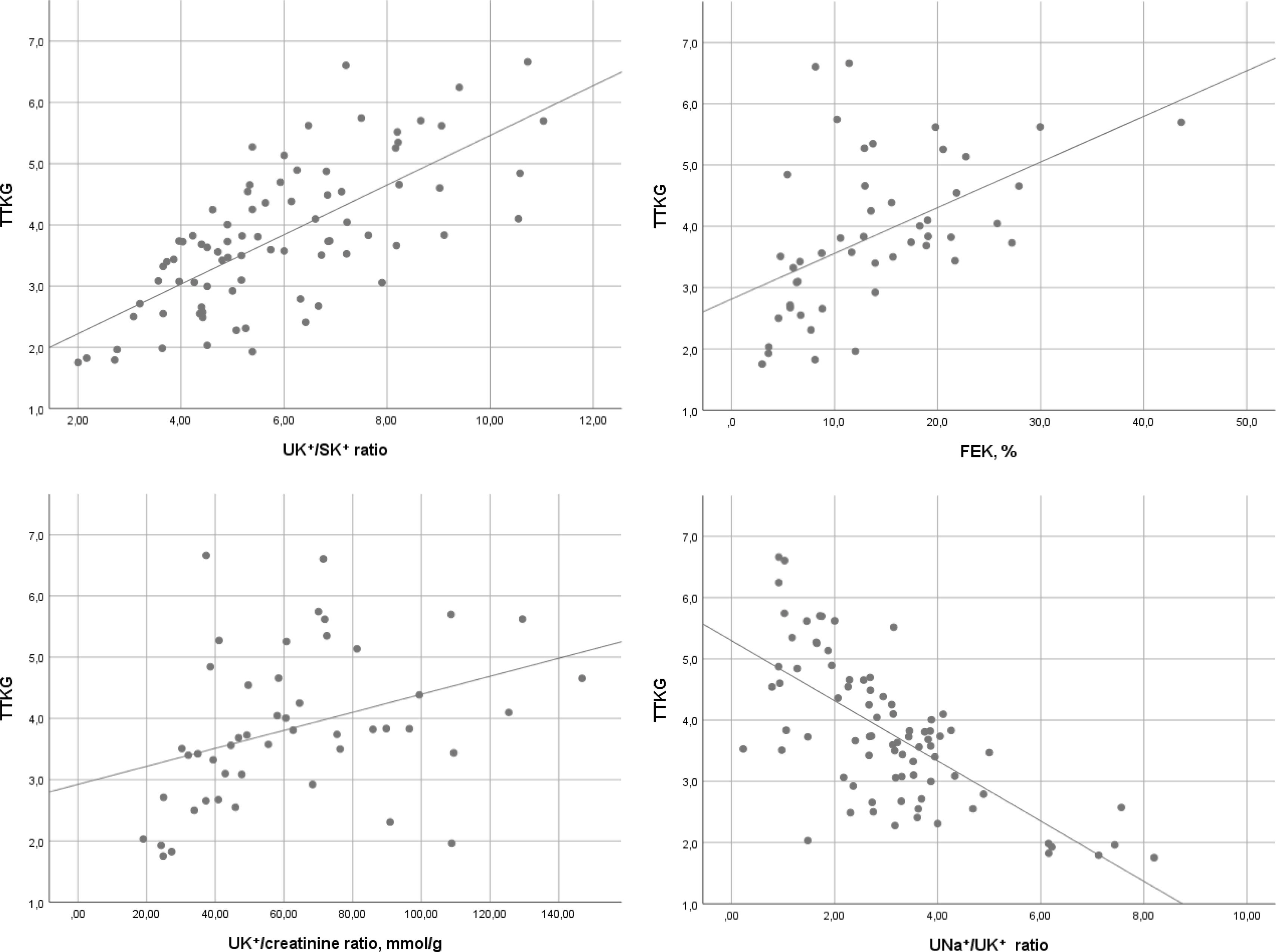
ResultsMean age, 74±12 years; mean TTKG, 3.8±1.2, and UK+/creatinine, 62±28mmol/g. Median UNa+/UK+, 2.78 [1.81–3.72]; UK+/SK+, 5.1 [4–6.8]; FEK, 12.9% [7–18.9]. TTKG showed a strong positive correlation with UK+/SK+ (ρ=0.706; p<0.001) and FEK (ρ=0.585; p<0.001), and a moderate correlation with UK+/creatinine (r=0.352; p=0.011). An inverse strong correlation was observed with UNa+/UK+ (ρ=−0.655; p<0.001).
ConclusionsUK+/SK, UK+/creatinine, FEK, and UNa+/UK+ seem to be practical alternatives for assessing renal potassium excretion in hyperkalemia associated with hypoaldosteronism. Further studies are warranted to validate these findings in larger and diverse cohorts.
El gradiente transtubular de potasio (TTKG) es una herramienta ampliamente aceptada para evaluar la actividad mineralocorticoide en contextos de hiperpotasemia. Sin embargo, su utilidad clínica se ve limitada por la frecuente indisponibilidad de la osmolalidad urinaria. La identificación de índices urinarios alternativos, más accesibles, podría mejorar la precisión diagnóstica en casos de sospecha de hipoaldosteronismo.
MétodosSe analizaron parámetros urinarios y plasmáticos en pacientes con hiperpotasemia por hipoaldosteronismo evaluados en una unidad especializada. Se calcularon el TTKG y diversos índices urinarios alternativos, como la razón potasio urinario/potasio sérico (UK+/SK+), el potasio urinario/creatinina (UK+/Cr), la excreción fraccional de potasio (FEK) y la razón sodio/potasio urinarios (UNa+/UK+), y se analizó su correlación entre ellos.
ResultadosLa edad media fue de 74±12 años. El TTKG medio fue 3,8±1,2; UK+/Cr: 62±28mmol/g. La mediana de UNa+/UK+ fue 2,78 [1,81-3,72], UK+/SK+: 5,1 [4-6,8] y FEK: 12,9% [7-18,9]. El TTKG mostró una correlación positiva fuerte con UK+/SK+ (ρ=0,706; p<0,001) y con FEK (ρ=0,585; p<0,001), y una correlación moderada con UK+/Cr (r=0,352; p=0,011). Se observó una correlación negativa fuerte con UNa+/UK+ (ρ=−0,655; p<0,001).
ConclusionesLos índices UK+/SK+, UK+/Cr, FEK y UNa+/UK+ se perfilan como alternativas prácticas al TTKG para evaluar el manejo renal del potasio en hiperpotasemia asociada a hipoaldosteronismo. Se requieren estudios adicionales en cohortes más amplias y diversas para validar la aplicación clínica de estos resultados.
Hypoaldosteronism is a condition marked by reduced aldosterone activity in the distal nephron, causing decreased urinary potassium (UK+) excretion, often with increased urinary sodium (UNa+) excretion and reduced hydrogen ion excretion.1 This leads to hyperkalemia, hypovolemic hyponatremia, and hyperchloremic metabolic acidosis.2
The prevalence of acquired hypoaldosteronism is largely unknown, but Wilczynski et al. reported it may cause 10–80% of hyperkalemia cases.3 Since hyperkalemia is common, hypoaldosteronism is likely frequent in clinical practice.
Accurate diagnosis of hypoaldosteronism requires a solid understanding of mineralocorticoid physiology. Aldosterone is a key regulator of renal potassium excretion in patients with preserved kidney function. Therefore, hypoaldosteronism should be suspected when persistent hyperkalemia occurs in a patient with a glomerular filtration rate (GFR) >20–30mL/min/1.73m2, as this is the threshold beyond which renal potassium clearance declines and mineralocorticoid activity becomes the key driver of its excretion.1,3 In this setting, concurrent with hyperkalemia, urinary potassium excretion should be evaluated to determine whether renal elimination is appropriate. In cases of impaired potassium excretion, hypoaldosteronism is highly probable.
To assess mineralocorticoid activity in this previous context, only a few urinary parameters are routinely considered as markers of mineralocorticoid activity – most notably the transtubular potassium gradient (TTKG)4 and the urinary sodium-to-potassium ratio (UNa+/UK+).5–7 Among these, TTKG is the most extensively studied and widely used.5,8–12 TTKG is closely associated with potassium homeostasis, where higher mineralocorticoid activity results in higher TTKG values5,8,9 and lower activity in lower values.5,8,9,12 Recently, the original authors of the TTKG calculation advised caution when interpreting it in conditions affecting the renal urea cycle, such as hypovolemia, as values may be higher than expected.13 However, this situation would not significantly impact its use in hypoaldosteronism, where, despite hypovolemia, a low TTKG value would make the presence of hypoaldosteronism even clearer. Thus, a TTKG value <7 in the presence of hyperkalemia is considered indicative of hypoaldosteronism,9,14,15 a criterion supported by a recent study,2 where all patients with hypoaldosteronism exhibited TTKG values below this threshold. Other parameters, such as the fractional excretion of potassium (FEK),16 the urinary potassium-to-creatinine ratio (UK+/creatinine),17 and the urinary-to-serum potassium ratio (UK+/SK+) may have potential diagnostic value, but current evidence supporting their use in this setting is limited or absent.
The calculation of TTKG requires measurements of serum and urinary electrolytes and osmolality during hyperkalemia. However, in routine clinical practice, urine biochemistry, especially osmolality, is often not measured,18,19 likely due to difficulties in interpretation or unfamiliarity with its clinical value,18,20 or due to technical20/logistical21 constraints. Consequently, when TTKG cannot be calculated, the utility of alternative urinary markers becomes particularly relevant. The aim of this study is to determine whether other urinary indices such as FEK, UK+/creatinine, UNa+/UK+, and UK+/SK+ can serve as reliable surrogates for TTKG in the evaluation of hypoaldosteronism-induced hyperkalemia.
MethodsStudy design and populationWe conducted a retrospective study of adult patients with hypoaldosteronism managed by the Endocrinology and Nutrition Department of Hospital Clínico San Carlos (Madrid, Spain), either during hospitalization or at the outpatient clinic, between 2012 and 2019. The study design and specific methodological details have been described previously.2,22 The study adhered to the principles of the Declaration of Helsinki and was approved by the Ethics Committee of Hospital Clínico San Carlos (Code 20/714-E_BS; December 14, 2020). The requirement for written informed consent was waived.
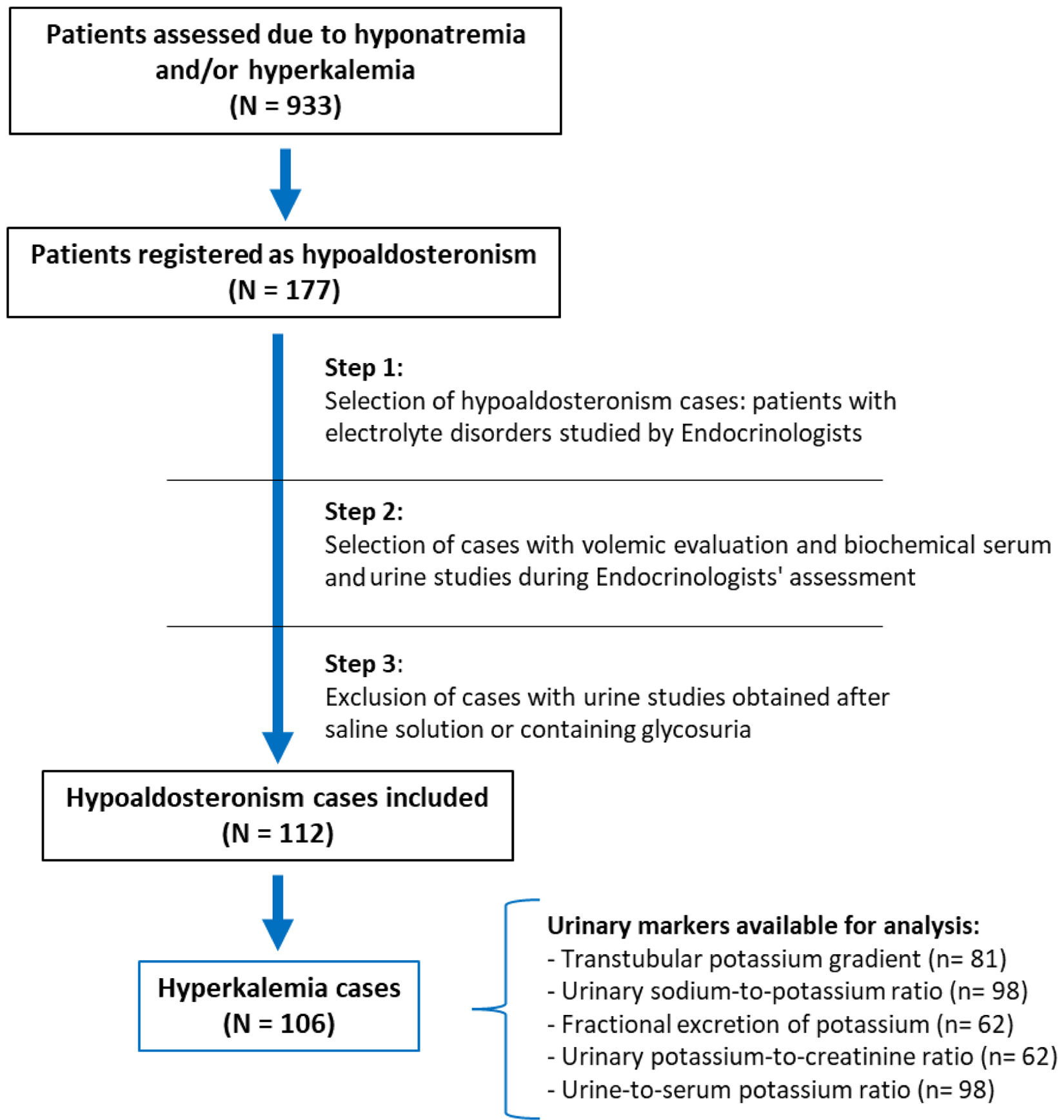
Briefly, the study collected demographic, clinical (comorbidities and concomitant treatments) and biochemical (serum and urinary) data related to the diagnostic evaluation of cases with hypoaldosteronism. The current study focused on cases with hyperkalemia, and urinary markers of potassium homeostasis were assessed. Additionally, we collected data from the moment of blood aldosterone and renin (direct renin concentration) evaluation, when available. The flowchart of cases selection is shown in Fig. 1.

In detail, hypoaldosteronism diagnosis was based on criteria exposed in Table 1, and performed in Step 1 of the above-mentioned flowchart. Transitory or permanent hypoaldosteronism due to adrenal surgery or direct optimal medical therapy for primary aldosteronism were excluded, as were pregnant patients, and those with hypervolemia of any cause, oliguric renal failure or GFR levels <30mL/min/1.73m2.
Criteria used for diagnosis of hypoaldosteronism.
| A. Non-spurious persistent hyperkalemia:At least two laboratory tests with hyperkalemia performed on different daysIn addition to:Absence of an external potassium loadGlomerular filtration rate >30mL/min/1.73m2 |
| B. Absence of a prior history of hypokalemia |
| C. An adequate therapeutic response:Improvement of the electrolyte disorder with mineralocorticoid replacement therapy and/or volume and salt repletion with isotonic saline, hypertonic saline, or increased water and salt intake. |
| Diagnosis required the presence of A, together with B and C. |
Hyperkalemia was defined as a serum potassium level (SK+) ≥5mmol/L. Spurious hyperkalemia was ruled out. All patients had GFR levels >30mL/min/1.73m2. Serum and urinary parameters were used to calculate TTKG, UNa+/UK+, FEK, UK+/creatinine, and the ratio of UK+/SK+ using the formulas shown in Table 1 of supplementary data.
Hyponatremia was defined as serum sodium (SNa+) levels ≤135mmol/L following correction for glycemia.23 Hypovolemia was defined as the presence of the maximum height of the internal jugular pulse (HIJP) at or below the sternal angle with the patient reclined at 0–30°, in addition to at least two of the following data: thirst, orthostatic symptoms/signs, blood pressure ≤90/60mmHg, heart rate ≥90bpm, decreased eye tone on palpation, distal venous filling of the upper limbs underneath the diaphragmatic line in a sitting position, and/or a rise in serum creatinine accompanying the descent in SNa.24,25 If HIJP was not measured, hypovolemia was determined by the presence of at least three of the above-mentioned symptoms/signs. Patients without symptoms/signs of hypovolemia and with a HIJP at 1–3cm above the sternal angle were classified as euvolemic.
Laboratory methodsAs per the routine clinical protocol, blood and urine samples are collected early in the morning, while fasting, before 09:00 a.m., and are processed by the laboratory the same morning. All biochemical variables used in the analysis were confirmed to have been obtained from morning-processed samples.
Serum and urinary electrolytes were assessed using the AU5800® Analyzer (Beckman Coulter) via indirect potentiometry (inter-assay coefficient of variation <1%), or by direct whole blood gas analysis when applicable. Plasma and urinary osmolality were measured by freezing point depression using the A2O® osmometer (Advanced Instruments, Inc.). Aldosterone and renin were measured in the hospital's central laboratory using commercially available radioimmunoassay kits.
Statistical analysisCategorical variables were expressed as frequencies and percentages; continuous variables as mean±SD (standard deviation) or median [IQR: interquartile range], depending on distribution. Ranges and 95% confidence intervals (95%CI) were reported when relevant. Normality was assessed using the Kolmogorov–Smirnov or Shapiro–Wilk tests.
Comparisons of categorical variables were performed using chi-squared or Fisher's exact tests. For quantitative variables, parametric tests (Student's t-test or ANOVA) or non-parametric tests (Mann–Whitney U or Kruskal–Wallis) were applied as appropriate. To explore the main objective of determining whether urinary indices could serve as surrogate markers for TTKG, we assessed their correlation using Spearman's test (ρ value) when non-parametric, and Pearson's test (r value) when parametric. A two-sided p-value <0.05 was considered statistically significant. Analyses were performed using SPSS version 25 (IBM Corp., NY, USA).
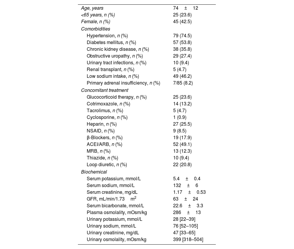
ResultsA total of 106 cases with hyperkalemia were evaluated. The mean age was 74±12 years. Forty-five (42.5%) patients were women. Hypovolemic hyponatremia was present in 55 of all patients (51.9%), and metabolic acidosis in 34 (59.6%) of the 57 cases with evaluated acid–base status. General characteristics of the studied cohort are shown in Table 2.
Characteristics of all cases included.
| Age, years | 74±12 |
| <65 years, n (%) | 25 (23.6) |
| Female, n (%) | 45 (42.5) |
| Comorbidities | |
| Hypertension, n (%) | 79 (74.5) |
| Diabetes mellitus, n (%) | 57 (53.8) |
| Chronic kidney disease, n (%) | 38 (35.8) |
| Obstructive uropathy, n (%) | 29 (27.4) |
| Urinary tract infections, n (%) | 10 (9.4) |
| Renal transplant, n (%) | 5 (4.7) |
| Low sodium intake, n (%) | 49 (46.2) |
| Primary adrenal insufficiency, n (%) | 7/85 (8.2) |
| Concomitant treatment | |
| Glucocorticoid therapy, n (%) | 25 (23.6) |
| Cotrimoxazole, n (%) | 14 (13.2) |
| Tacrolimus, n (%) | 5 (4.7) |
| Cyclosporine, n (%) | 1 (0.9) |
| Heparin, n (%) | 27 (25.5) |
| NSAID, n (%) | 9 (8.5) |
| β-Blockers, n (%) | 19 (17.9) |
| ACEI/ARB, n (%) | 52 (49.1) |
| MRB, n (%) | 13 (12.3) |
| Thiazide, n (%) | 10 (9.4) |
| Loop diuretic, n (%) | 22 (20.8) |
| Biochemical | |
| Serum potassium, mmol/L | 5.4±0.4 |
| Serum sodium, mmol/L | 132±6 |
| Serum creatinine, mg/dL | 1.17±0.53 |
| GFR, mL/min/1.73m2 | 63±24 |
| Serum bicarbonate, mmol/L | 22.6±3.3 |
| Plasma osmolality, mOsm/kg | 286±13 |
| Urinary potassium, mmol/L | 28 [22–39] |
| Urinary sodium, mmol/L | 76 [52–105] |
| Urinary creatinine, mg/dL | 47 [33–65] |
| Urinary osmolality, mOsm/kg | 399 [318–504] |
ACEI: angiotensin-converting enzyme inhibitors, ARB: angiotensin-II receptor blockers, MRB: mineralocorticoid receptor blockers, NSAID: non-steroidal anti-inflammatory drugs, GFR: glomerular filtration rate.
The TTKG was calculable in 81 of 106 cases (76.4%). The UNa+/UK+ and UK+/SK+ ratios could be determined in 98 cases (92.4%), whereas both the fractional excretion of potassium (FEK) and urinary potassium-to-creatinine ratio (UK+/creatinine) were calculable in 62 cases (58.5%). All cases showed TTKG values <7 and UK+/creatinine values <150mmol/g. In addition, 61 of 62 cases (98.4%) had FEK values ≤30%, 91 of 98 (92.9%) had UNa+/UK+ ratios >1, and 94 of 98 (95.9%) had UK+/SK+ ratios ≤10. Descriptive statistics for these parameters are shown in Table 3.
Descriptive analysis of urinary potassium indices.
| Mean±SD | 95% CI of mean | Median [IQR] | Min–max | |
|---|---|---|---|---|
| TTKG | 3.8±1.2 | 3.5–4.1 | 3.7 [2.9–4.5] | 1.8–6.7 |
| UNa+/UK+ | 2.97±1.55 | 2.85–3.28 | 2.78 [1.81–3.72] | 0.23–8.2 |
| UK+/SK+ | 5.5±2.1 | 5.1–5.9 | 5.1 [4–6.8] | 2–11 |
| FEK, % | 13.6±7.7 | 11.7–15.6 | 12.9 [7–18.9] | 3–43.7 |
| UK+/creatinine, mmol/g | 62±28 | 55–69 | 57 [41–76] | 19–147 |
TTKG: transtubular potassium gradient; UNa+/UK+: urinary sodium-to-potassium ratio; UK+/SK+: urinary-to-serum potassium ratio; FEK: fractional excretion of potassium; UK+/creatinine: urinary potassium-to-creatinine ratio.
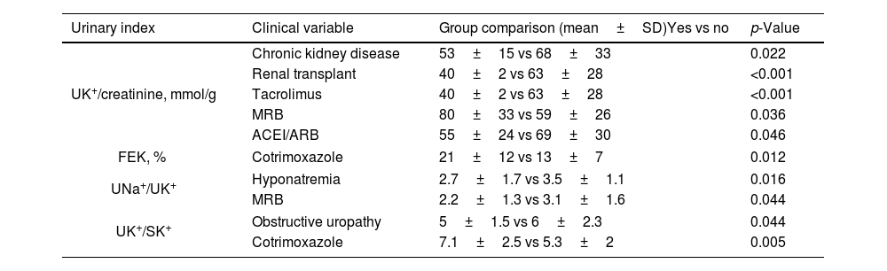
In the subgroup analyses stratified by sex, comorbidities, ongoing treatments, and the presence of concomitant hyponatremia or metabolic acidosis, TTKG values remained consistent across all groups. In contrast, the UK+/creatinine ratio was significantly influenced by several clinical variables, while FEK, UNa+/UK+, and UK+/SK+ showed more modest but still notable variations, as shown in Table 4.
Clinical variables significantly affecting each urinary potassium index.
| Urinary index | Clinical variable | Group comparison (mean±SD)Yes vs no | p-Value |
|---|---|---|---|
| UK+/creatinine, mmol/g | Chronic kidney disease | 53±15 vs 68±33 | 0.022 |
| Renal transplant | 40±2 vs 63±28 | <0.001 | |
| Tacrolimus | 40±2 vs 63±28 | <0.001 | |
| MRB | 80±33 vs 59±26 | 0.036 | |
| ACEI/ARB | 55±24 vs 69±30 | 0.046 | |
| FEK, % | Cotrimoxazole | 21±12 vs 13±7 | 0.012 |
| UNa+/UK+ | Hyponatremia | 2.7±1.7 vs 3.5±1.1 | 0.016 |
| MRB | 2.2±1.3 vs 3.1±1.6 | 0.044 | |
| UK+/SK+ | Obstructive uropathy | 5±1.5 vs 6±2.3 | 0.044 |
| Cotrimoxazole | 7.1±2.5 vs 5.3±2 | 0.005 | |
UK+/creatinine: urinary potassium-to-creatinine ratio; FEK: fractional excretion of potassium; UNa+/UK+: urinary sodium-to-potassium ratio; UK+/SK+: urinary-to-serum potassium ratio; ACEI: angiotensin-converting enzyme inhibitors; ARB: angiotensin-II receptor blockers; MRB: mineralocorticoid receptor blockers.
The TTKG was directly correlated with the UK+/SK+ (ρ=0.706, p<0.001), the UK+/creatinine (r=0.352, p=0.011), and the FEK (ρ=0.585; p<0.001), while inversely correlated with the UNa+/UK+ (ρ=−0.655, p<0.001). Dispersion graphics of these analyses are shown in Fig. 2.
Urinary indices and blood aldosterone
Aldosterone and renin levels were measured in 82 cases, coinciding with hyperkalemia in 52 cases. In these cases, median aldosterone and renin concentrations were 82pg/mL (IQR, 40–152) and 17pg/mL (IQR, 8–46), respectively. Aldosterone showed no significant correlation with serum potassium (SK+; ρ=−0.040, p=.783), transtubular potassium gradient (TTKG; ρ=0.167, p=.316), urinary-to-serum potassium ratio (UK+/SK+; ρ=0.049, p=.744), urinary potassium-to-creatinine ratio (UK+/creatinine; ρ=−0.083, p=.647), or fractional excretion of potassium (FEK; ρ=0.107, p=.533).
DiscussionThis study demonstrated that alternative urinary indices calculable without the need for osmolality measurements – such as FEK, UK+/creatinine, the ratio of UK+/SK+, and UK+/SK+ – showed significant correlation with TTKG during hyperkalemia in the context of hypoaldosteronism. These findings suggest that such markers possibly serve as reliable surrogate indicators for TTKG in clinical scenarios similar to those evaluated in our study.
First and foremost, to test our hypothesis, it is important to note that – beyond the consistently low TTKG values observed in all patients – the other urinary indices of potassium excretion also indicated an inadequate renal response to hyperkalemia, as expected based on the clinical diagnosis of hypoaldosteronism. Strikingly, the values of these parameters – except for UK+/creatinine – were either unaffected or only mildly influenced by different clinical variables. Most importantly, none of them exhibited a clinically relevant change in response to these variables. Specifically, the UK+/creatinine ratio was inappropriately low in all cases, falling below the expected threshold of 150mmol/g during hyperkalemia.17 All but one patient showed a reduced FEK, despite values >30% being typically expected in the setting of adequate renal potassium excretion.16 The UNa+/UK+ ratio – an additional but less frequently used marker of mineralocorticoid activity – was often >1, whereas values <1 are usually anticipated when the mineralocorticoid axis is intact.5–7 Moreover, the UK+/SK+ ratio – a simple and previously unexplored parameter conceptually reflecting urinary potassium in relation to concurrent serum potassium – was <10 in more than 95% of cases. Although still unvalidated in the literature, this marker may hold potential value for assessing renal potassium handling. In fact, Rabelink et al.7 observed that in healthy subjects, as an adequate response of mineralocorticoid activity, the UK+/SK+ ratio increased following a marked rise in potassium intake. Taken together, these findings support our hypothesis that alternative urinary indices may provide insights comparable to TTKG in assessing renal potassium excretion and mineralocorticoid activity.
Former studies have evaluated the FEK,16,26 the UK+/creatinine ratio,17,18 and the UNa+/UK+5–7 ratio as indices of renal potassium handling. The first two as markers of renal excretion, and the latter as a marker of mineralocorticoid activity. However, despite their physio-pathological relevance, none of these parameters have been thoroughly studied as a diagnostic tool in hyperkalemic patients, much less in hypoaldosteronism, and are not consistently included in the diagnostic algorithm for hyperkalemia. With the exception of the recent Spanish multisocietal consensus,27 which briefly mention this aspect, no formal clinical practice guidelines exist on the diagnostic approach to hyperkalemia, with most recommendations stem from narrative reviews or dedicated to treatment management. Nevertheless, our results showing a significant correlation between these markers with TTKG support the potential utility of all these indices, alongside TTKG, in the evaluation of hyperkalemia in patients with suspected hypoaldosteronism. Although additional longitudinal studies are needed to validate their clinical applicability and performance.
Unfortunately, studies specifically evaluating the same aims as ours are scarce, limiting meaningful comparisons. Musso et al. reported a negative correlation between TTKG and creatinine clearance in adult patients without hyperkalemia,28 a finding similar to the well-known inverse relationship between FEK and GFR in normokalemic individuals.26 This supports the idea that TTKG and FEK behave in parallel, as observed in our study. In fact, a direct correlation between TTKG and FEK has previously been described in non-hyperkalemic adults with chronic kidney disease receiving Olmesartan,29 and in healthy children.30 Both studies also found a correlation between TTKG and the UNa+/UK+ ratio. However, to our knowledge, no study has evaluated the correlation between TTKG and each of the urinary indices examined in our work in the setting of hyperkalemia due to hypoaldosteronism.
In contrast to our hypothesis suggesting that any of the evaluated parameters may aid in diagnosing hypoaldosteronism, Chacko et al.5 reported that TTKG, FEK, and the UNa+/UK+ ratio failed to do so. They observed overlapping values between pharmacologically induced hyper- (2mg/day fludrocortisone) and hypoaldosteronism (200mg/day spironolactone) in a crossover study with 8 healthy subjects. However, each intervention was administered for only 4 days, with a 2-week washout period, which may have been insufficient for full electrolyte adaptation. Moreover, the use of healthy volunteers could have introduced compensatory renal mechanisms in potassium handling that are absent in chronically ill patients with hypoaldosteronism who spontaneously develop hyperkalemia. Therefore, these findings may not be directly extrapolable to real-world clinical settings. However, further prospective investigation is warranted to confirm the diagnostic utility of the above-mentioned indices.
Interestingly, not all patients in our study had a full set of urinary measurements. The least frequently recorded indices were those requiring urinary creatinine. In contrast, TTKG was available in most patients. This diverges from prior reports highlighting the underuse of urine osmolality in the routine clinical practice,18–20 and likely reflects the clinical routinary approach at our Endocrinology Department. In this setting, urinary osmolality is routinely measured to calculate TTKG whereas urinary creatinine is less widely requested, possibly due to the limited validation of creatinine-based indices for investigating endocrinological causes of hyperkalemia, namely hypoaldosteronism. Consequently, these indices are not usually incorporated in endocrinology-focused clinical management. This situation highlights the other side of the coin and further reinforces the rationale of our study: to evaluate the potential utility of all these urinary indices by exploring their correlation with TTKG in a real-life clinical cohort.
It is not unexpected that aldosterone levels did not correlate with TTKG or with other urinary markers of potassium excretion, given that all patients in our cohort had hypoaldosteronism. In individuals with intact mineralocorticoid function, aldosterone typically shows a direct correlation with renal potassium secretion parameters.6,30 However, in the presence of impaired aldosterone production or action, this physiological relationship is lost. While it is rare in endocrinological practice for the direct measurement of a hormone to provide limited diagnostic value, our findings illustrate precisely that in hyperkalemic patients with hypoaldosteronism, measuring aldosterone alone may be insufficient. In such cases, assessing the renal response through urinary indices may offer a more practical and informative approach to evaluating mineralocorticoid activity.
Diagnosing electrolyte disturbances such as hyperkalemia is often challenging – particularly when the evaluation occurs after the acute episode has resolved and key biochemical data were not collected. In such cases, clinicians may only have access to limited urinary parameters (e.g., urinary ions alone, or combined with creatinine or osmolality). In instances of previously resolved hyperkalemia not associated with significant reductions in GFR – where hypoaldosteronism is considered a likely cause, as highlighted by Spanish experts27 – the identification of other indices beyond TTKG, calculable from the available biochemical data, becomes clinically relevant.
To illustrate a potential application of our findings, consider a scenario in which urinary osmolality was not measured, making TTKG calculation impossible. In such cases, alternative indices – such as FEK, UK+/creatinine, UNa+/UK+, and/or UK+/SK+ – can still be calculated. If one or more of these markers fall within a range suggestive of impaired potassium excretion, they may help support or reinforce clinical suspicion of hypoaldosteronism. Therefore, our study provides a novel contribution by demonstrating that simpler and more readily available urinary parameters can offer insights comparable to those of TTKG in evaluating renal potassium handling, in hyperkalemia related to hypoaldosteronism. Nonetheless, these findings should be interpreted with caution, as further studies are needed to compare these indices across contrasting states of renal potassium handling to properly validate their diagnostic value.
The main limitations of our study are related to its retrospective design. Only a few markers were available in a subset of patients, introducing a potential selection bias and decreasing the statistical power for certain analyses. The fact that multiple mechanisms (drugs and comorbidities) potentially involved on hypoaldosteronism genesis were present in a several number of cases, as shown in Table 1, could also interfere with results, although findings were consistent across all clinical parameters explored. Another factor that could have influenced the results is potassium intake, which was not recorded. On the other hand, the main strengths of this study lie in the meticulous case selection, diagnostic homogeneity, and its representativeness of real-world clinical practice. Moreover, to our knowledge, this is the first study to evaluate the utility of alternative urinary potassium excretion markers as potential surrogates for the TTKG in cases of hypoaldosteronism with hyperkalemia.
Overall, these results support the possible clinical utility of simple urinary ratios to estimate renal potassium handling when TTKG is unavailable. While TTKG remains a valuable parameter and should be considered in hyperkalemia evaluation, our findings suggest that indices such as FEK, UK+/creatinine, UK+/SK, and UK+/SK+ offer practical and physiologically relevant alternatives. Nonetheless, further prospective studies across diverse clinical contexts – including Intensive Care, Internal Medicine, Nephrology, and Primary Care – are needed to validate these results.
Author's contributionsConceptualization: JGR-S, IR; Methodology: JGR-S, ALCP, MPDMN, MARH; Validation: JGR-S, ISR; Formal analysis: JGR-S; Investigation: JGR-S; Writing – original draft preparation: JGR-S, IR; Writing – review & editing: JGR-S, ALCP, MPDMN, MARH, IR; Supervision and reviewing: IR, ALCP, MPDMN, MARH; Resources: JGR-S, ALCP, IR. All authors have read and agreed to the published version of the manuscript.
Ethical approvalThe study was approved by the Ethical Committee of the HCSC (Cod. 20/714-E_BS, December 14th, 2020).
Consent to participateWritten informed consent was waived given the registry's anonymized and retrospective nature.
Consent for publicationWritten informed consent was waived given the registry's anonymized and retrospective nature.
FundingThis research received a grant from Sociedad de Endocrinología, Nutrición y Diabetes de la Comunidad de Madrid (SENDIMAD) through the “Beca de Ayuda a la Investigación SENDIMAD 2020” awarded on November 25th, 2020 in Madrid, Spain. During the period of data collection, JGRS was employed as a research fellow by the Foundation for Biomedical Research at Hospital Clínico San Carlos (Reference No. INV-15-2019).
Conflicts of interestNone declared.
The following are the supplementary data to this article: