Patients with hypoparathyroidism are at a higher cardiovascular risk (CVR) due to mechanisms that are not yet well understood. In patients with renal failure, fibroblast growth factor 23 (FGF23) has been associated with an increased calcification and arterial stiffness (AS), leading to an increased CVR. In hypoparathyroidism—due to the situation of hyperphosphatemia—an increase in FGF23 is also probable. There is a paucity of research examining the relationship between FGF23 levels and AS in patients with hypoparathyroidism. The present study aims to assess AS, by measuring aortic pulse wave velocity (aPWV), and FGF23 levels in patients with hypoparathyroidism vs age and sex matched healthy controls.
Material and methodsWe conducted a cross-sectional descriptive study, and collected demographical data of patients and treatment characteristics (calcium and vitamin D doses). Furthermore, we measured aPWV using the SphygmoCorXCEL® device and assessed FGF23 and all other parameters of calcium–phosphorus metabolism in a blood and 24h-urine samples.
Results and discussionA total of 20 subjects with hypoparathyroidism (85% postoperative) and 16 healthy matched controls were included. The mean age was 58.6 years (SD, 12.2). The mean course of the disease was 16.31 years (3–44). Regarding the general characteristics, the most remarkable finding was that patients had an estimated glomerular filtration rate (eGFR) significantly lower than controls (72.5 [SD, 21.25] vs 95.3 [SD, 12.21]ml/min/1.73m2; p<0.001). FGF23 levels were clearly elevated in subjects with hypoparathyroidism (255.2 [35.9–1487.4] vs 61.9 [27.8–134.8]RU/mL; p<0.05). A trend toward higher AS was observed in subjects with hypoparathyroidism (aPWV 8.4 [5.2–17.3] vs 6.9 [5.1–10.8]m/s; p=0.08), as well as a positive correlation between aPWV and time of disease progression (r=0.679; p<0.01). A negative correlation was observed in subjects with hypoparathyroidism between aPWV and eGFR (−0.519; p<0.05). We found no significant correlation between aPWV and FGF23 or with phosphate levels or calcium supplements or calcitriol doses used in replacement treatment.
ConclusionsIn subjects with hypoparathyroidism there is a marked increase in FGF23 levels and a trend toward a higher AS, which correlates with disease duration. However, these findings may be conditioned by an impaired renal function that this pathological process entails. A causal relationship could not be established between the elevated levels of FGF23 and AS.
Los pacientes con hipoparatiroidismo presentan un riesgo cardiovascular (RCV) incrementado debido a mecanismos todavía no conocidos completamente. En pacientes con insuficiencia renal, el factor de crecimiento de fibroblastos 23 (FGF23) se ha asociado con un aumento de la calcificación y rigidez arterial (RA), contribuyendo así a un mayor RCV. En el hipoparatiroidismo, dada la presencia habitual de hiperfosfatemia, es probable que también se observe un incremento en los niveles de FGF23. Sin embargo, la relación entre FGF23 y la rigidez arterial en esta población ha sido poco estudiada. El objetivo de este estudio es evaluar la rigidez arterial, mediante la medición de la velocidad de onda de pulso aórtica (VOPa), y los niveles plasmáticos de FGF23 en pacientes con hipoparatiroidismo en comparación con controles sanos emparejados por edad y sexo.
Material y métodosEstudio descriptivo transversal en el que se recogieron datos demográficos y características del tratamiento (dosis de calcio y vitamina D), se midió la VOPa utilizando el dispositivo SphygmoCorXCEL® y se determinaron los niveles de FGF23, así como otros parámetros del metabolismo calcio-fósforo en muestras de sangre y de orina simple y de 24 horas.
Resultados y discusiónSe incluyeron 20 sujetos con hipoparatiroidismo (85% postquirúrgico) y 16 controles sanos emparejados. La edad media fue de 58,6 años (DE 12,2). El tiempo medio de evolución de la enfermedad fue de 16,31 años (rango 3-44). Entre las características generales, se observó que los pacientes presentaban una tasa de filtración glomerular estimada (TFGe) significativamente menor que los controles (72,5 [DE 21,25] vs 95,3 [DE 12,21] ml/min/1,73 m2; p <0,001). Se objetivaron niveles de FGF23 claramente elevados en pacientes con hipoparatiroidismo (255,2 [rango 35,9–1487,4] vs 61,9 [27,8–134,8] RU/mL; p <0,05). Se evidenció una tendencia a una mayor rigidez arterial en los sujetos con hipoparatiroidismo (VOPa 8,4 [5,2–17,3] vs 6,9 [5,1–10,8] m/s; p=0,08), así como una correlación positiva entre VOPa y duración de la enfermedad (r=0,679; p <0,01). Además, se observó una correlación negativa entre VOPa y TFGe en este grupo (-0,519; p <0,05). No se encontró correlación significativa entre VOPa y los niveles de FGF23, ni con los niveles de fosfato, ni con las dosis de suplementos de calcio o calcitriol empleados en el tratamiento sustitutivo.
ConclusionesEn pacientes con hipoparatiroidismo existe un aumento marcado de los niveles de FGF23 y una tendencia a una mayor rigidez arterial, que se correlaciona con la duración de la enfermedad. Sin embargo, estos hallazgos podrían estar condicionados por el deterioro de la función renal asociado a esta patología. No se pudo establecer una relación causal entre los niveles elevados de FGF23 y la rigidez arterial.
Hypoparathyroidism is a rare disorder characterized by hypocalcemia and undetectable or inappropriately low parathyroid hormone (PTH) levels. Although hyperphosphatemia is widely observed, its severity can vary.
The estimated prevalence is about 24 per 100,000 inhabitants and in 80% of cases is due to an adverse effect of thyroid and parathyroid surgery. The remaining causes are autoimmune (associated or not with mutations in the AIRE gene), genetic (including DiGeorge syndrome, or mutations in CASR, GCM2, among others), or idiopathic. Attempts to develop hormone replacement therapy with recombinant PTH are under development but the standard treatment for hypoparathyroidism remains the administration of calcium supplements to prevent hypocalcemia and acting as phosphorus chelators along with calcitriol or other vitamin D analogs.
The pathophysiology of the disease and its current treatment leads to the development of complications such as renal lithiasis and tissue calcifications, including vascular vessels.1,2 A certain analogy can be stablished between the vascular deleterious effect caused by hypoparathyroidism and that observed in chronic renal disease (CKD). In the latter, there is also a trend toward hyperphosphatemia—due to the inability to excrete phosphate—and hypocalcemia—due to the final hydroxylation step of vitamin D is compromised.
Fibroblast growth factor 23 (FGF23) is increased in patients with CKD, mainly due to hyperphosphatemia and is a well- established CVR factor.3 Ix et al. observed FGF23 levels were associated with all-cause mortality and incidental heart failure, associations that were particularly strong among people with CKD. They also noted that hyperphosphatemia and Vitamin D deficiency are more strongly associated with subclinical cardiovascular disease (CVD) in individuals with vs without CKD.4
In 2015, Underbjerg et al. conducted a nationwide controlled historic follow- up study in Denmark and observed that patients with non-surgical hypoparathyroidism had significantly more CKD (hazard ratio [HR], 6.01; 95%CI, 2.45–14.75) and a significantly higher CVD risk (HR, 1.91; 95%CI, 1.29–2.81) vs controls.5 Later, in 2018, Vadiveloo et al. documented an increased mortality and CVD risk (HR, 2.18 [1.41–3.39]) in non-surgical hypoparathyroidism patients in a population-based study in Scotland. They observed that renal failure and death were associated with elevated serum calcium concentrations.6 Meena et al.7 investigated vascular damage associated with hypoparathyroidism by measuring intima-media thickness in the carotid (CIMT), renal, and aortic arteries, demonstrating that it was significantly increased in patients with spontaneous idiopathic hypoparathyroidism (SIH) vs healthy controls.7 In addition, Agarwal et al. observed that patients with SIH seemed to have more CVD assessed by coronary calcium score measured by multidetector computer tomography (MDCT), while low serum calcium levels could be a predisposing factor for this increased risk.8
The measurement of AS, directly by estimating the aPWV between the femoral and carotid arteries or indirectly by analyzing the pulse wave at the aortic root and the calculation of the Augmentation Index (AIx), has become a major and very useful parameter in cardiovascular medicine. In fact, the velocity at which the pulse wave propagates through the arterial tree is related to the intrinsic elasticity of its wall. In stiffer arteries, this velocity is higher and, when measured over the aorta, its value as an independent prognostic factor for cardiovascular events has been widely established. For all of this, the measurement of aPWV is considered the gold-standard technique for measuring AS.9–11 It is estimated that by each 1m/s of aPWV increase, the risk of a vascular event rises about 15%.10 Therefore, the measurement of AS may be useful to identify patients at high risk of cardiovascular events.9–12
The relationship between AS and hypoparathyroidism remains largely unknown. A Danish study has recently used the measurement of AS to assess the vascular impact of non-surgical hypoparathyroidism vs pseudohypoparathyroidism, showing that patients with hypoparathyroidism have higher AS than patients with pseudohypoparathyroidism.13 In addition, a study by Pamuk et al.8 conducted in a Turkish population compared 42 patients with hypoparathyroidism (postoperative or spontaneous) with 60 healthy controls matched for age, sex, and body mass index (BMI). They observed higher central and peripheral systolic (SBP) and diastolic blood pressure (DBP), and higher aPWV in the hypoparathyroidism group (PWV, 6.2m/s [4.7–10.2] vs the control group 5.9m/s [4.3–8.5; p=0.02]. Age and phosphorus levels turned out to be independent predictors of PWV.14
PTH is the fundamental hormone in human calcium and phosphorus metabolism. However, we are increasingly aware of the important role that FGF23 also plays. FGF23 is produced by osteocytes and osteoblasts. Both PTH and FGF23 are phosphaturic hormones and regulate phosphate homeostasis by inhibiting phosphate absorption in intestine and reabsorption in the kidneys through decreasing membrane expression of sodium- phosphate cotransporters NpT2a and NpT2c. PTH increases 1α-hydroxylase activity increasing active vitamin D levels and calcium absorption in intestine and reabsorption in the kidney, whereas FGF23 decreases 1α hydroxylase activity, thereby decreasing the absorption of calcium. PTH and FGF23 regulate the synthesis of each other. Higher serum phosphate increases the synthesis of FGF23 and lowers serum calcium, which will trigger synthesis of PTH.15–20
Hypoparathyroidism, due to hyperphosphatemia and regular treatment with calcitriol is another situation where FGF23 should very plausibly be increased. A study in patients with postoperative transient hypoparathyroidism documented transient elevation of FGF23 reactive to hyperphosphatemia.21 Another study published in 2022 showed that the phosphaturic effects of PTH and FGF23 are interdependent and both are required to adequately regulate renal phosphate handling.22 However, still there are very limited data on the role of FGF23 in the vascular injury present in chronic hypoparathyroidism.
The measurement of AS in patients with hypoparathyroidism and its comparison with healthy controls may be useful to better quantify and characterize the vascular deleterious effect of the disease. The potential relationship between AS values and variables such as the type of treatment or time of disease progression may help to optimize therapeutic management and to identify subjects at higher CVR who would benefit from other diagnostic and therapeutic measures. The study of the correlation of AS with levels of soluble FGF23 may help to improve our knowledge of the pathophysiology of vascular damage in hypoparathyroidism.
Materials and methodsWe conducted a cross-sectional descriptive study. Patients with hypoparathyroidism (either postoperative or spontaneous) treated at our endocrinology department and age- and sex-matched healthy controls were recruited for the study. Inclusion criteria were age≥18 years, spontaneous hypoparathyroidism of any duration or a 1-year history of postoperative hypoparathyroidism, active treatment with calcium supplements and vitamin D analogs, and agreement to participate in the study. Patients with eGFR levels<30ml/min/1.73m2 or previous CVD were excluded.
The research project was reviewed and approved by the hospital Drug Research Ethics Committee. This study was conducted in accordance with Good Clinical Practice (GCP) guidelines and in full compliance with the ethical principles outlined in the Declaration of Helsinki. All participants gave their prior written informed consent to participate in the study.
To perform sample size calculation, we rely on data from the aPWV of subjects with hypoparathyroidism from the study by Underbjerg13 [7.6±1.6m/s] and healthy population data from the study of AS in subjects with type 1 diabetes mellitus by Llauradó et al.23 [men, 6.3 (5.7–6.7)m/s; women, 6.0 (5.3–6.7)m/s], assuming a common standard deviation of 1m/s, an α error of 5%, and a β error of 20%. It was estimated that at least 16 subjects with hypoparathyroidism and 16 controls would be needed to demonstrate a difference in aPWV of 1m/s across both groups.
The following variables were collected: demographic data, weight, body mass index (BMI), blood pressure, treatment (with particular focus, in the case of hypoparathyroidism, on the type and daily dose of calcium, as well as on 25-hydroxyvitamin D and 1,25-dihydroxyvitamin D levels) and degree of compliance. CVR factors (diabetes, hypertension, dyslipidemia, obesity and tobacco consumption) and complications of hypoparathyroidism.
AS was assessed using the SphygmoCorXCEL® device (Atcor Medical, Australia) by the same operator. aPWV was determined by sequential applanation tonometry at the carotid and femoral arteries(m/s), gated to three-lead electrocardiography. Central pulse wave analyses was used to calculate Aix (%) as a surrogate marker of AS.
A blood sample was collected and analyzed in the same laboratory with determination in serum of fasting glucose (mg/dL), calcium (mg/dL), phosphorus (mg/dL), magnesium (mg/dL), albumin (g/dL), ionic calcium (mg/dL), PTH (pg/mL), 25-(OH) vitamin D (ng/mL), 1,25-(OH) vitamin D (pg/mL), creatinine (mg/dL), sodium (mmol/L), potassium (mmol/L), total cholesterol (mg/dL), HDL-cholesterol (mg/dL), and triglycerides (mg/dL). FGF23 (RU/mL) levels were measured in plasma from the collected blood sample.
A 24h urine sample was collected to measure 24h calcium (mg/24h). LDL-cholesterol (mg/dL) was calculated using Friedewald's formula, calcium×phosphate product value and estimated glomerular filtration (eGFR) rate according to CKD-EPI. All patients showed acceptable biochemical control with their regular treatment.
Statistical analysis was performed using the free software R (version 4.0.5, R Commander interface). For descriptive statistics, quantitative variables with a normal distribution were expressed as mean and standard deviation (SD). For quantitative variables without a normal distribution, results were expressed as median and range. Normality of quantitative variables was assessed using the Kolmogorov–Smirnov test. Qualitative variables were expressed as percentages. Inferential comparisons were drawn using parametric and non-parametric tests. For normally distributed quantitative variables, independent samples t-tests were conducted. For non-normally distributed quantitative variables, the Wilcoxon rank-sum test was used. Categorical variables were compared using the Chi-squared test. Statistical significance was defined as a p-value<0.05.
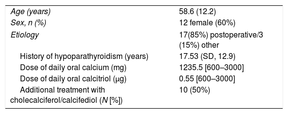
ResultsA total of 20 subjects with hypoparathyroidism and 16 healthy controls, matched for age and sex, were consecutively recruited for the study. The characteristics of the patients with hypoparathyroidism are shown in Table 1. The group included 12 women and 8 men, with a mean age of 58.6 years (SD, 12.2). The predominant etiology of hypoparathyroidism was postoperative (85%), and the mean course of the disease, 16.3 years (SD, 11.11). All patients were on oral calcium, predominantly calcium carbonate, at a median calcium dose of 1235.5mg/day [600–3000mg]. Nearly all patients (18/20) were treated with calcitriol at a median dose of 0.55mg/day [0–1]. Additionally, 50% of the patients were on cholecalciferol or calcifediol.
Clinical characteristics of subjects with hypoparathyroidism.
| Age (years) | 58.6 (12.2) |
| Sex, n (%) | 12 female (60%) |
| Etiology | 17(85%) postoperative/3 (15%) other |
| History of hypoparathyroidism (years) | 17.53 (SD, 12.9) |
| Dose of daily oral calcium (mg) | 1235.5 [600–3000] |
| Dose of daily oral calcitriol (μg) | 0.55 [600–3000] |
| Additional treatment with cholecalciferol/calcifediol (N [%]) | 10 (50%) |
Data express percentages, mean (SD) or median (range).
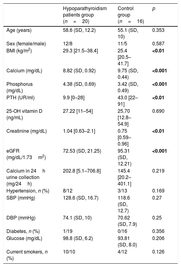
A comparison of general characteristics between patients and controls is specified in Table 2. As expected, there were no significant differences in age and sex. However, a significant difference was found in BMI, which was higher in subjects with hypoparathyroidism. Parameters of calcium–phosphorus metabolism were consistent with the presence or absence of disease, with patients with hypoparathyroidism showing lower calcium, higher phosphorus, and lower PTH plasma levels. There were no significant differences in serum levels of 25OH vitamin D. 24-h urinary calcium excretion was slightly higher in subjects with hypoparathyroidism but did not reach statistical significance. A statistically significant difference, however, was observed in relation with renal function, with subjects diagnosed with hypoparathyroidism showing higher serum creatinine and therefore poorer eGFR levels. No significant differences were observed in other CVR factors, such as SBP, DBP, diagnosis of hypertension, lipid parameters, glucose, diagnosis of diabetes, tobacco consumption.
Clinical characteristics of study population.
| Hypoparathyroidism patients group (n=20) | Control group (n=16) | p | |
|---|---|---|---|
| Age (years) | 58.6 (SD, 12.2) | 55.1 (SD, 10) | 0.353 |
| Sex (female/male) | 12/8 | 11/5 | 0.587 |
| BMI (kg/m2) | 29.3 [21.5–38.4] | 25.4 [20.5–41.7] | <0.01 |
| Calcium (mg/dL) | 8.82 (SD, 0.92) | 9.75 (SD, 0.44) | <0.001 |
| Phosphorus (mg/dL) | 4.38 (SD, 0.69) | 3.42 (SD, 0.49) | <0.001 |
| PTH (UR/ml) | 9.9 [0–28] | 43.0 [22–91] | <0.01 |
| 25-OH vitamin D (ng/mL) | 27.22 [11–54] | 25.70 [12.8–54.9] | 0.690 |
| Creatinine (mg/dL) | 1.04 [0.63–2.1] | 0.75 [0.59–0.96] | <0.01 |
| eGFR (mg/dL/1.73m2) | 72.53 (SD, 21.25) | 95.31 (SD, 12.21) | <0.001 |
| Calcium in 24h urine collection (mg/24h) | 202.8 [5.1–706.8] | 145.4 [20.2–401.1] | 0.219 |
| Hypertension, n (%) | 8/12 | 3/13 | 0.169 |
| SBP (mmHg) | 128.6 (SD, 16.7) | 118.6 (SD, 12.7) | 0.27 |
| DBP (mmHg) | 74.1 (SD, 10) | 70.62 (SD, 7.9) | 0.25 |
| Diabetes, n (%) | 1/19 | 0/16 | 0.356 |
| Glucose (mg/dL) | 98.8 (SD, 6.2) | 93.81 (SD, 8.0) | 0.206 |
| Current smokers, n (%) | 10/10 | 4/12 | 0.126 |
Data express percentages, mean (SD) or median (range) BMI, body mass index; PTH, parathormone; eGFR, estimated glomerular filtration rate; SBP, systolic blood pressure; DPB, diastolic blood pressure.
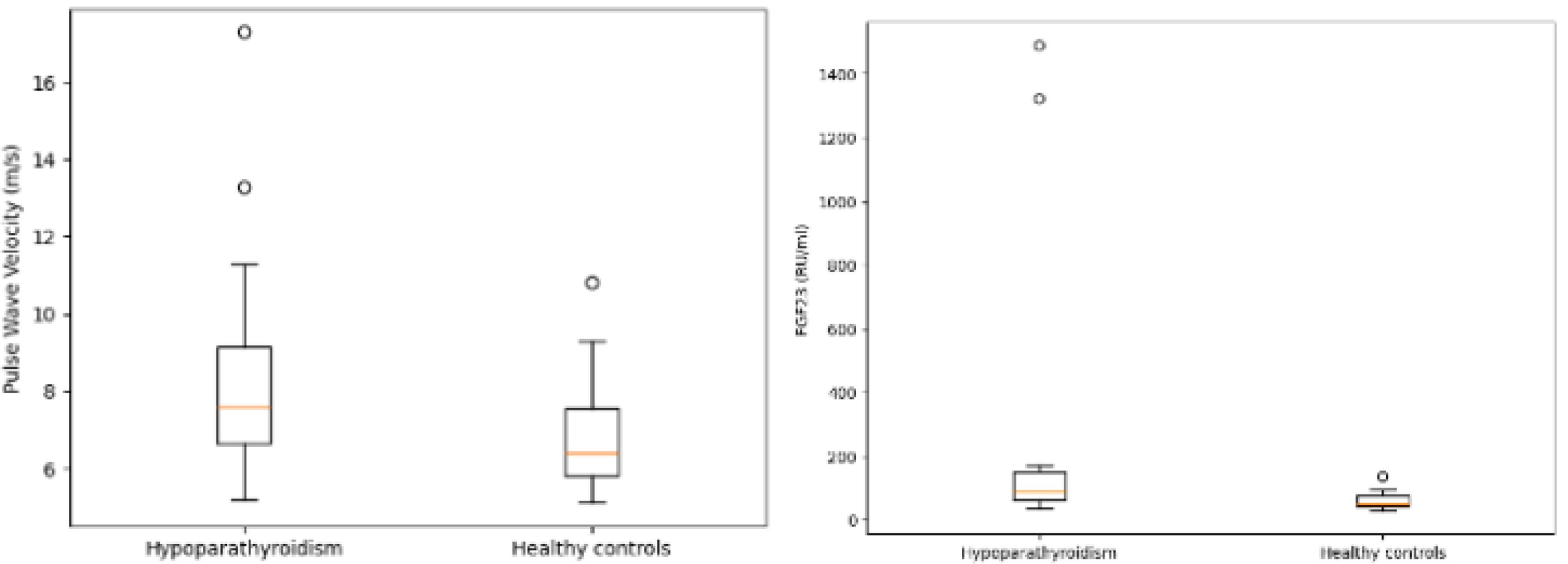
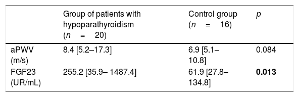
When analyzing the variables that make up the primary objectives of the study, significantly higher levels of FGF23 (>4 times) were evident in subjects with hypoparathyroidism vs healthy controls (255.2 vs 61.9RU/mL); p<0.05). aPWV showed a trend toward being higher in subjects with hypoparathyroidism, reaching the limit of statistical significance (8.4 vs 6.9m/s; p=0.08). Other surrogate markers of AS, such Aix, did not show significant differences. These comparisons between patients and healthy controls are shown in Table 3 and Fig. 1.
Comparison of PWV and FGF23 between patients with hypoparathyroidism and healthy groups.
| Group of patients with hypoparathyroidism (n=20) | Control group (n=16) | p | |
|---|---|---|---|
| aPWV (m/s) | 8.4 [5.2–17.3] | 6.9 [5.1–10.8] | 0.084 |
| FGF23 (UR/mL) | 255.2 [35.9– 1487.4] | 61.9 [27.8–134.8] | 0.013 |
Data expressed median [range] aPWV, aortic pulse wave velocity; FGF 23, Fibroblast Growth Factor.

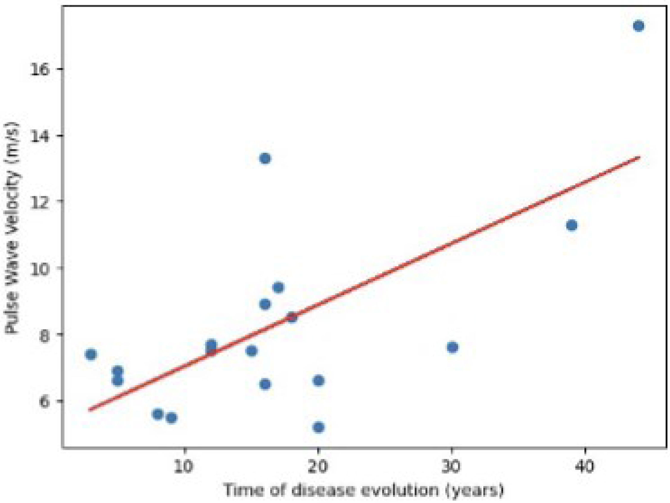
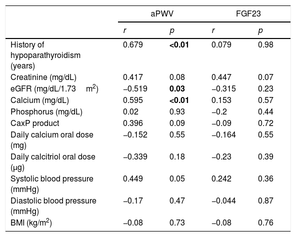
We analyzed the correlation between the most important clinical variables and FGF23 and aPWV levels in subjects with hypoparathyroidism. The most notable findings were a positive correlation between aPWV and hypoparathyroidism duration (r=0.679; p<0.01) (Fig. 2) and a negative correlation between eGFR and aPWV (r=−0.510; p=0.03). A certain positive correlation between calcium levels and aPWV was also evident, although more difficult to interpret (r=0.595; p<0.01). No significant correlation was found between FGF23 and aPWV, nor was a significant correlation found between either of these 2 variables and phosphorus levels or the calcium–phosphorus product. Correlations are shown in Table 4.

Pearson correlation of aPWV and FGF23 with main clinical characteristics in the group of patients with hypoparathyroidism.
| aPWV | FGF23 | |||
|---|---|---|---|---|
| r | p | r | p | |
| History of hypoparathyroidism (years) | 0.679 | <0.01 | 0.079 | 0.98 |
| Creatinine (mg/dL) | 0.417 | 0.08 | 0.447 | 0.07 |
| eGFR (mg/dL/1.73m2) | −0.519 | 0.03 | −0.315 | 0.23 |
| Calcium (mg/dL) | 0.595 | <0.01 | 0.153 | 0.57 |
| Phosphorus (mg/dL) | 0.02 | 0.93 | −0.2 | 0.44 |
| CaxP product | 0.396 | 0.09 | −0.09 | 0.72 |
| Daily calcium oral dose (mg) | −0.152 | 0.55 | −0.164 | 0.55 |
| Daily calcitriol oral dose (μg) | −0.339 | 0.18 | −0.23 | 0.39 |
| Systolic blood pressure (mmHg) | 0.449 | 0.05 | 0.242 | 0.36 |
| Diastolic blood pressure (mmHg) | −0.17 | 0.47 | −0.044 | 0.87 |
| BMI (kg/m2) | −0.08 | 0.73 | −0.08 | 0.76 |
eGFR, estimated glomerular filtration rate; SBP, systolic blood pressure; DPB, diastolic blood pressure; FGF23, Fibroblast Growth Factor 23, BMI, body mass index.
The results obtained in our study indicate that FGF23 is significantly increased in subjects with hypoparathyroidism compared to age- and sex-matched healthy controls. Furthermore, these subjects showed a trend toward increased AS that correlated positively with disease duration and negatively with glomerular filtration rate. However, our study did not reveal a significant correlation between FGF23 and AS that would have suggested a role for this hormone in the physiopathology of AS and therefore of CVR.
AS is an increasingly important variable in cardiovascular medicine, serving as a risk factor for vascular events. Subjects with hypoparathyroidism have a higher risk of these events through mechanisms that are not yet fully understood. Studies evaluating AS in patients with hypoparathyroidism vs healthy controls are scarce. The most notable, in our opinion, is that of Pamuk et al.,14 which showed a significant increase in aPWV as well as in other parameters indicative of AS. Our study provides new data in this regard. Although the difference in aPWV between subjects with hypoparathyroidism and healthy controls is at the limit of statistical significance (p=0.08), the values do not appear to occur at random (8.4m/s in subjects with hypoparathyroidism vs 6.9m/s in healthy controls). Of note, aPWV values in our study are markedly higher than those observed in the study by Pamuk et al., both in patients and controls. Although these discrepancies could be influenced by the different devices used for measurement (Mobil- O-Graph PWA/ABPM device vs SphygmoCorXCEL®), we believe that it is fundamentally due to the fact that our sample of patients and matched controls is older and, above all, disease progression is longer and show more impaired renal function. Therefore, AS significantly correlated positively with the duration of hypoparathyroidism and negatively with glomerular filtration rate. Overall, these findings suggest that AS is an important contributing factor to the increased CVR of patients with hypoparathyroidism throughout the course of the disease.
As mentioned in the introduction, FGF23 is a hormone secreted by osteocytes and osteoblasts that plays a crucial role in the physiological regulation of phosphorus-calcium metabolism. Its increase in pathological conditions, such as CKD can lead to deleterious effects on the cardiovascular system.2,3 In hypoparathyroidism, the status of FGF23 and its involvement in the development of CVD have been poorly studied to this date. Given the predisposition to chronic hyperphosphatemia, a sustained elevation in serum phosphate levels is expected.
A study evaluated FGF23 levels in patients with transient postoperative hypoparathyroidism and indeed showed a significant elevation of FGF23.21 Our study demonstrates a marked elevation of FGF23 in subjects with chronic hypoparathyroidism of different etiology (postoperative and spontaneous). It could be expected that FGF23 would be more elevated in patients with a more unsatisfactory treatment of hypoparathyroidism (with greater hyperphosphatemia or greater elevation of the calcium×phosphate product). All our patients were on calcium and vitamin D since, in our setting, there is currently no availability of any standard PTH preparation for the replacement therapy of hypoparathyroidism. Subjects had biochemically acceptable therapeutic targets (calcium 8.82mg/dL, phosphorus 4.38mg/dL, 24-h urinary calcium 202.8mg) and despite this, showed serum FGF23 levels>4 times higher vs controls (255.2 vs 61.9pg/mL).
However, the role of this increase in FGF23 in the generation of CVD in subjects with hypoparathyroidism remains uncertain. In our study, no significant correlation was shown between FGF23 and aPWV (r=0.272; p=0.32) or with SBP or DBP. Of note, the role of the kidney damage caused by hypoparathyroidism and its treatment to this date (with calcium supplements and vitamin D), both in AS and FGF23 levels. In our study, patients showed a significantly lower glomerular filtration rate vs controls (72.5 vs 95.3ml/min/1.73m2), which could condition the differences observed in aPWV and FGF23 values. However, in the study by Pamuk et al.,14 there were no significant differences in renal function between patients and controls and a significant increase in aPWV was evident in subjects with hypoparathyroidism. In any case, it is necessary to reiterate the negative impact of hypoparathyroidism and its regular treatment with calcium supplements and Vitamin D on kidney health. We would like to emphasize the need for solutions that enable optimal replacement of the hormonal deficit without harming the renal parenchyma. It would also be of interest to measure the urinary microalbumin/creatinine ratio in these subjects, to see if it would have any predictive value for future deterioration of glomerular filtration and to better understand the pathophysiology of renal damage, beyond hypercalciuria, nephrocalcinosis, and the appearance of kidney stones.
The present study has some limitations. As a descriptive cross-sectional study, it provides a limited degree of evidence. Furthermore, although the sample size was small, it should be noted that hypoparathyroidism is a rare disease and that the study included the number of participants required by the formal sample size calculation. Furthermore, the differences in renal function between the 2 groups may act as a confounding factor for the findings in aPWV and FGF23. The very small sample size also limited the possibility of conducting a multivariate analysis. Nevertheless, this is a study that provides interesting data in an area with limited available information and that, for the first time, explores these two variables of interest (AS and FGF23) together in subjects with hypoparathyroidism.
Further studies, probably longitudinal ones, are needed to better characterize the impact of hypoparathyroidism on cardiovascular health. In any case, it is evident that patients with this disease must be managed carefully to minimize CVR factors and kidney damage, and that we need the availability of more physiological replacement therapies for this hormone deficiency.
ConclusionsIn patients with hypoparathyroidism, an increase in FGF23 and a trend toward higher AS are observed, which correlates with the years of disease duration. However, our findings may be conditioned by the deterioration in renal function that this pathological process entails. A causal relationship could not be established between the elevated levels of FGF23 and AS. Further investigations are required to completely understand the pathophysiological mechanisms involved.
Declaration on the use of generative AI and AI-assisted technologies in the writing processThe authors declare that no generative AI tools were employed in the creation of the content of this manuscript. AI-based tools, specifically DeepL and ChatGPT, were used solely for the purposes of grammatical review and enhancement of linguistic clarity in English.
FundingNone declared.
Conflicts of interestNone declared.