


To analyze the anatomic characteristics of the left atrium and pulmonary veins in individuals undergoing ablation for atrial fibrillation and to identify possible anatomic factors related with recurrence.
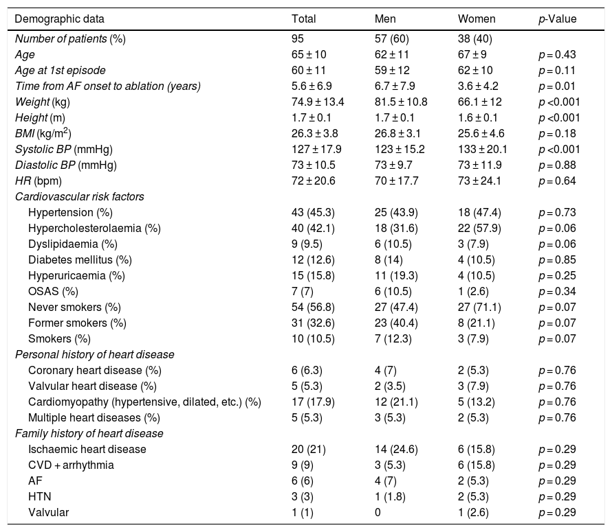
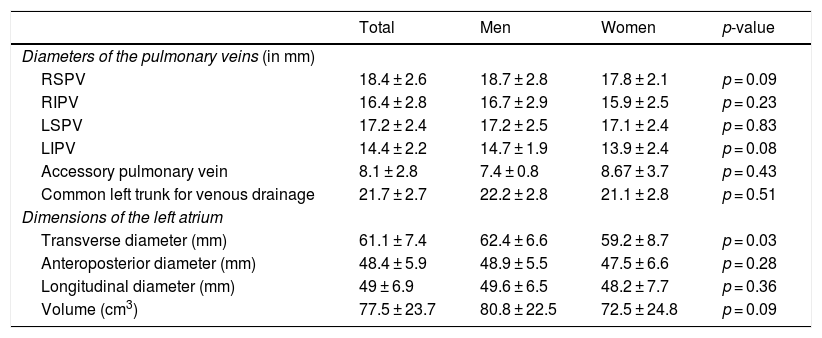
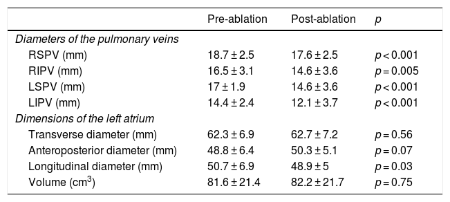
Material and methodsWe retrospectively reviewed the CT angiography studies done to plan radiofrequency ablation for atrial fibrillation in 95 patients (57 men; mean age, 65 ± 10 y). We reviewed the anatomy of the pulmonary veins and recorded the diameters of their ostia as well as the diameter and volume of the left atrium. We analyzed these parameters according to the type of arrhythmia and the response to treatment.
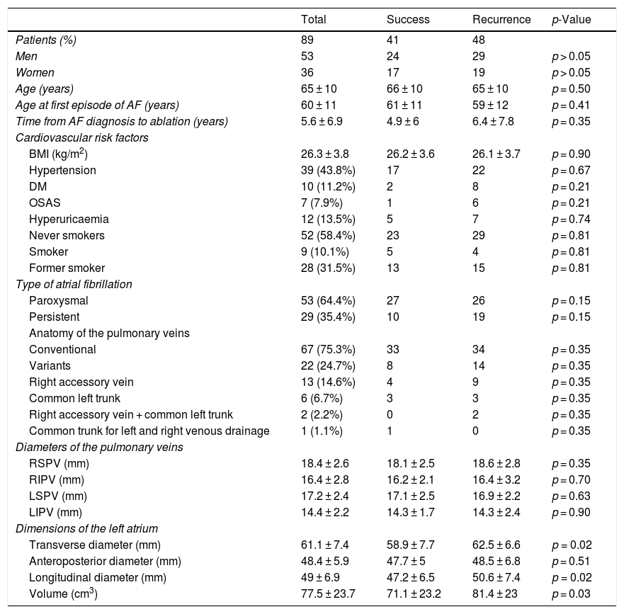
ResultsIn 71 (74.7%) patients, the anatomy of the pulmonary veins was normal (i.e., two right pulmonary veins and two left pulmonary veins). Compared to patients with paroxysmal atrial fibrillation, patients with persistent atrial fibrillation had slightly larger diameter of the left pulmonary veins (left superior pulmonary vein 17.9 ± 2.6 mm vs. 16.7 ± 2.2 mm, p = 0.04; left inferior pulmonary vein 15.3 ± 2 mm vs. 13.8 ± 2.2 mm, p = 0.009) and larger left atrial volume (91.9 ± 24.9 cm3 vs. 70.7 ± 20.3 mm3, p = 0.001). After 22.1 ± 12.1 months’ mean follow-up, 41 patients had sinus rhythm. Compared to patients in whom the sinus rhythm was restored, patients with recurrence had greater left atrial volume (81.4 ± 23.0 mm3 vs. 71.1 ± 23.2 mm3, p = 0.03). No significant differences in pulmonary vein diameters or clinical parameters were observed between patients with recurrence and those without.
ConclusionThe volume of the left atrium is greater in patients with persistent atrial fibrillation and in those who do not respond to ablation.
Definir las características anatómicas de la aurícula izquierda y las venas pulmonares (VP) en sujetos sometidos a ablación de fibrilación auricular (FA) e identificar posibles factores anatómicos relacionados con la recurrencia.
Material y métodosSe estudiaron de manera retrospectiva los estudios de angio-TC de 95 pacientes (57 hombres, edad media 65 ± 10 años), realizados para planificación de ablación por radiofrecuencia de FA. Se revisó la anatomía de las VP y se recogieron los diámetros de sus ostium y los diámetros y volumen de la aurícula izquierda. Estos parámetros fueron comparados con el tipo de arritmia y la respuesta al tratamiento.
ResultadosLa anatomía de las venas pulmonares fue normal (dos venas pulmonares derechas y dos izquierdas) en la mayoría de los pacientes (74,7%). Los pacientes con FA persistente presentaron diámetros ligeramente superiores de las venas pulmonares izquierdas (VPSI de 17,9 ± 2,6 mm vs. 16,7 ± 2,2 mm, p = 0,04; VPII de 15,3 ± 2 mm vs. 13,8 ± 2,2 mm, p = 0,009) y mayor volumen de la aurícula izquierda (91,9 ± 24,9 cm3 vs. 70,7 ± 20,3 mm3, p = 0,001) que los sujetos con FA paroxística. Tras un seguimiento medio de 22,1 ± 12,1 meses, el 43% de los pacientes presentaba ritmo sinusal. Los pacientes con recurrencia mostraron mayor volumen de la aurícula izquierda (81,4 ± 23 mm3 vs. 71,1 ± 23,2 mm3, p = 0,03). No se objetivaron diferencias significativas en los diámetros de las VP ni en los parámetros clínicos estudiados en ambos grupos.
ConclusiónEl volumen de la aurícula izquierda es mayor en pacientes con FA persistente y en pacientes que no responden al procedimiento de ablación.

