


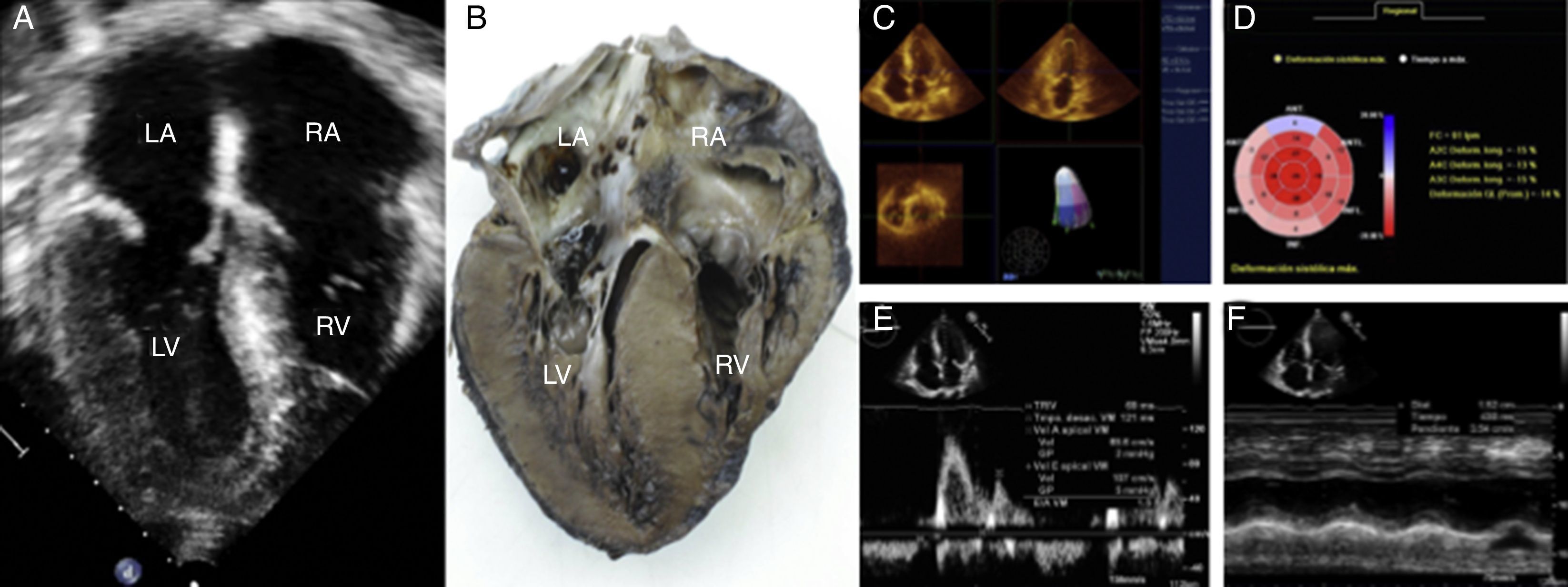
64-year-old man who came to our institution with a history of exertional dyspnea, fatigue and loss weight of 10kg in six months, without previous history of cardiac disease. Clinical examination showed regular tachycardia, raised jugular venous pressure, and bilateral leg edema. Heart auscultation revealed mild apical systolic murmur. Two-dimensional transthoracic echocardiography showed biatrial dilatation, normal end-diastolic and end-systolic left ventricular diameters with symmetrical thickening of the walls, and “granular sparkling” appearance of the interventricular septum, suggesting amyloid infiltration. These images have had a very good correlation with the corresponding specimen (Fig. 1A and B). The papillary muscles, atrioventricular valves, aortic valve and interatrial septum were also thicker, best visualized by three-dimensional echocardiography. A mild mitral, aortic and tricuspid regurgitation was found at color Doppler. Left ventricular systolic function was normal with an ejection fraction (3-D) of 57% (Fig. 1C), but with signs of subclinic systolic dysfunction with longitudinal deformation of −14% (Speckle tracking), Fig. 1D. Pulsed wave Doppler of LV inflow showed restrictive diastolic dysfunction of left ventricle, with E/A 1.5, deceleration time 121ms, and isovolumic relaxation time 69ms, without respiratory variability (Fig. 1E), with E/e′ ratio of 19.9, suggesting elevated left ventricular filling pressure. The right ventricle had normal cavity size but the systolic function was mildly depressed (TAPSE=15mm), Fig. 1F. Also a mild posterior pericardial effusion and a moderate left pleural effusion were visualized. Diagnosis was confirmed by fat and bone marrow biopsy.
 Two-dimensional transthoracic study showing symmetric thickening of the left ventricular walls and granular sparkling of the interventricular septum. (B) Anatomic specimen with a very good correlation with the echocardiographic image in A. (C) 3D echo with normal left ventricular ejection fraction. (D) Diminished left ventricular longitudinal deformation. (E) Left ventricular diastolic disfunction type III. (F) Mildly depressed right ventricular systolic dysfunction.")
(A) Two-dimensional transthoracic study showing symmetric thickening of the left ventricular walls and granular sparkling of the interventricular septum. (B) Anatomic specimen with a very good correlation with the echocardiographic image in A. (C) 3D echo with normal left ventricular ejection fraction. (D) Diminished left ventricular longitudinal deformation. (E) Left ventricular diastolic disfunction type III. (F) Mildly depressed right ventricular systolic dysfunction.
Cardiac amyloidosis (CA) is observed in about 50% of patients with primary amyloidosis. The optimal diagnostic workup for patients with suspected CA includes a combination of medical history, cardiac imaging (two and three dimensional echocardiography and speckle tracking)1,2 and histopathological examination.
This case demonstrates how the integration of the new echocardiographic techniques, such as 3-dimensional echocardiography and speckle tracking and anatomo-echocardiographic correlation has added useful information for the assessment of cardiac biventricular function.
Ethical responsibilitiesProtection of people and animalsThe authors declare that in this research have not been conducted experiments on humans or animals.
Confidentiality of dataThe authors declare that they have followed the protocols of the workplace for the publication of patient data.
Right to privacy and informed consentThe authors have obtained the informed consent of patients and/or subjects referred to in the article consent. This document is in the possession of the corresponding author.
FundingNo endorsement of any kind is received to conduct this article.
Conflict of interestThe authors declare no conflict of interest.