


The purpose of this in vitro study was to determine the apical seal of three different obturation techniques and correlated to the chemical composition of five gutta-percha cone brands. Five brands were used: Dentsply; Tanari; Konne; Obtura Spartan and Analytic Endodontics. One hundred and twenty human upper central incisors were instrumented using a pressureless crown-down technique and irrigated with 0.9% saline solution. The experimental groups were obturated using the Continuous Wave of Condensation and with a shaped single cone technique. The control group was obturated using the lateral condensation. All specimens were stored in 100% humidity for 1 week, coated with nail varnish, except for the apical 2mm, and suspended in India ink for 10 days. Teeth were decalcified, rendered transparent, and linear dye penetration was measured. The results showed significantly greater dye penetration between lateral condensation and shaped single cone and wave of condensation. Could be concluded that the best technique was the wave of condensation and the brands Obtura and Tanari had the best results.
O objetivo do presente estudo foi determinar o selamento apical de três diferentes técnicas de obturação na qual foram utilizadas 5 diferentes marcas de cone de guta-percha. As marcas utilizadas foram: Dentsply, Tanari, Konne, Obtura Spartan e Analytics Endodontics. Cento e vinte incisivos centrais superiores foram instrumentados utilizando uma técnica crown-down sob irrigação constante de solução salina 0.9%. Os grupos foram obturados utilizando duas diferentes técnicas: a) técnica da onda contínua de condensação com auxílio do aparelho System B ou b) técnica do cone único. No grupo controle foi utilizado condensação lateral. Todos os espécimes foram armazenados por 7 dias em 100% de umidade. Decorridos os 7 dias os espécimes foram cobertos com esmalte de unha, deixando livre os 2mm apicais, e colocados em tinta nanquim por mais 7 dias. Os dentes foram então descalcificados, diafanizados e a penetração do corante foi mensurada. Os resultados mostraram uma maior penetração do corante nas técnicas de condensação lateral e na técnica do cone único do que na onda contínua de condensação.
Pode ser concluído que a melhor técnica de obturação foi a onde contínua de condensação associada aos cones da marca Obtura e Tanari.
The hermetic sealing of the root canal system is one of the major objectives in successful endodontic therapy. Recently, a number of filling techniques based on heated gutta-percha have been introduced with the aim of enhancing three-dimensional filling of the root canal. These include warm vertical condensation, low-temperature and high-temperature thermoplasticized gutta-percha, thermoplasticized gutta-percha as a coating on a flexible carrier, thermal compaction and warm lateral condensation.1–4
The concept of thermoplastic compaction is based entirely on the heat softening of gutta-percha combined primarily with the vertical compaction.4 The shape of nonstandardized cones provides the necessary bulk of the gutta-percha for the vertical compaction.4 Clinicians have noticed that cones of various brands may have a different behaviour during compaction. There are also differences in the flow and in the quality of seal with gutta-percha from different manufactures.5,6
These differences could be explained by the great chemical heterogeneity that is found among gutta-percha cones. Brittleness, stiffness, tensile strength, and radiopacity have been shown to depend primarily on the organic and inorganic components.7 The composition of gutta-percha points is approximately 14.5–22% gutta-percha polymer and 37–84.3% zinc oxide.8,9 The particular percentages of components vary with manufacturer. Previous studies reported that different brands may have quite different chemical compositions.7–9 It is evident that since the cones differ in their composition, they may differ in their physical properties, which could interfere in the apical sealing. These differences may be related to errors and misinterpretations in root canal filling studies. This is evident since different brands cones may provide different results in several tests, as they have different chemical composition. Thus the aim of this study was to correlate the chemical composition of five gutta-percha cone brands with apical sealing ability using different obturation techniques and also to correlate chemical composition with the manufacturer. The null hypothesis tested was that there were no differences in the apical sealing ability in canals filled with different gutta-percha cones brands and endodontic filling techniques.
Materials and methods120 permanent maxillary incisors with straight root were used for this study. The apical foramen was breached with a size 20 K-file. Working length was determined by subtracting 1mm from the length when the top of the K-file appeared at the apical foramen. The canals were instrumented using Flexofiles (Dentsply-Maillefer, Ballaigues, Switzerland) until a size #50 reached the working length. The coronal part of each canal was widened with Gates-Glidden. Finally a stepback preparation was executed with circumferential filling. After each file the canal was irrigated with 2ml of 2% chlorhexidine gel and final irrigation with 5ml of distilled water.10 Before obturation the canals were dried with absorbent paper points and the foramen was breached with a size 20 K-file.
Thirteen groups were randomly formed, eleven experimental groups with ten teeth each, and two control groups with five teeth each. The positive control group was instrumented and not obturated. In the negative control group the whole root was covered with two full nail varnish layers. The endodontic sealer used was the Endomethasone (Septodont, Saint-Maur, France). In all tested groups the sealer was taken to the canal with the main gutta-percha cone.
Groups (1–5) were obturated with Continuous Wave of Condensation with System B and five commercially available gutta-percha cones. The brands used are listed in Table 1. A medium nonstandardized gutta-percha cone was properly fitted. A medium plugger was selected to within 5–7mm from the canal terminus. The heat source was set to 200°C, the canal was thoroughly dried, and the medium cone was selected using a calibration ruler, corresponding to a #50 file. The plug tip was driven through the master cone with a single motion to a point 5mm short the working length. While pressure on the plug was maintained, the button on the heating system was released and the plug was slowed in its binding position, pressure was maintained on the plug until the apical mass of the gutta-percha has set (5–10s). Then the switch was reactivated for a short burst of heat (1s) to release the plug and the surplus of gutta-percha. Then, the coronal portion of the canal was backfilled. This was done with the same system with modified temperatures (100°C).
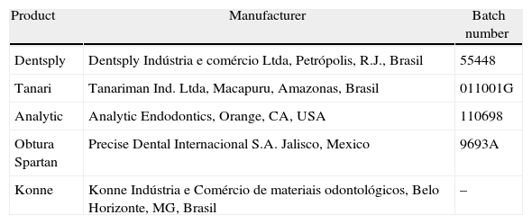
Dental gutta-percha cone size medium selected for study.
| Product | Manufacturer | Batch number |
| Dentsply | Dentsply Indústria e comércio Ltda, Petrópolis, R.J., Brasil | 55448 |
| Tanari | Tanariman Ind. Ltda, Macapuru, Amazonas, Brasil | 011001G |
| Analytic | Analytic Endodontics, Orange, CA, USA | 110698 |
| Obtura Spartan | Precise Dental Internacional S.A. Jalisco, Mexico | 9693A |
| Konne | Konne Indústria e Comércio de materiais odontológicos, Belo Horizonte, MG, Brasil | – |
Groups (6–10) were obturated with Shaped Single Cone and five commercially available gutta-percha cones. The medium cone was calibrated and fitted in the same manner as in lateral condensation. Based on the De Deus11 hydraulic compression technique a modification was proposed in this study: the modelling of the gutta-percha cone tip. After the calibration of the accessory cone tip (#50 file), the root canal was fulfilled with chlorhexidine gel 2% used as lubricant.11 The cone was inserted and removed from the canal ten times pressing the point of the cone against the apical stop until noticing its adaptation represented by the resistance to the removal. The canal was flushed with saline, dried with paper points and the obturation was managed with this shaped single cone. The sealer was applied, the cones were seated and a hand-held plug adapted at the entrance of the canal was heated and used to cut the gutta-percha excess. Then, another cold hand-held plug that penetrated from 1 to 2mm inside the canal was used for the obturation vertical hydraulic compression for 15s.14 In Group 11 the lateral condensation (n=10) was performed.
After obturation, the teeth were stored in an incubator at 37°C and 100% humidity for 1 week to allow the complete set of sealer. The root surface, except the apical 2mm, was coated with two layers of nail varnish. The teeth were immersed in Indian ink (Pelikan, Fort Madison, USA) under 600-mmHg vacuums for 40min and then kept for 10 days in an incubator at 37°C. A final wash with tap water to remove excess ink was accomplished. The teeth were cleaned of varnish and wax with a scalpel blade. The teeth were decalcified in flasks containing 50ml of 5% hydrochloric acid solution at 37°C, changing the solution every 24h, where they remained for 3 days. After decalcification, the teeth were washed in water for 12h, dehydrated in ascending concentration of alcohol and cleared in 98% methyl salicylate.12
Apical microleakage was measured blindly by one skilled evaluator under a stereomicroscope with a magnification of X20 (Lambda Let, Hong Kong, China). The experimental teeth were viewed for its 4 surfaces, buccal, mesial, lingual and distal plane to ensure the dimensional accuracy. Ink penetration was then measured to within 0.01mm from the terminus of the root canal preparation to the maximum coronal point penetration with a computer-imaging program (Imagelab 2.3, São Paulo, Brazil). Each cleared specimen was examined three times by the same observer with a intraobserver kappa of 0.916. Data were analysed by using the ANOVA. The statistical difference level was set at p<0.05. The Kruskal–Wallis test was then applied.
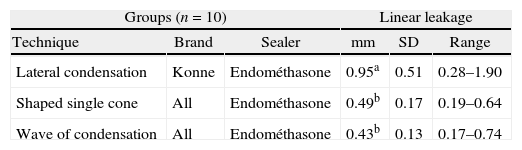
ResultsAll experimental groups demonstrated leakage. The negative control showed no ink penetration, while positive control showed total ink penetration. The results showed significantly greater dye penetration between lateral condensation and thermoplastic techniques. There was no statistical difference (p>0.05) between wave of condensation and shaped single cone (Table 2). It was observed when lateral condensation was compared with wave of condensation there was a significant difference (p<0.05) depending on the brands that was used (Table 3).
Mean apical leakage (mm), standard deviation (SD) and range independent of the gutta-percha cone brand used.
| Groups (n=10) | Linear leakage | ||||
| Technique | Brand | Sealer | mm | SD | Range |
| Lateral condensation | Konne | Endométhasone | 0.95a | 0.51 | 0.28–1.90 |
| Shaped single cone | All | Endométhasone | 0.49b | 0.17 | 0.19–0.64 |
| Wave of condensation | All | Endométhasone | 0.43b | 0.13 | 0.17–0.74 |
Different letters indicate statistically significant difference (p<0.05).
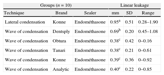
Mean apical leakage (mm), standard deviation (SD) and range of wave of condensation and lateral condensation techniques.
| Groups (n=10) | Linear leakage | ||||
| Technique | Brand | Sealer | mm | SD | Range |
| Lateral condensation | Konne | Endométhasone | 0.95a | 0.51 | 0.28–1.90 |
| Wave of condensation | Dentsply | Endométhasone | 0.69b | 0.20 | 0.45–1.08 |
| Wave of condensation | Obtura | Endométhasone | 0.38c | 0.42 | 0–0.16 |
| Wave of condensation | Tanari | Endométhasone | 0.38c | 0.21 | 0–0.61 |
| Wave of condensation | Konne | Endométhasone | 0.39c | 0.36 | 0–0.92 |
| Wave of condensation | Analytic | Endométhasone | 0.40c | 0.22 | 0–0.85 |
Different letters indicate statistically significant difference (p<0.05).
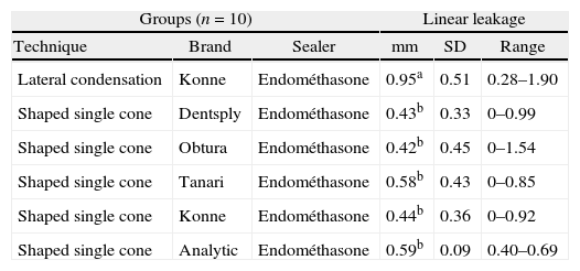
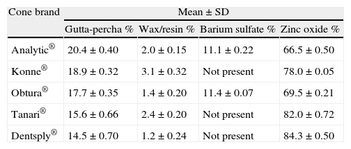
There was no significant difference (p>0.05) between lateral condensation and wave of condensation when Dentsply cone was used. A significantly greater dye leakage (p<0.05) was observed when Dentsply cone was used with wave of condensation when compared with the others brands (Table 3). The leakage was not influenced by the brand when the shaped single cone technique was used (p>0.05). These results can be observed in Table 4. Table 5 shows the amount of gutta-percha polymer ranges from 14.5 to 20.4% and zinc oxide from 66.5 to 84.3%.
Mean apical leakage (mm), standard deviation (SD) and range of shaped single cone and lateral condensation techniques.
| Groups (n=10) | Linear leakage | ||||
| Technique | Brand | Sealer | mm | SD | Range |
| Lateral condensation | Konne | Endométhasone | 0.95a | 0.51 | 0.28–1.90 |
| Shaped single cone | Dentsply | Endométhasone | 0.43b | 0.33 | 0–0.99 |
| Shaped single cone | Obtura | Endométhasone | 0.42b | 0.45 | 0–1.54 |
| Shaped single cone | Tanari | Endométhasone | 0.58b | 0.43 | 0–0.85 |
| Shaped single cone | Konne | Endométhasone | 0.44b | 0.36 | 0–0.92 |
| Shaped single cone | Analytic | Endométhasone | 0.59b | 0.09 | 0.40–0.69 |
Different letters indicate statistically significant difference (p<0.05).
Composition of gutta-percha endodontic filling materials.
| Cone brand | Mean±SD | |||
| Gutta-percha % | Wax/resin % | Barium sulfate % | Zinc oxide % | |
| Analytic® | 20.4±0.40 | 2.0±0.15 | 11.1±0.22 | 66.5±0.50 |
| Konne® | 18.9±0.32 | 3.1±0.32 | Not present | 78.0±0.05 |
| Obtura® | 17.7±0.35 | 1.4±0.20 | 11.4±0.07 | 69.5±0.21 |
| Tanari® | 15.6±0.66 | 2.4±0.20 | Not present | 82.0±0.72 |
| Dentsply® | 14.5±0.70 | 1.2±0.24 | Not present | 84.3±0.50 |
SD=standard deviation.
A hermetic three-dimensional obturation of the root canal system is one of the conditions to achieve a long-term successful root canal treatment.2 This procedure is normally carried out using gutta-percha and an endodontic sealer. Endomethasone is a zinc oxide-sealer with good physicochemical and biological properties.13,14 Based on these good properties it was the selected sealer for the present study. The option of using only one sealer was an attempt to standardize the results and minimize the bias that sealer could have on the results. Whilst some filling techniques use cold gutta-percha cones, others involve thermomechanical compaction of the material.3,4 With the development of these thermomechanical techniques, a resurgent interest in the nonstandardized cones has appeared.
During the obturation, when the sealer layer is thick, the results of shrinkage and consequently the leakage of the teeth may be greater.15–18 Thus the large sealer layer produced by lateral condensation technique may explain the poor apical seal that was observed in this work. These results are in agreement with previous study that showed a higher leakage in the lateral condensation technique when compared to Thermafill, Vertical condensation and System B.17 Under the conditions of the present study the mean value of apical penetration for wave of condensation was significantly lower than cold lateral condensation and shaped single cone technique. These results could be explained as the vertical pressure in the apical direction allows the gutta-percha, when plasticized, a better penetration yielding a better apical sealing.2 The backfill method with a single cone is easier and fast technique, however empty spaces were noted in the area between the apical filling and the coronal filling. Previous reports showed better results when the backfill was done using devices like BeeFill 2 in 1 and the System B/Obtura II.17,19,20
Dentsply and Tanari cone brands showed relative low percentages of gutta-percha. These low percentages of gutta-percha could decrease the material flow resulting in poor plasticity,9 allowing a poor apical sealing as observed in this study. Also the sealing ability of the cone, when submitted to heat, was influenced by the amount of gutta-percha. The results revealed a significant difference between lateral condensation with a standardized cone and lateral condensation with an accessory cone (shaped single cone). These results are probably influenced by the better adaptation of the medium cone at the final 5mm apical third. Lateral condensation poor seal could be explained by empty spaces presents between the filling and the sealer layer. In agreement with the present study, previous research reported better results using standardized cones then accessory cones when the obturation technique was the lateral condensation.21
Chlorhexidine gluconate has been recommended as a root irrigant and studies have demonstrated its broad-spectrum antimicrobial action, substantivity, and low grade of toxicity.22,23 However the inability of chlorhexidine to dissolve pulp has been a problem. Kennedy et al.24 stated that the lowest degree of infiltration in the apical region is obtained when the smear layer is removed from dentinal walls by increasing the contact surface between dentin and filling material. The association of 17% EDTA and 2% chlorhexidine gel was able to remove the smear layer.10 Due to its viscosity the gel seems to compensate the chlorhexidine's inability to dissolve pulp tissue by promoting a better mechanical cleansing of the root canal and removing dentin debris as well as remaining tissues. Our results show low degree of microleakage similar with the finds of teeth irrigated by sodium hypochlorite.23 The chlorhexidine gel (viscous form) does not interfere with the sealing ability of the sealer, being soluble and removable with a final flush of 5ml distilled water.25
The teeth were cleared to assess dye leakage. This process was found to be simple and inexpensive, and avoided the hazards of sectioning and/or reduces radiopacity used in other methods of examining root fillings. In addition, adaptation of the filling material to the canal wall could be observed and the specimens could be photographed. To date the relationship between dye penetration and the success of endodontic treatment is not clear, so the clinical relevance of the results of laboratory studies should be interpreted with caution.
ConclusionThe results obtained in the present study justify the rejection of the null hypothesis set out previously, i.e., that there are no differences in the apical sealing ability in canals filled with different gutta-percha cones brands and endodontic filling techniques. However, we recall that even the groups that showed relatively better sealing properties, were not able to completely block the ink penetration. As such, it is not possible to directly correlate the amount of leakage to the clinical outcomes of endodontic treatments. Thus, clinical studies are required to confirm the relevance of the present results.
Ethical disclosuresProtection of human and animal subjects. The authors declare that the procedures followed were in accordance with the regulations of the responsible Clinical Research Ethics Committee and in accordance with those of the World Medical Association and the Helsinki Declaration.
Confidentiality of data. The authors declare that no patient data appears in this article.
Right to privacy and informed consent. The authors declare that no patient data appears in this article.
Conflicts of interestThe authors have no conflicts of interest to declare.