


To examine the value of 18F-fluorodeoxyglucose positron emission tomography/computed tomography (FDG-PET/CT) for the detection of bone metastasis in breast cancer patients and assess whether whole body bone scan (BS) with 99mTc-methylene diphosphonate provides any additional information.
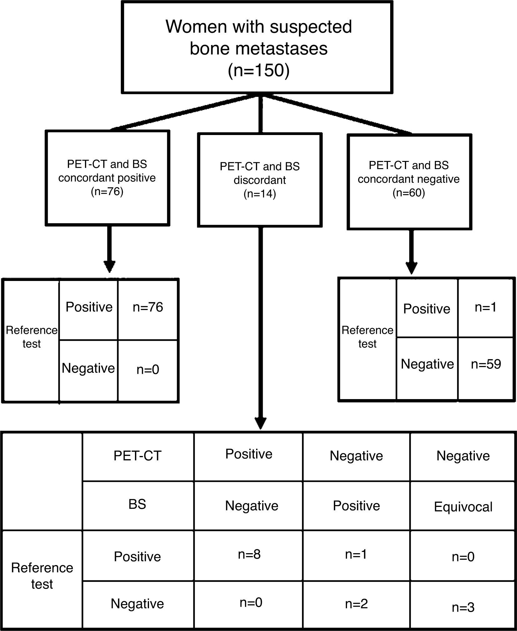
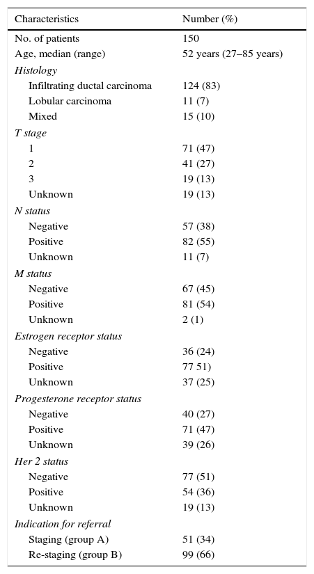
Material and MethodsStudy group comprised 150 patients, mean age 52 years (range 27–85) with breast cancer, suspected of having bone metastases. All patients had undergone both FDG-PET/CT and BS with or without single photon emission tomography/computed tomography (SPECT/CT) within a period of 6 weeks. The final diagnosis of bone metastasis was established by histopathological findings, additional imaging, or clinical follow-up longer than 10 months. Cancer antigen 15-3 (CA15-3) and carcinoembryogenic antigen (CEA) were measured in all patients.
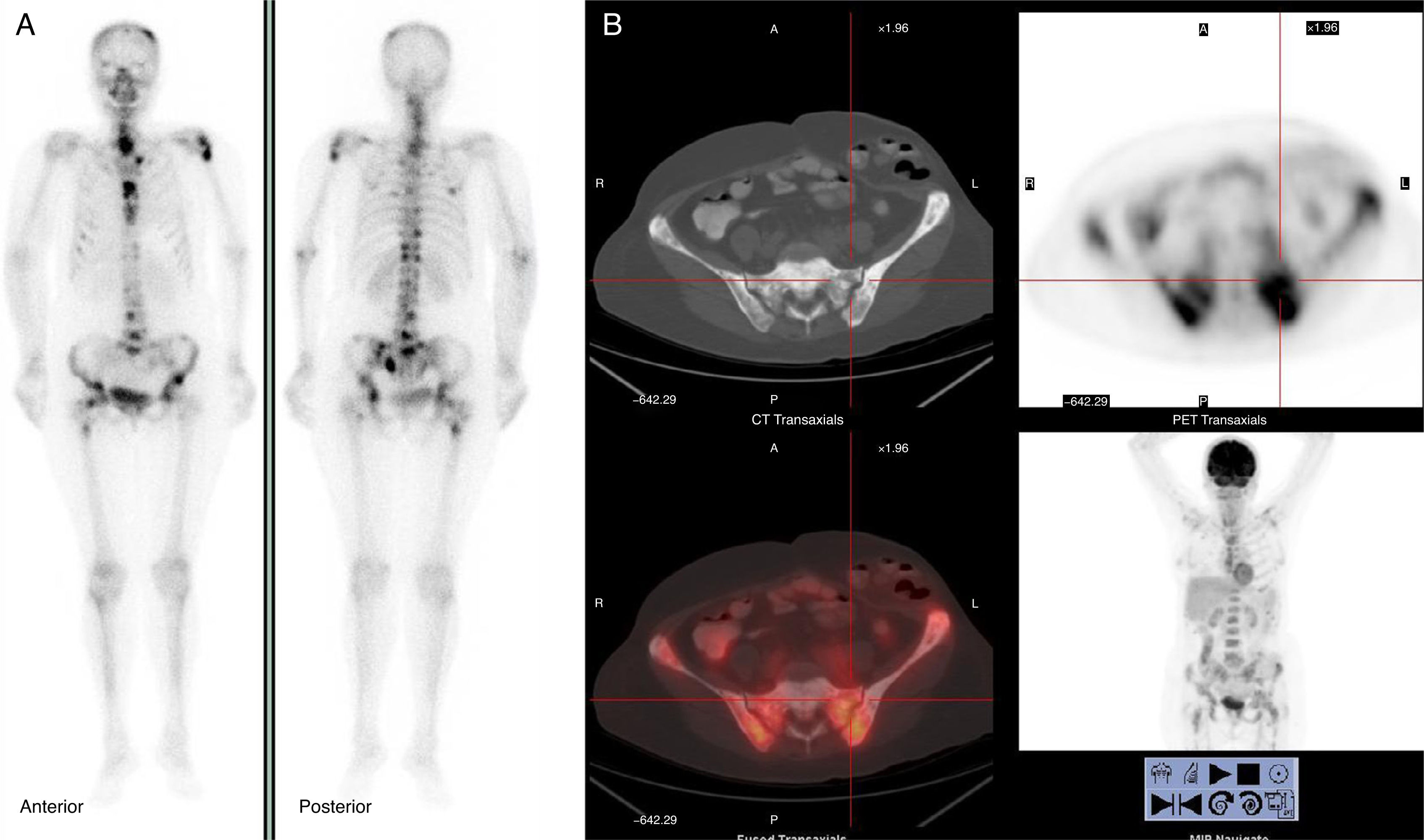
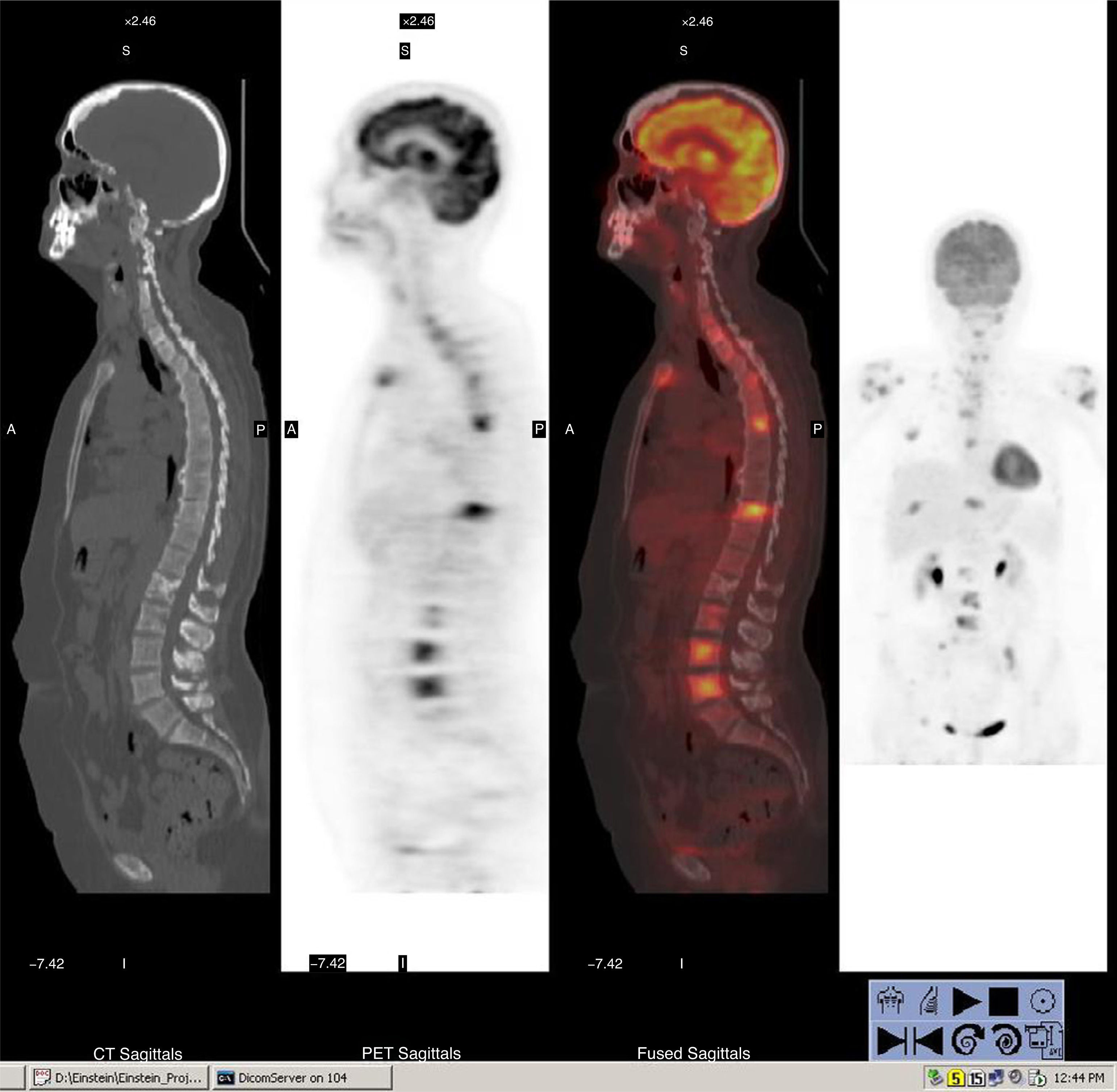
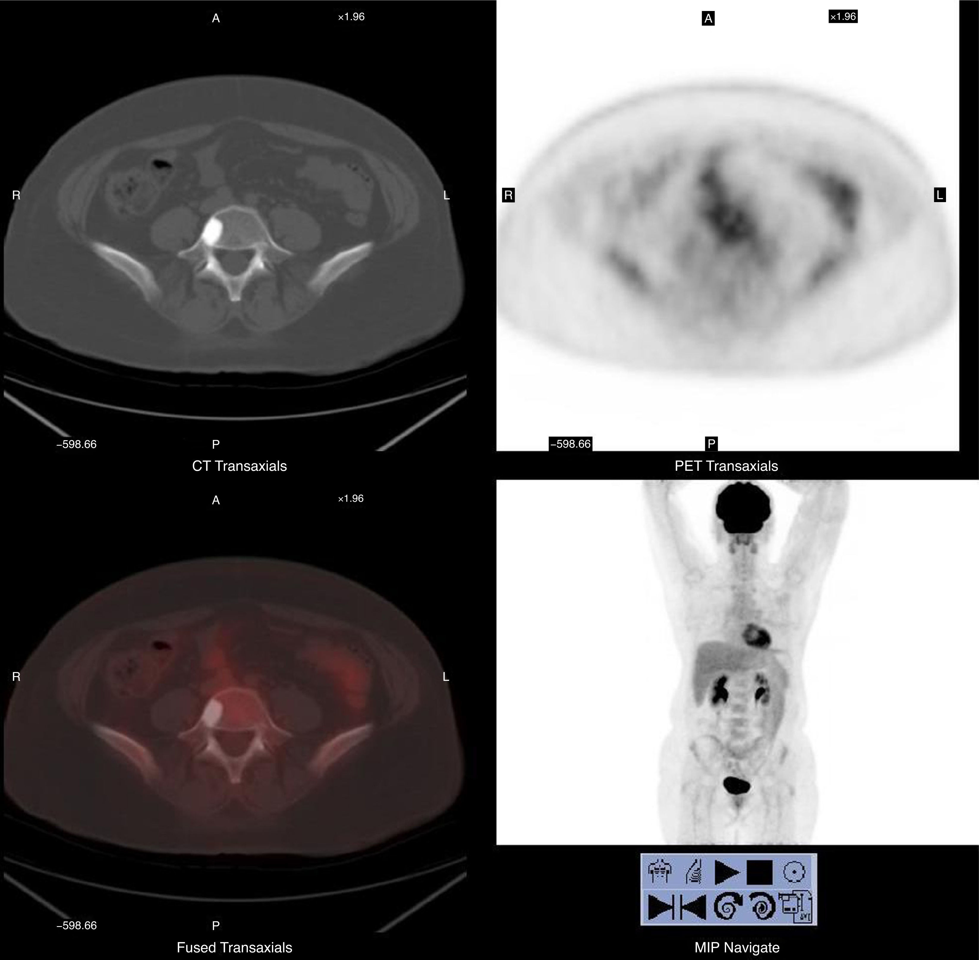
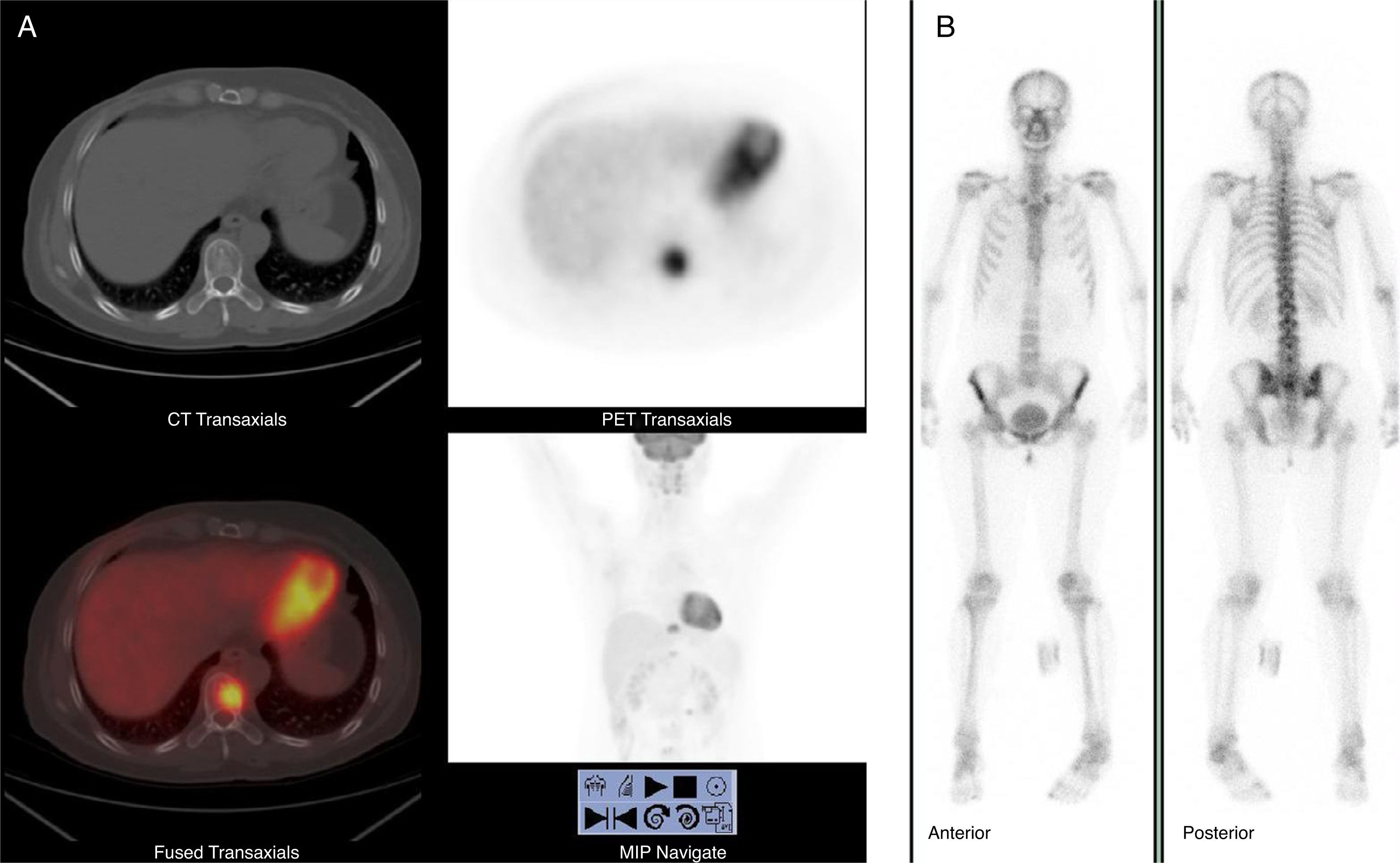
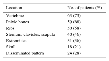
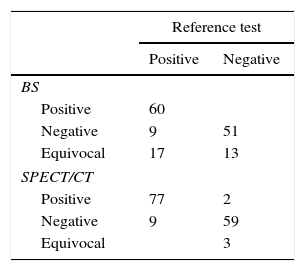
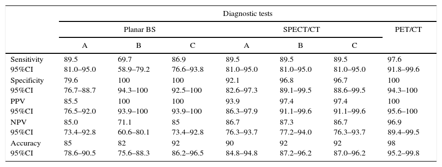
ResultsHistologically 83%, 7% and 10% had infiltrating ductal, lobular and mixed carcinoma respectively. Confirmed bone metastases were present in 86 patients (57.3%) and absent in 64 (42.7%). Mean CA15-3 and CEA values in patients with bone metastases were 74.6ng/mL and 60.4U/mL respectively, compared to 21.3ng/mL and 3.2U/mL without metastases (p<0.001). The sensitivity of FDG-PET/CT for the detection of bone metastases was 97.6% compared to 89.5% with SPECT/CT. In 57 patients, FDG-PET/CT correctly identified additional pulmonary, hepatic, nodal and other soft tissue metastases, not detected by BS.
ConclusionOur findings suggest that FDG-PET/CT is superior to BS with or without SPECT/CT.
Para examinar el valor de la 18F-fluorodeoxiglucosa Tomografía por Emisión de Positrones y de la Tomografía Axial Computerizada (FDG-PET/TAC) para la detección de metástasis óseas en pacientes con cáncer de mama y evaluar si el rastreo gammagráfico óseo (GO) con 99mTc-difosfonato de metileno proporciona cualquier información adicional.
Material y métodosEl grupo de estudio estuvo integrado por 150 pacientes, media de edad 52 años (rango 27-85) con cáncer de mama y sospecha de metástasis óseas. Todas las pacientes tenían FDG-PET/TAC y GO con o sin SPECT/TAC en un plazo de 6 semanas. El diagnóstico final de metástasis óseas fue establecido por los resultados histopatológicos, los estudios adicionales de imagen o el seguimiento clínico superior a 10 meses. En todas las pacientes se determinaron los niveles séricos de CA 15-3 y antígeno carcinoembrionario (CEA).
ResultadosHistológicamente, 83%, 7% y 10% tenían carcinoma ductal infiltrante, lobular y mixto respectivamente. Las metástasis óseas confirmadas estuvieron presentes en 86 pacientes (57.3%) y ausentes en 64 (42.7%). Los valores medios de CA15-3 y CEA en pacientes con metástasis óseas fueron 74.6 ng/mL y 60.4 U/mL, respectivamente, en comparación con 21,3 ng/mL y 3,2 U/mL sin metástasis (p <0,001). La sensibilidad de la FDG-PET/TAC para la detección de metástasis óseas fue del 97,6% en comparación con el 89,5%, para la SPECT/TAC. En 57 pacientes, la FDG-PET/TAC identificó correctamente metástasis adicionales pulmonares, hepáticas, ganglios linfáticos y partes blandas, no detectadas por GO.
ConclusiónNuestros resultados sugieren que la FDG-PET/TAC es superior a GO con o sin SPECT/TAC.
Article
Revista Española de Medicina Nuclear e Imagen Molecular (English Edition)
