


The experimental porcine model is anatomically and physiologically similar to the human heart; it is easily reproducible and very useful to test new stent and balloon generations. This study was aimed at analyzing an experimental model to evaluate different coronary devices for percutaneous coronary intervention.
MethodsWe evaluated 131 juvenile commercial farm pigs, 109 were female, weighing 26.4±3.2kg. They were anesthetized and had mechanical ventilation and monitoring. Vascular access was obtained via the femoral artery by dissection or puncture. The coronary device was used after a selective catheterization of the coronary arteries with a JR 6F catheter. Animals were maintained on mechanical ventilation until recovery and were submitted to angiographic evaluation 7, 28, 90 and/or 180 days after the procedure. After euthanasia, the hearts were collected and submitted to macro and microscopic analysis.
ResultsSix drug-eluting stents, two drug-eluting balloons and two bare-metal stents were tested. Unplanned deaths were observed in 1.5% of the cases during the procedures and in 9.2% of the cases after the procedure, occurring within 12 hours to 6 days (2.3±1.6days). In addition to angiographic evaluations, intravascular ultrasound and optical coherence tomography were performed during the procedures in 20% and 60% of the cases, respectively. There was no death related to the use of the devices.
ConclusionsThe experimental percutaneous porcine model proved to be reproducible with similar outcomes and low mortality for the different devices tested and is an essential tool for the evaluation of new coronary devices.
Modelo Porcino para Avaliação e Desenvolvimento de Diferentes Dispositivos Coronários Baseados em Cateter: Ferramenta Pré-Clínica Fundamental
IntroduçãoO modelo experimental porcino tem grande similaridade anatômica e fisiológica com o coração humano, e é de fácil reprodutibilidade, sendo de grande valia para testar novas gerações de stents e balões. Este estudo teve como objetivo analisar o desempenho de um modelo experimental para intervenção coronária percutânea na avaliação de diferentes dispositivos coronários.
MétodosForam estudados 131 porcos juvenis de granja comercial, sendo 109 fêmeas, pesando 26,4±3,2kg, anestesiados, monitorados e ventilados mecanicamente, com acesso vascular obtido por via femoral (dissecção ou punção). Após a cateterização seletiva das artérias coronárias com cateter JR 6F, procedeu-se à utilização do dispositivo coronário a ser estudado. Os animais foram mantidos sob ventilação mecânica até a recuperação e submetidos a reestudos angiográficos 7, 28, 90 e/ou 180 dias após o procedimento. Após a eutanásia, os corações foram coletados e submetidos a análises macro e microscópicas.
ResultadosForam testados seis stents farmacológicos, dois balões farmacológicos e dois stents não farmacológico. O óbito intraprocedimento não planejado incidiu em 1,5% dos casos e, no pós-procedimento, em 9,2%, ocorrendo em um período de 12 horas a 6 dias (2,3±1,6 dias). Além das análises obtidas pela angiografia, foram realizados, durante os procedimentos, ultrassom intracoronário em 20% e tomografia de coerência óptica em 60%, não sendo observados óbitos relacionados ao emprego dessas ferramentas.
ConclusõesO modelo experimental porcino percutâneo mostrou ser reprodutível, com desempenho homogêneo entre os vários dispositivos e de baixa mortalidade, sendo ferramenta indispensável na investigação de novos dispositivos coronários.
The experimental porcine model is extremely useful to test new generations of stents and balloons, and is the most used model in the evaluation of coronary devices. The main advantages of this animal model are its anatomical and physiological similarities with human hearts.1 The distribution of the coronary arteries, myocardial supply by collaterals, the coagulation system, and platelet activity are very similar to those found in humans.2−4
Although in the healing response to vascular injury involves similar processes and phases, the time course of events in pigs is different from humans. Based on the periods of restenosis development after implantation of drug-eluting stents, a time ratio of 1 to 6 (animal to human) is usually observed.5−9
Although animal production is not expensive, the costs of handling and housing are high. In Brazil, investments in the public and private sectors of translational research are justified when the ultimate goal is the application of improved technologies, many of which are developed in the country, which should translate into benefits for patients with cardiovascular disease.
To participate in research, development, and innovation activities in the field of percutaneous coronary intervention, the subject of intense interest in many countries in the world, it is necessary to have well-established and reproducible animal models – as this has been a mandatory step in the effectiveness assessment of any new coronary device, as required by the United States (Food and Drug Administration [FDA]) and European (European Medicines Agency [EMEA]) regulatory agencies.10,11
This study aimed to analyze the performance of an experimental model for percutaneous coronary intervention in the evaluation of different coronary devices.
METHODSAll experiments were performed according to the protocol approved by the Institutional Research Ethics Committee. In this study, domestic pigs (Sus scrofa domestica, MS60, EMBRAPA) were used. All animals underwent clinical examination before the start of protocol, and only those with no signs of disease were used in the study.
The animals were kept in a local commercial farm, with free access to water and food during the protocols. They were acclimated in the Experimental Division of Instituto do Coração, Hospital das Clínicas, University of São Paulo (Incor-FMUSP), Brazil, for at least 24 hours before catheterization.
Anesthesia procedureThe animals were sedated before the procedure with a mixture of ketamine chloride (8mg/kg) and midazolam chloride (0.5mg/kg). After 10 to 20 minutes, an IV line was inserted (superficial ear vein). Anesthesia was induced with thiopental sodium (12.5mg/kg) and then orotracheal intubation was performed with a long straight blade laryngoscope (7 or 7.5-mm tube). Anesthesia was maintained with 1.5% to 2.5% isoflurane and 100% oxygen in the anesthetic equipment. Before the procedure, the pigs received an intramuscular injection of benzathine penicillin (1.2 million units), to prevent infections.
Platelet antiaggregants were started 24 hours before the procedure: 300 to 600mg of acetylsalicylic acid (ASA) and 75 to 150mg of clopidogrel. The maintenance dose was 100mg and 75mg for ASA and clopidogrel, respectively.
Cardiac catheterization and invasive and noninvasive monitoringThe pigs were placed in the supine position under general anesthesia. Oxygen saturation was monitored through the tail of the animal, and the heart rate and rhythm by a heart monitor. After inguinal asepsis, a 6F vascular sheath was introduced by direct visualization into the common femoral artery through puncture or dissection, followed by the administration of 10,000 units of heparin, while maintaining invasive blood pressure monitoring.
The right and left coronary arteries were selectively cannulated using a therapeutic 6F Judkins catheter (Philips, Eindhoven, the Netherlands) monitored by fluoroscopy provided by digital angiography Philips BV Pulsera (Philips, Eindhoven, the Netherlands) equipment. Intracoronary injection of nitroglycerin at a dose of 200mg and subsequent injection of iodinated contrast for the acquisition of the initial angiography was administered. The angiographic projection of choice for both the left and right coronary arteries was the left anterior oblique 60° projection.
In case of acute ventricular arrhythmia, bolus doses of lidocaine hydrochloride (2.5 to 12mg/kg) were administered. When ventricular fibrillation was detected, electrical cardioversion was performed by applying 200 to 300J, with the paddles placed against the anterior chest wall.
Tested devices, restudy, and euthanasiaThe devices tested were drug-eluting balloons, drugeluting stents, and bare-metal stents. The angiographic study was performed at seven, 28, 90, and 180 days, depending on the current protocol.
The animals, under deep anesthesia, were euthanized with a lethal dose of potassium chloride. A left paramedian thoracotomy was performed and the pericardium was opened. The vena cava, pulmonary artery, and aorta were carefully clamped and severed, and then the heart was removed. Then, the hearts were washed with clean water and a solution of 10% formalin was infused under pressure of 100mmHg at the aortic root, for coronary perfusion for 30 minutes. The arterial segment containing the stent was dissected, removed, and placed in a solution of 10% formalin, and submitted to histological processing.
Use of adjunct intravascular imaging methodsPrior to the methods of intravascular imaging, intracoronary nitrate (10-20mg of isosorbide mononitrate, or 150 to 400μg of nitroglycerin) was administered to promote epicardial vasodilation and prevent arterial spasm.
The intravascular ultrasound (IVUS) was performed using an Atlantis 40MHz catheter connected to an iLab console (both from Boston Scientific Inc., United States). The acquisition was performed during automatic retreat of 0.5mm/s.
At the optical coherence tomography (OCT), an OCT M2 system (LightLab Imaging, Westford, United States) was used with automated retreat of the ImageWire™ image catheter (LightLab Imaging, Westford, United States) at the rate of 1 to 2mm/s, after previous proximal occlusion of the coronary artery with Helios® balloon (LightLab Imaging) and saline solution infusion.
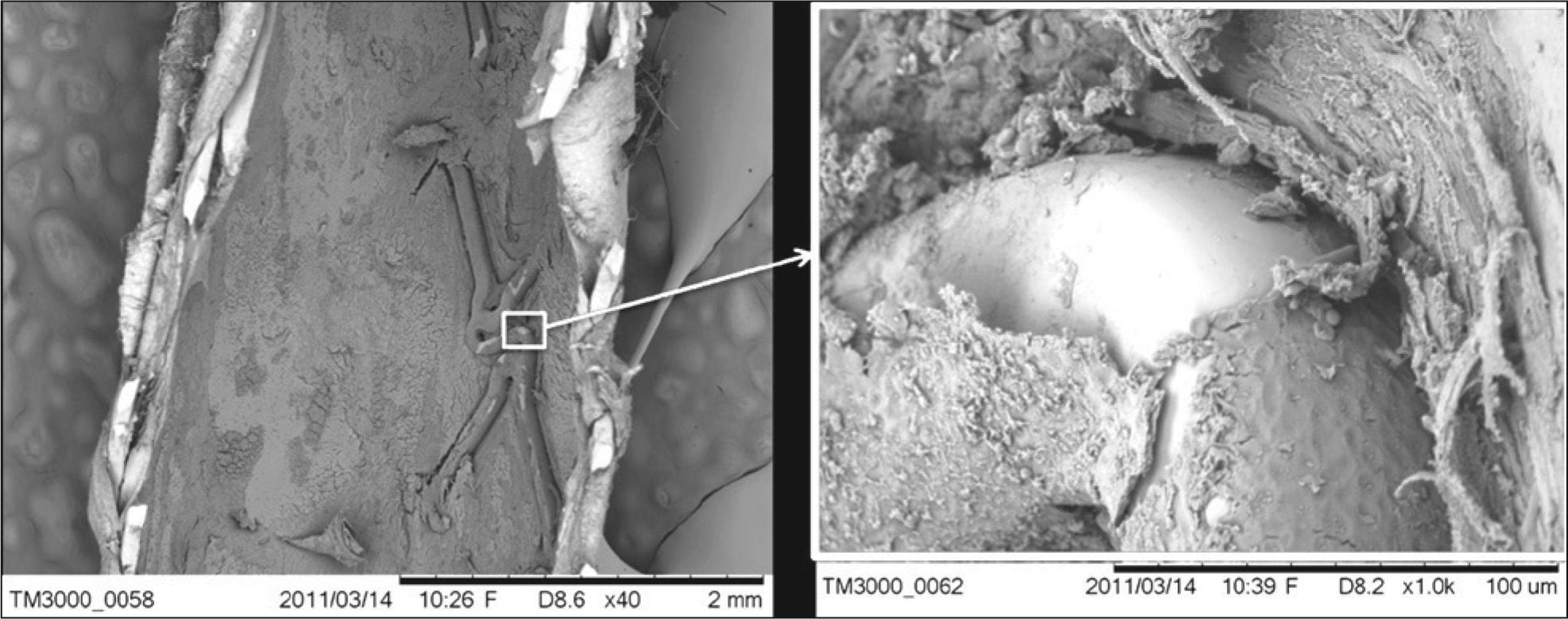
Scanning electron microscopyThe coronary artery segment containing the stent was dissected, removed, and subjected to a longitudinal section, with one half sent to pathology and the other half to specific processing and further analysis in a scanning electron microscope.
The hemi-segment containing the stent was immersed in modified Karnovsky solution for 12 hours at 4°C, washed in buffer solution of sodium cacodylate, post-fixed in a 1% buffered osmium tetroxide solution, and dehydrated in an increasing series of alcohol solutions, up to absolute alcohol. The drying of the samples was performed to the critical point in a Balzers CPD 030 apparatus.
The samples were metallized with gold ions in a Balzers SCD 040 apparatus and analyzed with a JSM 7401S scanning electron microscope (JEOL – Japan), which takes pictures with a magnification of 25×. The images were stored and submitted to analysis using Adobe® Photoshop® software, version 7.0 (Adobe Systems Inc.) and ImageJ version 1.42q for Windows (NIH – Bethesda, United States), and areas of the exposed stent struts (mm2) were measured and the percentages of endothelization for each stent type were calculated (Figure 1).
Histopathological analysis
The sections were stained with hematoxylin-eosin and Verhoeff staining for elastic fibers and subsequently analyzed qualitatively and quantitatively by pathologists and a specialized technician. At the quantitative analysis, the modified Kornovski et al. inflammation scores,12 the fibrin score,13 and the Schwartz injury scores were used.10,14
Quantitation of the area of the lumen, stent, internal elastic lamina, external elastic lamina area, and neointimal thickness on the struts and inter-strut was performed using Leica Qwin software (Leica Microsystems, Wetzlan, Germany).
RESULTSA total of 131 juvenile pigs obtained from a commercial farm were studied from 2006 to 2012, of which 109 were female, weighing 26.4±3.2kg. The animals were maintained on mechanical ventilation until recovery and submitted to restudy after seven, 28, 90, and 180 days.
Six drug-eluting stents, two drug-eluting balloons, and two bare-metal stents (Table 1) were tested. Unplanned intra-procedural death was 1.5%, and post-procedure death was 9.2%, occurring from 12 hours up to six days (2.3±1.6days). In addition to the analysis obtained by angiography, IVUS and OCT were performed in 20% and 60% of the animals during procedures, respectively, with no deaths observed during the use of these tools (Figure 2).
Description of coronary devices studied using the porcine model
| Studied devices | Date | n |
|---|---|---|
| Nitric oxide-eluting stent | April 2006 | 9 |
| National cobalt-chromium stent | August 2006 | 10 |
| Supralimus vs. sirolimus stent | March 2007 | 3 |
| Pre-assembled national cobalt-chromium stent | September 2007 | 3 |
| Bare-metal stents of different models | July 2008 | 5 |
| National sirolimus-eluting stent | October 2008 | 6 |
| National drug-eluting stent with external vs. internal and external drug coating | May 2009 | 14 |
| Sirolimus vs. paclitaxel-eluting stents | December 2009 | 7 |
| Nitric oxide-eluting stent | March 2010 | 4 |
| Sirolimus-eluting stent | June 2010 | 16 |
| Everolimus vs. sirolimus-eluting stent | August 2010 | 14 |
| National cobalt-chromium stent vs. national sirolimus-eluting stent 180 days | September 2010 | 4 |
| Comparison of stents with clot vs. thrombus | November 2010 | 4 |
| National cobalt-chromium stent vs. sirolimus-eluting stent in overlapping | December 2010 | 3 |
| National cobalt-chromium stent vs. national sirolimus-eluting stent 90 days | December 2010 | 7 |
| Nanomagnetic stent | June 2011 | 4 |
| Sirolimus-eluting stent | September 2011 | 10 |
| National stent with new design | September 2012 | 8 |
 Optical coherence tomography; (B) Histological analysis; (C) overlapping images, aiming to choose the cross-section of optical coherence tomography analogous to the histological picture.")
The present study reflects the results of a pioneering center for pre-clinical validation in Brazil, specifically organized and equipped for the scientific evaluation of devices and procedures for percutaneous coronary intervention. The knowledge gained from the porcine model over the years, among other devices, has allowed the authors to participate in the development of the first stent of Brazilian design, using technological innovations in its creation.15−19 These results were obtained thanks to the implementation of several complex processes, which began with the design of a new endovascular device and continued through the steps of producing prototypes, validation of preclinical tests, (mechanical and biological in vitro and in vivo) and, finally, tests of safety and efficacy in humans.20
The histopathological analysis is part of this process, which remains the current standard for evaluation of devices to be studied. Such histological processing of vessels with stents is one of the most limiting factors in the process, due to the risk of damage to tissues adjacent to stent struts. The inclusion in methacrylate resin and micrometric sectioning of specimens containing metal are the main difficulties that restrict the histological processing of stents to a few centers worldwide.
Currently, it is recommended that these models, previously used as benchmark tests to evaluate the effectiveness of new devices, should focus specifically on the safety aspects.21 For that purpose, the stent performance should be evaluated sequentially. Therefore, it is essential to have intravascular imaging methods that allow the outcome assessment at different moments of the vascular healing process.
IVUS, despite providing a volumetric estimate of all the neointimal tissue, has little precise correlation with histological findings.22,23 Therefore, this is not the best technique to assess the presence of fibrin or thrombus, nor does it have enough spatial resolution to verify whether the stent struts are coated or denuded.
OCT, on the contrary, showed a high correlation with histological findings24,25 due to its high axial resolution, allowing for a proper categorization of the stent struts as denuded or coated.25,26 Heterogeneous images with low light intensity signal, visible at OCT, correlated with areas of low cellularity, with presence of fibrin and proteoglycans.19,27,28
Currently, the evaluation at 28 days is recommended to verify the occurrence of neointimal hyperplasia, and at least once more, to analyze the long-term effects. The second evaluation (after three to six months) depends on the time when the healing of the treated segment and drug release were completed. It is noteworthy that, for drug-eluting stents, the FDA recommended a six-month interval after stent implantation to obtain preclinical data; more recently, it has extended this recommendation to one year after implantation.10,11
CONCLUSIONSThe percutaneous porcine experimental model showed to be reproducible, with homogeneous behavior among the different tested devices and low mortality, representing an indispensable tool in the investigation of new coronary devices.
SOURCES OF FUNDINGThis study is part of the National Program for the Development of Vascular Endoprosthesis (stents) (Programa de Desenvolvimento Nacional de Endopróteses Vasculares – PDNS), initiated in 2004–2005. It has the support of the Science, Technology, and Strategic Input Secretariat (Secretaria de Ciência, Tecnologia e Insumos Estratégicos – SCTIE)/Department of Science and Technology (Departamento de Ciência e Tecnologia – DECIT) of the Brazilian Ministry of Health, the National Council of Scientific and Technological Development (Conselho Nacional de Desenvolvimento Científico e Tecnológico – CNPq), and the Sponsor of Studies and Projects (Financiadora de Estudos e Projetos – FINEP) of the Brazilian Ministry of Science and Technology. The development of the laser stent cutting process had the support of the Foundation for Research Support of the State of São Paulo (Fundação de Amparo à Pesquisa do Estado de São Paulo [FAPESP], form of support: Technological Innovation – Innovative Research in Small Businesses [Pesquisa Inovativa na Pequena e Microempresa – PIPE]).
CONFLICTS OF INTERESTCelso Kiyochika Takimura, Francisco Rafael Martins Laurindo, and Pedro Alves Lemos Neto are scientific consultants for Scitech Medical Prod. Ltda. The other authors declare to have no conflicts of interest.