Buscar en
GE - Portuguese Journal of Gastroenterology
Toda la web
Inicio GE - Portuguese Journal of Gastroenterology Successful Endoscopic Removal of a Kerh-Tube Placed in an Iatrogenic Esophageal ...
Journal Information
Vol. 23. Issue 2.
Pages 119-121 (March - April 2016)
Share
Share
Download PDF
More article options
Visits
3230
Vol. 23. Issue 2.
Pages 119-121 (March - April 2016)
Endoscopic Snapshot
Open Access
Successful Endoscopic Removal of a Kerh-Tube Placed in an Iatrogenic Esophageal Perforation: Push to Pull
Remoção Endoscópica de um Tubo Kerh Colocado Após Perfuração Esofágica Iatrogénica: Push to Pull
Visits
3230
Rosa Coelho
Corresponding author
rosacoelhoabrantes@hotmail.com

Corresponding author.
, Alexandre Sarmento, Guilherme Macedo
Gastroenterology Department, Centro Hospitalar São João, Porto, Portugal
This item has received

Under a Creative Commons license
Article information
Full Text
Bibliography
Download PDF
Statistics
Figures (2)
Additional material (1)
Full Text
1Case description

Esophageal perforation is a rare complication during cervical surgery associated with high morbidity and mortality.1–3 Currently, the treatment of esophageal perforations remains controversial.1 The use of modified T-tube (Kerh-tube) repair can be a therapeutic option associated with a low mortality rate.4

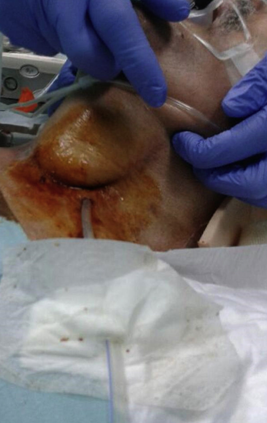
The authors report a 68-year-old man with a cervical spinal cord lesion submitted to an anterior cervical surgery when a plate fixation removal complicated with an iatrogenic cervical esophageal perforation that was immediately diagnosed. A T-tube was inserted into the esophagus following debridement of adjacent wall with its inferior portion extending into the stomach. The esophageal wall was closed loosely about the tube with sutures placed in the nearest healthy submucosa (Figs. 1 and 2A and B). Two weeks later, an upper endoscopy was performed in order to remove the T-tube. Using a polypectomy snare (10mm, Olympus®) the distal end of the Kerh-tube was grabbed. The external (cutaneous) part of the T-tube was then cutted and the entire tube was distally pulled into the esophagus. Endoscopic extraction of the Kerh-tube was then successfully performed. An orifice, with 6mm of diameter, localized 18cm from the upper incisor teeth was observed after drain removal. As it was located in the beginning of the esophagus it was decided not to perform an endoscopic clip closure. Seven days later, a cervical computed tomography (Fig. 2C) confirmed the healing of the esophagus perforation and the remaining orifice. The patient started oral intake without complications.

Figure 1.

Image of the T-tube (Kerh-tube) placed in the cervical esophagus.

(0.12MB).
Figure 2.

Computed tomography showing the T-tube placed in the cervical esophagus (arrows). (A) Axial view. (B) Coronal view. Cervical computed tomography confirmed the healing of the esophagus perforation the remaining orifice. Coronal view. (C).

(0.23MB).

This is the first case reported in the literature of a successful endoscopic removal of a T-tube in the esophagus. This case highlights the safety, simplicity and efficacy of this procedure avoiding a more invasive technique. Due to the T-tube characteristic form, the authors recommend that before its removal the drain must be pulled down to the esophagus to minimize the risk of esophageal laceration.

Conflict of interest

The authors declare that there is no conflict of interest.

Financial support

The authors declare that did not receive any financial support for this work.

Appendix A
Supplementary data

The following are the supplementary data to this article:

(37.25MB)

Upper endoscopy video showing T-tube removal pulling the distal end of the drain into the esophagus using a polypectomy snare.

References
[1]
W.G. Jones 2nd, R.J. Ginsberg.
Esophageal perforation: a continuing challenge.
Ann Thorac Surg, 53 (1992), pp. 534-543
[2]
C.J. Brinster, S. Singhal, L. Lee, M.B. Marshall, L.R. Kaiser, J.C. Kucharczuk.
Evolving options in the management of esophageal perforation.
Ann Thorac Surg, 77 (2004), pp. 1475-1483
[3]
R.F. Gaudinez, G.M. English, J.S. Gebhard, J.L. Brugman, D.H. Donaldson, C.W. Brown.
Esophageal perforations after anterior cervical surgery.
J Spinal Disord, 13 (2000), pp. 77-84
[4]
P.A. Linden, R. Bueno, S.J. Mentzer, L. Zellos, A. Lebenthal, Y.L. Colson, et al.
Modified T-tube repair of delayed esophageal perforation results in a low mortality rate similar to that seen with acute perforations.
Ann Thorac Surg, 83 (2007), pp. 1129-1133
Copyright © 2015. Sociedade Portuguesa de Gastrenterologia
Article options
Tools
Supplemental materials
es en pt

¿Es usted profesional sanitario apto para prescribir o dispensar medicamentos?

Are you a health professional able to prescribe or dispense drugs?

Você é um profissional de saúde habilitado a prescrever ou dispensar medicamentos