


The objective of this study was to standardize and validate the dot-blot test for the serological diagnosis of bovine brucellosis, compare the results with those found in the 2-mercaptoethanol (2-ME) and complement fixation test (CF), and estimate the relative sensitivity and specificity of the dot-blot compared to these tests. Fifty bovine blood serum samples were used for the test standardization, and 1315 samples were used for evaluation and comparison between the tests; the results were compared using the Kappa indicator. At the end of standardization, it was established as optimal for the antigen obtained from Brucella abortus B19 after passing through a microorganism rupture process, the blood serum samples diluted at 1:100, and the conjugate at 1:30,000. The comparison of the dot-blot results with 2-ME showed Kappa index of 0.9939, sensitivity of 99.48%, and specificity 99.91%, with CF, Kappa index of 0.8226, sensitivity 100% and specificity 95.32%. Using the combination of the test results 2-ME and CF to establish the true condition of the animal, the dot-blot showed relative sensitivity of 100%, and relative specificity of 99.91%. The evaluated test proved to be effective and reliable, besides being easy to handle and interpret the results.
Brucellosis is an infectious disease of chronic character that affects animals, causing great losses to livestock. It is also a public health problem, for being a zoonosis of occupational character, transmitted by contact with contaminated fetal membranes with the causative agent Brucella abortus, and foodborne by unpasteurized milk intake, fresh cheese and undercooked meat from animals with brucellosis.1,2
Due to the social and economic importance of this disease, the Ministry of Agriculture, Livestock and Supply of Brazil (MAPA) has set up a control and eradication program, which defined as official tests for the diagnosis of bovine and buffalo brucellosis: Milk ring test (MRT) used for monitoring the health condition and as a diagnostic tool in epidemiological surveillance systems, Rose Bengal test (RB) as a screening test and 2-mercaptoethanol (2-ME) and complement fixation (CF) as confirmatory tests in addition to the fluorescence polarization assay (FP).3
However, there are some difficulties concerning these tests, such as the need for highly trained staff, the use of labile reagents that need to be constantly prepared, and titrated, toxic reagents. These facts highlight the need to develop new diagnostic techniques in order to collaborate for the control and eradication of brucellosis.4
The aim of this study was to standardize and validation the dot-blot technique for the serological diagnosis of bovine brucellosis, and compare the results obtained by this technique with the ones found in the official tests: complement fixation and 2-mercaptoethanol, and also to estimate the relative sensitivity and relative specificity of dot-blot in comparison with official diagnostic tests used in the study.
Materials and methodsFifty blood sera samples from cattle of various breeds, male and female, from different properties in the North, Northeast and Southeast regions of Brazil were used for test standardization, samples previously tested and with the same results in all official tests and more 1315 samples were used for validation of the tests 2-ME, CF and dot-blot.
Standardization of dot-blotFrom an immunological point of view, Brucella antigens can be divided into two major groups: proteins and the LPS.5 With the intention of evaluate what is the best antigen for the present study were tested the two antigens, the Brucella abortus sample B19 obtained from the commercial vaccine sold in Brazil after undergoing a microorganism rupture process, by the method of freezing for 5min in liquid nitrogen and thawing for 5min in water bath at 37°C, twenty times in a row, and quantified at 3.1μg/μL of protein, the reference antigen for dot-blot tests. The second antigen tested was lipopolysaccharide extract obtained from the Brucella abortus strain S996 quantified at 0.52μg/μL, the LPS was tested for being one of the most important antigen combated by immunoglobulins, it is extremely immunogenic and commonly detected in serological tests.3
In order to standardize and establish the ideal amount of antigen used, tests were performed with 0.5μg, 1μg, 1.5μg and 2μg of the antigens tested. The quantity that showed the best results for the two antigens tested was 2μg, concentration which allowed the visibility of the color reaction with excellent sharpness, and was the value established for the membrane sensitization.
For the technique standardization, 50 control samples of bovine blood serum were used, 17 positive with different titrations (weak, medium and strong), and 33 negative in the 3 tests: Rose Bengal test, 2-ME and CF.
The technique was developed according to the previously described methodology.7 The test started by cutting the nitrocellulose membrane (code N-9888, Sigma-Aldrich, St. Louis, MO, USA) in two formats, square and circle, with the interest to establish the shape that would enable better solution homogenization, and economy of material when making the cuts. To perform the membrane cutting, scissors were used for the square format, and a hole punch for the circle format, both properly sanitized and handled with gloves. To make the cuts, materials of easy acquisition and manipulation were used, for the purpose of facilitating the technique.
Each nitrocellulose membrane was sensitized with 2μg of antigen, manipulated with forceps and gloves to prevent contamination, and placed on a surface of hydrophobic material. As for the support on which to carry out the reactions, two types of plate were evaluated, the polyacrylamide plate with 96 wells, and the cell culture plate with 24 wells and a flat bottom.
The sensitized membranes were blocked for 12h with 300μL TBS (20mM Tris, 500mM NaCl, pH 7.5) with addition 0.05% Tween 20 and 5% powder milk into each well of the plate with 24 wells and 200μL into each well of the plates, with 96 wells to minimize the occurrence of non-specific reactions,8 leaving the plate stirring at 4°C.
After incubation, the blocking solution was removed, and 500μL of the serum to be tested, was pipetted into each well of the plate, with 24 wells and 200μL into each well of the plate with 96 wells, diluted in the proportions 1:25, 1:50 and 1:100 in TBS 0.05% Tween 20 with powder milk at 5%. The material was then incubated for 2h at room temperature under constant agitation.
After the incubation was finished, the wells were washed with TBS 0.05% Tween 20. Then, it was evaluated which was the optimal number of washes 1, 2 or 3 times, each wash lasting 5min. Subsequently, 300μL of conjugate IgG of rabbit anti-total bovine IgG linked to alkaline phosphatase (code n. A0705, Sigma-Aldrich, St. Louis, MO, USA) was pipetted into each well of the plate with 24 wells and 200μL into each well of the plate with 96 wells, in the dilutions 1:4000; 1:10,000; and 1:30,000.
The material was incubated for 1h at room temperature. Conjugate was removed and three washes, 5min each, were performed using TBS 0.05% Tween 20. The bands were visualized by the addition of the enzyme substrate 5-bromo-4-chloro-3-indoyl phosphate/nitroblue tetrazolium chloride, following the manufacture's recommendations (code n. 170-6432, NBT-BCIP, Bio-Rad, Hercules, CA, USA).
The development lasted 5min, since this is the time in which the positive membranes distinctly stain, and the negative ones remain with the original color, followed by a 5min rinse in distilled water to remove any non-specific color reaction from substrate residue.
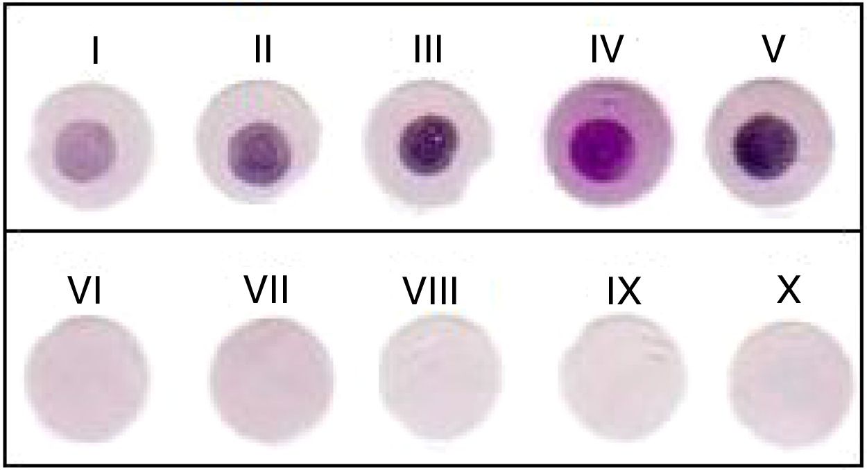
When reading the results, the interpretation was by visual observation of the membranes. The positive samples were standardized to contain a strong or weak purplish circle, surrounded by a circle without staining, and the negative ones without any staining, as shown in Fig. 1.
 showed positive results with different color tints, and in row 2 (samples VI, VII, VIII, IX, and X) membranes with negative results, without reaction.")
Results observed in the dot-blot test for the diagnosis of brucellosis in cattle. The membranes in row 1 (samples I, II, III, IV, and V) showed positive results with different color tints, and in row 2 (samples VI, VII, VIII, IX, and X) membranes with negative results, without reaction.
The 1315 samples were evaluated by the confirmatory tests: 2-ME and CF. The 2-ME test was performed according to the recommendations of Technical Manual PNCEB,3 and the CF test, was performed according to the methodology described in study techniques for brucellosis laboratories.9 The animals with positive results in these two tests were considered infected, and the animals with negative results in these two tests were considered uninfected.
Data analysisThe sensitivity and specificity values and confidence intervals (CI) were obtained following recommended criteria.10 The Z test was used to assess the significance of the kappa indicator following interpretation criteria,11 being established that the one-tailed null hypothesis kappa does not differ from zero. The calculations were performed using the package epiR, software R.
Results and discussionStandardization of dot-blot testThe shape that provided the best results for membrane was a circle, and among the tested antigens, the one obtained from Brucella abortus after undergoing a microorganism rupture process, provided the best result, which was the one used in the testing of 1315 samples in this study.
The standardized dot-blot test showed important characteristics for the feasibility of their use in routine diagnostic laboratories, such as the use of an easily produced antigen, high performance, and providing a great distinction for the visualization of the results of positive and negative samples.4 The support that allowed the best agitation, not demonstrating competition with the membrane, was the cell culture plate with 24 wells and a flat bottom.
The dilution that demonstrated the best results for serum, was 1:100 and for conjugate 1:30,000, and it was established the number of three washes per step. An important feature of the test is the fact that it does not require specific equipment nor generate any risk to the handlers health.
Comparison between the testsDue to the difficulties related to the isolation of the etiologic agent, the serological methods are best suited for the diagnosis of large herds. For effective control and eradication of brucellosis, it is necessary for the diagnosis to be safe, and of viable execution, and for this reason, the serologic tests are indicated.12 To verify the feasibility of using the dot-blot, the results were compared to the official Brazilian confirmatory tests 2ME and FC.
The comparison of the results of dot-blot with 2-ME showed an almost perfect agreement,11 with Kappa index of 0.9939 (CI 95%: 0.939–1.0484), sensitivity of 99.48% (CI 95%: 97.11–99.91) and specificity of 99.91% (CI 95%: 99.49–99.98), Z test (35.71; P=1.35×10−279); with CF the Kappa index of 0.8226 (CI 95%: 0.7690–0.8761) means an almost perfect agreement11; sensitivity of 100% (CI 95%: 97.42–100.0) and specificity of 95.32% (CI 95%: 94.03–96.47), Z test (30.1; P=2.29×10−199).
Using the combination of results of the 2-ME and CF to establish the true condition of the animal, the dot-blot showed relative sensitivity of 100% (CI 95%: 97.25–100.00) and relative specificity of 99.91% (CI 95%: 99.48–99.98). The combinations of results obtained in the tests are summarized in Table 1.
Combinations of resultsa obtained in the tests 2-mercaptoethanol (2-ME), complement fixation (CF) and dot-blot for serological diagnosis of bovine brucellosis.
| Number of serums | 2-ME | CF | Dot-blot | |
|---|---|---|---|---|
| 1086 | − | − | − | |
| 136 | + | + | + | |
| 52 | + | − | + | |
| 11 | − | A | − | |
| 12 | I | − | − | |
| 9 | I | + | + | |
| 4 | + | A | + | |
| 2 | I | A | + | |
| 1 | + | − | − | |
| 1 | − | − | + | |
| 1 | I | − | + | |
| Total | 1315 |
The results of this study have show almost perfect agreement between the official tests and the dot-blot (Tables 2 and 3), as well as in other studies13,14 when comparing the dot-blot to the Rose Bengal test and CF, and comparing the technique to agent isolation for diagnosis of infection by Brucella melitensis.
Comparison between the tests 2-mercaptoethanol (2-ME) and dot-blot for the serological diagnosis of bovine brucellosis using 2-ME as the gold standard in the comparison.
| 2-Mercaptoethanol | Total | ||
|---|---|---|---|
| Dot-blota | Positive | Negative | |
| Positive | 192 | 1 | 193 |
| Negative | 1 | 1097 | 1098 |
| Total | 193 | 1098 | 1291 |
Comparison between the tests complement fixation (CF) and dot-blot for the serological diagnosis of bovine brucellosis, using CF as the gold standard in the comparison.
| Complement fixation | Total | ||
|---|---|---|---|
| Dot-blota | Positive | Negative | |
| Positive | 145 | 54 | 199 |
| Negative | 0 | 1099 | 1099 |
| Total | 145 | 1153 | 1298 |
Studies performed with infected cattle, naturally and experimentally, indicated that the CF have drawbacks, such as the need for highly trained personnel, and the use of labile reagents, that need to be constantly prepared and tittered,4 is highly associated with the isolation of the etiological agent, with 98% specificity, detecting 93 negative animals of 95 that were free of the infection15 and also showing good sensitivity having the least amount of false negative reactions.16
In another study comparing the diagnostic tests for brucellosis17 the CF was the most efficient test, detecting the disease in 17 of 19 infected animals in the pre-vaccination tests and 22 of 23 infected animals in post vaccination tests. These results are due the high antibody detection sensitivity of the test, requiring only 5μg/mL of IgM and 10μg/mL of IgG1.18 Factors that attest the use of this test as a reference for the evaluation of another serological test (Table 3).19
Although the confirmatory serological tests individually present satisfactory results (Tables 2 and 3), one cannot disregard the occurrence of divergent results between them (Table 1), indicating the possibility of false-negative results in the confirmatory tests.20 In order to reduce this bias, the association of official confirmatory tests was used to establish the true condition of the animal in relation to brucellosis.19,21
The data from Table 4 demonstrates that the results obtained with dot-blot were excellent when compared to the true condition of the animal, established by the samples that obtained the same results in CF and 2-ME tests.
Dot-blot test results compared to the true condition of the particular animal determined by the combination of results in the tests 2-mercaptoethanol and complement fixation for the serological diagnosis of brucellosis.
| 2-ME+CF | Total | ||
|---|---|---|---|
| Dot-blota | Infected | Not infected | |
| Positive | 136 | 1 | 137 |
| Negative | 0 | 1086 | 1086 |
| Total | 136 | 1087 | 1223 |
Some serological tests have drawbacks, such as the use of large amounts of reagents, the use of toxic substances such as 2-ME test,4 and problems such as technical difficulties, prozone effect and anticomplementary sera, as CF.19 Thus, the use of an easily handled test like dot-blot, which uses no toxic or labile reagents, and gets a precise distinction between the positive and negative serum, generates a security in execution and interpretation of results.14
New diagnostic techniques are emerging however, and many have proved themselves impossible in routine use, because of the need of expensive equipment, and requiring highly trained teams, factors not necessary in the technique standardized in this study.
The dot-blot presents advantages over other primary binding assays. In this technique, the antigen adheres to the center of the nitrocellulose membrane, which ensures the use of an established antigen concentration, and its simplicity, precision and speed demonstrate that the assay can be used in the field for large scale diagnosis in eradication programs, such as the bovine brucellosis one.12
It is an attractive test for routine diagnosis, because the nitrocellulose membrane has a high affinity for proteins, being effective to correctly identify reactive individuals (positive) and non-reactive (negative) to an etiological agent.22
As reported in recent research,23 the study of brucellosis served and serves as the basis for some of the great advances in epidemiology, and the development and verification of good results of a diagnostic test provides a technique not only for the disease in question, but it also opens space for evaluation and standardization of the test for other species and other infections.
The dot-blot standardized in this study showed high relative sensitivity, high relative specificity, and had good agreement with the tests used for comparison, managing to detect antibodies against Brucella abortus in bovine serum. It is a viable test, easy to perform and interpret, as well as safe for the handler, proving suitable for the serological diagnosis of bovine brucellosis.
Conflicts of interestThe authors declare no conflicts of interest.