Sensory neural hearing loss (SNHL) is a disorder characterized by an important deterioration of the auditory function. Re-establishing normal ion homeostasis of the endolymph could be related to hearing recovery and it might be mediated by mineralocorticoids.
The main purpose of this preliminary, randomized controlled clinical trial was assessing the recovery of idiopathic sensory neural cochlear hearing loss (SNHL) by comparing the efficacy of 2 types of steroids versus vasodilators.
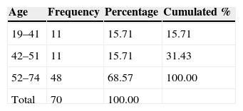
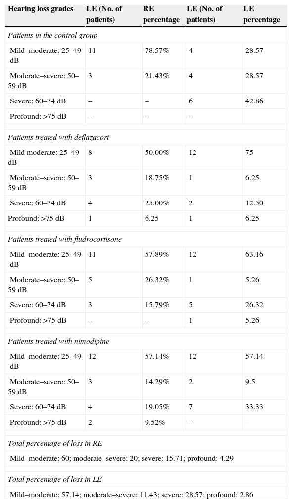
Materials and MethodsThe 3-month intervention involved 70 patients, allocated into 4 different groups: a control with no medication, consisting of 14 patients (8 men and 6 women); a vasodilator group of 21 patients (11 men and 10 women); a glucocorticoid group with 16 patients (10 men and 6 women); and a mineralocorticoid therapy group, consisting of 19 patients (11 men and 8 women). The level of hearing loss and its topography were estimated using Liminal Tone Audiometry (LTA) and Auditory Brainstem Response (ABR).
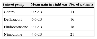
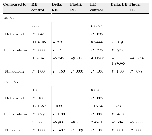
ResultsOur research found overall greater efficacy of mineralocorticoids versus glucocorticoids and vasodilators. There was better response in women than in men and it was higher from the left ear, regardless of patient gender.
ConclusionsThe hearing gain was significantly superior in the mineralocorticoid group, followed by the glucocorticoid group. However, the responses to vasodilators were lesser and of low statistical significance.
La hipoacusia neurosensorial (HNS) puede conducir a causar grave deterioro auditivo. Su recuperación funcional parece relacionarse con el control de la homeostasis iónica coclear experimentalmente dependiente de los mineralocorticoides.
El objetivo de este trabajo es valorar la eficacia terapéutica comparando 2 modalidades de corticoides frente a los vasodilatadores en pacientes con HNS idopática coclear (HNSIC).
Material y métodosEl ensayo dura 3 meses, se realiza en 70 pacientes asignados aleatoriamente en 4 grupos: grupo control, sin medicación, formado por 14 pacientes (8 varones y 6 mujeres); grupo tratado con vasodilatadores formado por 21 pacientes (11 hombres y 10 mujeres); grupo sometido a terapia glucocorticoidea formado por 16 pacientes (10 varones y 6 mujeres); y grupo sometido a terapia mineralocorticoidea formado por 19 pacientes (11 varones y 8 mujeres). La valoración del nivel de pérdida auditiva y su topografía se estiman mediante audiometría tonal liminal (ATL) y potenciales auditivos de tronco cerebral (PEATC).
ResultadosEncontramos mejor respuesta al tratamiento con los mineralocorticoides que con los glucocorticoides, siendo la respuesta más pobre para los vasodilatadores. Esta respuesta es mayor en las mujeres que en los hombres, y en general observamos mejor respuesta por parte del oído izquierdo, con independencia del sexo del paciente.
ConclusionesLas ganancias auditivas son significativamente mayores con los mineralocorticoides, seguidas por los glucocorticoides, mientras que con los vasodilatadores las respuestas son pobres y no significativas.
From an audiometric perspective, WHO considers hearing loss above 25dB to be hearing loss in the range of frequencies between 0.5 and 4.0kHz, word frequency range, most compromised in perceptive or sensorineural hypoacusis (SNHL), in general progressive and which can cause major hearing impairment in patients.1,2
SNHL results in hearing impairment that is not necessarily binaural or symmetrical, which could be explained, based on information gained in the last decade, by a dominance of cerebral specialization in hearing tasks, which is dependent on cochlear behaviour.3–6
Its etiopathogeny is multifactorial and includes both intrinsic factors (genetic predisposition), and extrinsic factors (infectious, autoimmune, vascular, toxic, degenerative, trauma, neoplastic, psychological, environmental, etc.).7–21 It is often impossible to know the exact cause which, by damaging the inner ear, eventually results in altering both cochlear transduction and the transmission of acoustic signals,1–6 and in these cases we classify the problem as idiopathic.
We believe that intrinsic SNHL meets etiopathogenic criteria similar to secondary SNHL, which have not been properly documented or expressed clinically, and that it might follow the same lesion topography.
Hearing impairment in SNHL used to be associated with vascular compromises which altered cochlear oxygenation and hence it was treated using vasodilators. Today it is argued that its pathogenesis lies in an alteration in the ionic concentration of the fluids of the inner ear, and that restoring this could be associated with a reversal of hearing loss.14,15
Corticosteroid receptors have been demonstrated in the inner ear, in the organ of Corti and the CNS22–24; this appears to show that these hormones could be involved in the metabolism of the hearing organ at a peripheral level as well as at a neuronal and neural level.
Both vasodilator therapy7–10 and corticosteroid therapy11–21 have been used as medical treatment for SNHL depending on its possible etiopathogeny. Due to improved knowledge of the action mechanism of corticosteroids on the ear, their pathogenic model25–28 and their side effects,29–32 they are currently the most supported therapy.
The objective of our study is to check whether adults with sensory neural cochlear hearing loss respond by recovering their hearing using different therapeutic options: vasodilators, glucocorticoids (already used in SNHL), and mineralocorticoids.
Materials and MethodsThis clinical trial has promoter code: FLUDRO-2007 and No. EUDRA-CT 2007-001742-41. The trial received a favourable opinion from the ethical committees of clinical research of the hospitals who participated in conducting it (registration No.: 18/07) and authorized by the AEMPS (Spanish agency of Medicines and Health Products) on 4 June 2009.
This clinical trial, which lasted for 3 months, included 70 patients aged between 19 and 74, 30 females and 40 males (Fig. 1 and Table 1). Their hypoacusis was asymmetrical and, in general, had gradually evolved over time, there was variability in the period of time over which the hearing dysfunction had become established and there was different age prevalence according to the gender studied (Figs. 2 and 3).
The patients we studied came from the 2 large university hospitals of the autonomous region (neither of these hospitals are reference centres for this pathological process) and they were assessed by the hospitals’ own health care practitioners, initially and at the end of the trial since they were participating as collaborators. Periodic control was performed at all times by the same practitioner.
All the patients had been diagnosed as having idiopathic SNHL, because their etiopathogeny could not be defined in their general clinical or otorolaryngological history (nonetheless, they all underwent–although they already had–a check by MRI scan contrasted with gadolinium for disease at the level of the pontine angle). In order to be able to participate in the trial they were required not to have taken treatment with vasodilators or steroids, at least in the 3 months prior to the start of the study, and to avoid, during the course of the trial, taking any medication which might cover or alter its results (antihypertensives, antiepileptics, etc.). They also had to be capable of giving their informed consent.
A non-treatment group was made the control group. After weighing up the technical complexity and the clinical inconvenience involved for the patient, a placebo group was not used. These opinions were, however, approved by the assessors of the clinical trial in relation to the system used.
The patients included in the study were randomly assigned into 4 study groups (Fig. 1): (a) a first non-treatment group or control group comprising 14 patients; (b) a second group treated with nimodipine® (vasodilator) at a dose of 30mg every 8h (active dose: 1.5mg/kg/day) comprising 21 patients; (c) a third group treated with deflazacort®31 (glucocorticoid) at a dose of 6mg every 12h, comprising 16 patients; and (d) a fourth group treated with fludrocortisone27 (mineralocorticoid) at a dose of 0.1mg/12h comprising 19 patients. We used concentrations which were within the minimum margins of effectiveness, according to the references consulted.
All the patients were assessed every 15 days in the first 2 months of the study and at the end of the clinical trial by liminar tonal audiometry (LTA) and auditory brainstem response (ABR). We confirmed with the ABR that the bioelectrical record profile corresponded to cochlear hypoacusis, rejecting patients who did not meet this criterion. We used the V wave to compare hearing grading in relation to LTA.
We also checked and monitored possible side effects of the drugs used, both on blood pressure and body mass index.
The data obtained are shown according to the ear analyzed, the gender and age of the patients and the drug used. More frequent follow-up was ruled out since the patients were not hospitalized and lived in different towns from the hospitals, with a view at all times to encourage completion of the study.
We established grading of hearing loss as follows: mild-losses<35dB; moderate-losses>35dB and <50dB; moderate-severe>50dB and <60dB; severe>60dB and <75dB and profound>75dB.
The degree of hearing loss was classified considering the mean between the magnitude obtained using LTA in conversational frequencies and that obtained with ABR.
The hearing gain achieved at the end of the study was evaluated individually for each ear, as the difference in dB obtained between the initial and final quantifying means (LTA and ABR).
The efficacy of the treatment was obtained crossing the results obtained comparatively with the different medications.
As this was a pilot trial we decided that, depending on the results we obtained, we would complete the study a posteriori with a greater number of patients, verbal discrimination techniques and blood hormone count (primarily aldosterone and cortisol).
We also decided that we should extend it to patients with idiopathic cochlear SNHL, assessing the dose and time period of treatment.
Sample SizeWhen we proposed to undertake this study we did not have sufficient data from literature to make a calculation of the sample size. It was suggested that an initial study be undertaken with the number of patients that we were able to recruit during the time period of the trial.
Study DesignWe did not make a double blind design due to the technical and legal difficulty in manufacturing 3 placebos and the complexity of administering them to the patient and completing the therapy; we counteracted any bias that might have occurred in evaluating the patients by using an objective assessment variable, ABR.
Statistical StudyUni- and bivariate descriptive statistics were made distributed by gender, age, affected ear and grade of hearing loss.
We made multiple comparison tests (Bonferroni) to check whether there really were significant differences between the different drugs used. This test, given the number of participants is statistically relevant.
ResultsSeventy patients were studied, aged between 19 and 74, with a presentation frequency, in both genders, of 68.57% above the age of 50 (Table 1).
In all the patients we checked the initial presentation of asymmetry in the amount of hearing loss (Table 2), with prevalence in losses graded as severe-profound in the left ear (31.43%) compared to the right (20%), whereas, by contrast, the right ear presented prevalence in the moderate–severe loss grading (80%) compared to the left ear (68.57%) (Table 2).
Mean Hearing Loss (Detailed and Total) Presented by the Patients in Both Ears.
Hearing loss grades | LE (No. of patients) | RE percentage | LE (No. of patients) | LE percentage |
---|---|---|---|---|
Patients in the control group | ||||
Mild–moderate: 25–49dB | 11 | 78.57% | 4 | 28.57 |
Moderate–severe: 50–59dB | 3 | 21.43% | 4 | 28.57 |
Severe: 60–74dB | – | – | 6 | 42.86 |
Profound: >75dB | – | – | – | |
Patients treated with deflazacort | ||||
Mild moderate: 25–49dB | 8 | 50.00% | 12 | 75 |
Moderate–severe: 50–59dB | 3 | 18.75% | 1 | 6.25 |
Severe: 60–74dB | 4 | 25.00% | 2 | 12.50 |
Profound: >75dB | 1 | 6.25 | 1 | 6.25 |
Patients treated with fludrocortisone | ||||
Mild–moderate: 25–49dB | 11 | 57.89% | 12 | 63.16 |
Moderate–severe: 50–59dB | 5 | 26.32% | 1 | 5.26 |
Severe: 60–74dB | 3 | 15.79% | 5 | 26.32 |
Profound: >75dB | – | – | 1 | 5.26 |
Patients treated with nimodipine | ||||
Mild–moderate: 25–49dB | 12 | 57.14% | 12 | 57.14 |
Moderate–severe: 50–59dB | 3 | 14.29% | 2 | 9.5 |
Severe: 60–74dB | 4 | 19.05% | 7 | 33.33 |
Profound: >75dB | 2 | 9.52% | – | – |
Total percentage of loss in RE | ||||
Mild–moderate: 60; moderate–severe: 20; severe: 15.71; profound: 4.29 | ||||
Total percentage of loss in LE | ||||
Mild–moderate: 57.14; moderate–severe: 11.43; severe: 28.57; profound: 2.86 |
dB: decibels; RE: right ear; LE: left ear.
This group comprised 14 patients, 8 males and 6 females (Table 1), we obtained the following results (Table 2):
In the right ear: mild–moderate hypoacusis in 11 people (78.57%) and moderate–severe in 3 (21.43%).
In the left ear: mild–moderate hypoacusis in 4 patients (28.57%), moderate–severe in 4 (28.57%) and severe in 6 (42.86%).
Despite the lack of medication we found changes in hearing gain of 0.5dB for the right ear and 0.9dB for the left ear (Table 3) between the beginning and the end of the trial.
Group of Patients Treated With NimodipineThis group comprised 21 people, 11 males and 10 females (Fig. 1) whose binaural hearing loss was as follows (Table 2):
The right ear hearing loss grading was: mild–moderate in 12 patients (57.14%), moderate–severe in 3 patients (14.29%), severe in 4 patients (19.05%) and profound in 2 patients (9.52%).
The grading in the left ear was: mild–moderate in 12 patients (57.14%), moderate–severe in 2 patients (9.52%) and severe in 7 patients (33.33%).
At the end of the study we found a mean increase in dB gains of 4.6 for the right ear and 3.3 for the left ear (Table 3).
Group of Patients Treated With DeflazacortThe group comprised 16 patients, 10 males and 6 females (Fig. 1) with graded binaural hearing loss (Table 2).
In the right ear: mild–moderate hearing loss in 8 patients (50%), moderate–severe in 3 patients (18.75%), severe in 4 (25%) and profound in one patient (6.25%).
We found the following grading in the left ear: mild–moderate in 12 patients (75%), moderate–severe in one patient (6.25%), severe in 2 (12.50%) and profound in one patient (6.25%).
At the end of the trial a gain was confirmed of around 6.6dB in the right ear and 9.0dB in the left ear (Table 3).
Group of Patients Treated With FludrocortisoneThis group comprised 19 patients, 11 males and 8 females (Fig. 1), with graded idiopathic SNHL (Table 2):
Right ear: mild–moderate in 11 cases (57.89%), moderate–severe in 5 cases (26.32%), and severe in 3 cases (15.79%).
In the left ear we found: mild–moderate loss in 12 patients (63.16%), moderate–severe in one case (5.26%), severe in 5 cases (26.32%) and profound in one case (5.26%).
At the end of the trial we found a mean hearing gain of around 9.4dB in the right ear and 12.6dB in the left ear (Table 2).
Final Evaluation of the Hearing Results ObtainedAt the end of the trial we were able to confirm the efficacy of the therapy by evaluating the changes in the patients’ hearing grading and these are shown in Tables 3 and 4.
Mean Hearing Gain Achieved in Each Ear at the End of the Trial, According to the Drug Used.
Patient group | Mean gain in right ear | No. of patients |
---|---|---|
Control | 0.5dB | 14 |
Deflazacort | 6.6dB | 16 |
Fludrocortisone | 9.4dB | 18 |
Nimodipine | 4.6dB | 21 |
Drug | Mean gain in left ear | No. of patients |
---|---|---|
Control | 0.9dB | 14 |
Deflazacort | 9.0dB | 16 |
Fludrocortisone | 12.6dB | 18 |
Nimodipine | 3.3dB | 21 |
dB: decibels.
Comparison of Final Hearing Gains According to Gender, Ear Studies and Drug Used. Bonferroni's Method.
Compared to | RE control | Defla. RE | Fludri. RE | LE control | Defla. LE | Fludri. LE |
---|---|---|---|---|---|---|
Males | ||||||
6.72 | 6.0625 | |||||
Deflazacort | P=.045 | P=.039 | ||||
11.4886 | 4.763 | 8.9444 | 2.8819 | |||
Fludricortisone | P=.000 | P=.21 | P=.279 | P=.952 | ||
1.6704 | –5.045 | –9.818 | 4.11905 | –1.94345 | –4.8254 | |
Nimodipine | P=1.00 | P=.160 | P=.000 | P=1.00 | P=1.00 | P=.078 |
Females | ||||||
10.33 | 8.080 | |||||
Deflazacort | P=.108 | P=.002 | ||||
12.1667 | 1.833 | 11.754 | 3.673 | |||
Fludricortisone | P=.029 | P=1.00 | P=.000 | P=.430 | ||
3.366 | –6.966 | –8.8 | 2.4761 | –5.6041 | –9.2777 | |
Nimodipine | P=1.00 | P=.407 | P=.109 | P=1.00 | P=.031 | P=.000 |
RE: right ear; LE: left ear; P: statistical significance.
We established the changes in these gains according to the ear analyzed (Fig. 4) and the gender of the patients (Fig. 5), their age group (Fig. 6) and the class of drug used (Fig. 7).
In all the groups we saw a greater hearing gain in the females than the males (Fig. 5). We also observed a greater hearing gain in the left ear (Fig. 7), except for in the nimodipine group.
Secondary EffectsWe found no repercussions of the medicine on body mass index, irrespective of the gender of those treated (Fig. 8).
We did see small changes in blood pressure with the corticosteroids in 8 of the patients, 3 treated with deflazacort and 5 treated with fludrocortisone. We normalized all of these patients’ blood pressure with hidrosaluretil (50mg/day) in 2 weeks. Only one patient of those treated with fludrocortisone withdrew their participation.
At the end of the trial we found no differences in blood pressure from the initial measurements (Fig. 9).
DiscussionOur trial is preliminary both in the use of a mineralocorticoid to assess its effect on hearing recovery in idiopathic SNHL and as a comparative randomized study between the 3 drugs that we studied.
Idiopathic SNHL is a chronic progressive condition (Fig. 2) and can result in severe hearing impairment.1,2 It is prevalent in adults over 50 (Table 1) and males (Figs. 1 and 3), in our experience.
The patients studied presented idiopathic SNHL with asymmetry in functional hearing loss (Table 2), this fact supports the theory that not all sensory neural topographies cause or lead to the same hearing effects.3–6 They depend on the number of affected cell populations, the damaged site, the duration of the damage and its repercussions on cerebral processing,5,6 responsibility for this asymmetry resides in the cochlea and/or the auditory tract.3,4
The idiopathic SNHL pathogenesis could be explained, like secondary SNHL, by alterations in cochlear hemodynamics7–10 given the prevalence of haemodynamic disorders in the adult population. In such a case, vasoactive therapy would be effective, as nimodipine is very useful due to its calcium-channel blocking action at cellular level.9,10 Nonetheless, our work demonstrates that this drug is not very effective in the reversal of hearing loss (Fig. 7).
The great wealth of corticosteroid receptors in the human ear22,28 and its neurones24 was discovered 25 years ago. The clinical application of these substances in diseases of the inner ear is based on this knowledge.
On demonstrating a greater concentration of these receptors in the cochlear stria vascularis, it was deduced that they acted directly on the endolymphatic ionic balance. Experimental work on animals genetically conditioned to have cochlear SNHL showed that the mineralocorticoid receptor is the main regulator of endolymphatic ionic balance through the Na-K ATPase enzyme, in turn dependent on the level of circulating steroids,23,24,26,27,29,32 prompting the suggestion that therapy with mineralocorticoids would be more effective than therapy with glucocorticoids in reversing hearing loss in the inner ear.27,33
Revision of the references makes it clear that glucocorticoids have been widely used in human beings in an attempt to resolve sensory neural hearing disorders,11–14 however we do not know the effect of mineralocorticoid therapy on the inner ear since we have not found any publications in this regard other than the abovementioned animal experiments.
These facts lead us to attempt to find out whether there was also any possibility of hearing loss reversal in cochlear SNHL using the same therapeutic methods as in secondary SNHL, comparing the results between vasodilators and corticosteroids, and putting them up against those obtained with mineralocorticoids, a therapy that has been supported by experiments but on which we have not been able to find any publications.
We confirmed in our trial that hearing loss reversal is indeed achieved in cochlear SNHL when it is treated medically. Hearing gain is greater with the corticosteroid than with nimodipine, and even better with the mineralocorticoid than the glucocorticoid, as we can see in Fig. 7 and Tables 3 and 4.
In all of the patient groups studied hearing gain was better in the left ear than in the right, except in the group treated with nimodipine; we could find no explanation for this. Neither are we able to explain the fact that the hearing gain was better in the women than in the men.
We corroborate the side effects of prolonged use of corticosteroids, even at low doses, essentially at cardiovascular level, in terms of their action on the substances involved in extracellular fluid regulation (vasopressin, auricular or atrial natriuretic peptide, etc.), in turn dependent on retained sodium and relating directly to volaemia and blood pressure,29,30,32 even though we used synthetic steroids, obtained through modifications of the chemical structure of the original hormone, which we know to have the same beneficial properties and fewer side effects.29,33 We used deflazacort® as the glucocorticoid and fludrocortisone as the mineralocorticoid, and the doses were guided by the references we consulted.27,29,32
These effects were confirmed through monitoring the blood pressure and body mass index of all the patients. On follow-up we did not observe any changes in body mass index (Fig. 8), but we did find blood pressure changes in 8 of the patients who underwent corticosteroid therapy: 3 treated with glucocorticoids (16%) and 5 treated with mineralocorticoids (26%). One of the patients who underwent the latter therapy withdrew from the study.
Nonetheless, we managed to normalize the blood pressure of all of the patients in barely 2 weeks using a thiazide, Hidrosaluretil®, at a dose of 50mg/day. The initial and final comparison of these parameters demonstrated no sustained harmful effects (Fig. 9). We believe that these effects can be influenced by systemic alterations in the patients and not manifest clinically, as they only presented in patients with blood pressure readings which were around the maximum normal range. As these facts are known, they should be taken into account in the sustained use of corticosteroids, even at low doses.
ConclusionsWe consider mineralocorticoid therapy to be the most effective in reversing hearing loss in patients with idiopathic cochlear SNHL, with predictable and controllable adverse effects.
We consider the maximum increase of 12dB obtained with this drug to be effective both functionally and clinically.
The left ear responds better than the right, and the women's ears responded better to this therapy than those of the men: it is not possible to provide a clinical explanation for this.
Use of this therapy gave statistically significant results greater than therapy with glucocorticoids and nimodipine which, according to our results, did not have any statistically significant effects on this disorder.
We intend to extend the study and complement it with a greater number of patients.
FundingThe work was financed through the Carlos III Health Institute's Convocatoria de proyectos de investigación clínica no comercial (Call for non- commercial clinical research projects) with drugs for human use (Call 2008) and co-financed with the help of the European Development Funds (FEDER).
Conflict of InterestThe authors have no conflict of interest to declare.
Please cite this article as: Campos-Bañales EM, López-Campos D, de Serdio-Arias JL, Esteban-Rodriguez J, García-Sáinz M, Muñoz-Cortés Á, et al. Estudio comparativo de la eficacia de los glucocorticoides, mineralocorticoides y vasodilatadores en la recuperación auditiva de pacientes que padecen hipoacusia neurosensorial idiopática de localización coclear. Ensayo clínico preliminar. Acta Otorrinolaringol Esp. 2015;66:65–73.