


The objective of this study was to compare the reliability of the measures of the computerized cephalometric program Nemoceph Nx, with the tracing done manually with digital lateral skull radiographs to 91% printed on photographic paper.
MethodsWe used 20 digital lateral radiographs of the skull taken from 20 patients with a Sirona brand direct digital ORTHOPHOS XG Plus cephalostat. Once the captured image was transferred directly to the same computer program (Nemoceph Nx) it was also printed for the tracing of 12 measures: 6 linear a nd 6 angular.
ResultsA comparison between the manual tracing and the program Nemoceph Nx measures was performed and we found no statistically significant differences (p > 0.05) between the two groups.
ConclusionsThe results show an excellent reliability for everyday use of the NX Nemoceph program for diagnosis using cephalometric digital radiography.
El objetivo de este estudio fue comparar la confiabilidad de las medidas del programa cefalométrico computarizado Nemoceph Nx con el trazado realizado manualmente con radiografías digitales laterales de cráneo, impresas 91% en papel fotográfico.
MétodosSe utilizaron 20 radiografías digitales de lateral de cráneo de 20 pacientes, tomadas con el aparato digital directo ORTHOPHOS XG Plus de la marca Sirona. Una vez capturada la imagen se pasaron directamente al programa computarizado Nemoceph Nx, mismas que también fueron impresas para realizar el trazado de 12 medidas: seis lineales y seis angulares.
ResultadosA la comparación entre el trazado realizado manualmente y las medidas del programa Nemoceph Nx no se encontraron diferencias estadísticamente significativas (p > 0.05) entre los dos grupos.
ConclusionesLos resultados demuestran una excelente confiabilidad para el uso cotidiano del programa Nemoceph Nx para realizar diagnósticos cefalométricos, pero usando radiografías digitales.
The computer development has had a tremendous influence on individuals and society in all aspects of daily life such as the medical area, the economy, education and communication just to mention a few. It has become an invaluable work tool, and orthodontics is not the exception because in the last 30 years there has been an expansion thr ough the development of cephalometric programs to make a diagnosis, a treatment plan and maintenance of the records in a digital form.1
The arrival of the digital systems for taking radiographs has been of great help to promote the use of the cephalometric programs in orthodontics in a more straightforward way. Earlier conventional X-ray had to be manipulated to turn them into a digital image through professional scanners to obtain a good image of the anatomical structures thus avoiding distortion for an excellent digital tracing.2
At the beginning of the decade of the 80's, digital cephalometry was created for use in orthodontics and maxillofacial surgery where the user reported to a geometry program the localization of the anatomical points on an X-ray. Since then many cephalometric programs have been developed to address the needs of the orthodontic patient and there is a wide variety of analysis for both lateral and posteroanterior computerized tracings, measurements as well as for analysis of plaster models.
In 1982 this kind of computer programs began to be used in orthodontics and maxillofacial surgery thus allowing a simulation of the effects from dental decompensation to maxillary, mandibular and chin skeletal movements.3,4
With the objective of verifying the reliability of growth predictions with digital cephalometric programs for orthodontics, there have been studies where cases of class II division I malocclusion were analyzed and growth predictions were performed and they were all treated with the Frankel activator (miofunctional appliance). Once orthopedic therapy was completed, the treatment was compared with the prediction. It was concluded that it is efficient to use predictions in orthodontics and orthopedics through digital programs. In this study it was not specified what kind of radiographs were used.5
Authors such as Thomas J. and Jessica M. have made comparisons between cephalometric program measurements to determine the reliability of one of these: (ACAS AND OSPES), were purchased and they used 15 radiographs, obtaining as a conclusion that ACAS is more reliable for soft profile measurements traced in orthodontics and orthognathic surgery than the OSPES. They do not mention the type of X-ray that was used.6
In this regard, Tourne L. emphasizes that the use of digital images is more efficient for tracing but describes processing limitations to perform the digital image and makes a critical review of its major applications in the field of orthodontics (i.e, growth and surgical prediction). He also shows that digital techniques are less accurate than the ones made manually.7 There have been other studies carried out with the program (Quick Ceph II), where growth was evaluated prior to treatment and compared post-treatment with X-rays. They studied 30 patients growth VTO, manually and in the software. Ten measurements were used in the radiographs. As a result, it was demonstrated that growth prediction with softwarecephalometric tracings provided a good graphical representation with an accuracy of 4 out of 10 variables and in the growth prediction with manual tracing it was 3 out of to 10 variables. As conclusion, they emphasized that software tracing is reliable to perform a VTO; the computer offers the benefits of a faster access to information and greater accuracy in the location production, as well as its use for patient education. This study was carried out with conventional radiographs that were manipulated into a digital format.8
Cohen digitized conventional radiographs and conducted superimpositions with digital radiographs of the same patient showing that it is less accurate to try to convert conventional radiographs into digital ones because linear measurements changes occur when tracing them. If begun with digital radiography, it is desirable to finish with these same rays to obtain a good superimposition and be able to verify the posttreatment changes.9
The use of digital radiography has many advantages such as higher sharpness which facilitates the placement of anatomic points. We can perform faster cephalometric diagnosis and offer several diagnosis but the disadvantage is that some orthodontists do not know how to use cephalometric programs and for this reason they refuse to change; coupled with this, the cost of the cephalometric programs is high and some orthodontists think that there is no point in investing in a program so expensive if they can do it manually. However it is a good financial investment and the trend in orthodontics is a paper-free office.10
Materials and methodsThey were carried out randomly in 20 patients; 20 samples of digital radiographic lateral headfilms in the Radiology Department of the Postgraduate Studies and Research Division, UNAM, with the direct digital appliance XG Plus ORTHOPOS Sirona brand. It was operated with 71 kilovolts (kV) and a milliamperage (mA) of 15 with an exposure time of 11 seconds. The images were also printed to 91% on medical paper because in a pilot study it was observed that in this percentage gave us a 1:1 ratio. Subsequently, two groups of cephalometry were created; group 1 (n=20) with digital images in the program Nemoceph Nx and group 2 (n=20) with printed digital images. Anatomical structures were drawn in the Nemoceph Nx cephalometric program, in which the digital image was transferred directly to the 91% without any manipulation. The program showed multiple analysis, but only 6 linear measures and 6 elating and angular measurements were used. The digital tracings were printed and superimposed over the printed image in order to reposition the anatomical landmarks thus avoiding a greater margin for error.
Then the linear and angular tracings of these headfilms were measured (1) facial convexity, (2) SL distance, (3) Pg to NaB distance, (4) posterior cranial base length, (6) upper facial height, (7) facial depth, (8) maxillary depth, (9) SNA, (10) SNB, (11) ANB, (12) angle of the saddle. To determine if there were statistically significant differences of the variables measured with the software Nemoceph Nx and the manual tracings a statistical analysis was performed using t (Student).
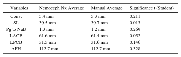
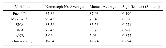
ResultsThe averages of each of the variables measured with the Nx Nemoceph program and with the manual technique were the following respectively: facial convexity5.4 mm(SD=2.5)and 5.3 mm(SD=2.5); SL distance 39.5 mm (SD=7.4) and 39.7 mm (SD=7.4); Pg to NaB distance 1.3 mm (SD=1.1) and 1.2 mm (SD=1.1); length of the anterior cranial base 61.6 mm (SD=5.3) and 61.4 mm (SD=5.1); length of the posterior cranial base 31.5 mm (SD=4.4) and 31.6 mm (SD=4.3); upper facial height 112.7 mm (SD=10.6) and 112.7 mm (SD=10.4); facial depth 87.8° (SD=2.7) and 87.9° (SD=2.7); maxillary depth 93.4° (SD=3.5) and 93.4° (SD=3.4); SNA 83.5° (SD=4.0) and 83.5° (SD=4.0); SNB 78.4° (SD=3.2) and 78.9° (SD=3.0); ANB 5.0° (SD=2.1) and 5.0° (SD=2.2) and sella turcica angle 126.4° (SD=5.4) and 126.4° (SD=5.4). Despite the fact that the values do not show significant differences (p > 0.05) it was determined that tracings with the cephalometric program Nemoceph Nx have greater accuracy in comparison with manually traced radiographs (Tables IandII)
The advantages and disadvantages of digital radiography and its reliability with the cephalometric programs mentioned have been discussed, but previous studies have been carried out in conventional radiography that have been manipulated to convert them into digital format in this regard. Cohen9 mentioned that despite the use of a scanner for the professional handling of a conventional radiography when trasnfering it to digital, there is a difference in linear measurements that can slightly modify the diagnosis. The advantage of using digital radiography is that there is 0% of error by transferring the program to perform some cephalometric tracings, and with the advantage of having a good sharpness to visualize anatomical structures the patients.
ConclusionsDigital records are being used more today according to a «paperless» model of storage. Cephalometric radiographs have received much attention on the part of software developers in its attempt to design the ideal program for cephalometric analysis. According to the present study the Nemoceph Nx program may be used with any reliability to make tracings cephalometric measurements. There is no significant difference in the manual or digital measurement of the linear and angular cephalometric measures, however the layout with the program Nemoceph Nx is more accurate. The main advantage forusing a software is the speed with which it performs this procedure, however, this model still presents a digital error of prediction, i.e. remains a representation of two dimensions of an anatomical structure of three dimensions.