


The mastitis subclinical and clinical in cows caused by fungi has been increased specially by yeast of the genus Candida.
ObjectiveTo identify what yeasts were present in milk samples obtained from mammary glands of healthy cows, and others suffering subclinical or clinical mastitis.
MethodsFrom a total of 1,095 milk samples 342 were from mammary glands of healthy dairy cows, 383 with subclinical mastitis, and 370 with clinical mastitis, were taken, in the states of Querétaro, Hidalgo, Puebla and Mexico City (Distrito Federal) in the so called Mexican High Plateu. The clinical status of the mammary glands was determined by clinical examination and the California Mastitis Test. Yeasts identification was carried out by morphology and biochemical methods.
ResultsTwenty different species of Candida were identified out of 282 (25.75%) milk samples. The most frequently identified species in the healthy cows and cows with clinical mastitis groups were Candida glabrata and Candida krusei. On the other hand, samples from the subclinical mastitis group showed a diversity of Candida species, including Candida zeylanoides, Candida norvegica, Candida viswanathii, Candida guilliermondii, and Candida tropicalis. Candida albicans was isolated only in 11 (3.9%) samples from the clinical and subclinical mastitis groups.
ConclusionsThese results suggest the possible role that Candida species other than C. albicans may play in mycotic mastitis in cows.
Las mastitis subclínicas y clínicas en bovinos por hongos se han incrementado principalmente por levaduras del género Candida.
ObjetivoConocer las levaduras presentes en la leche de glándulas mamarias de bovinos clínicamente sanos, y de aquellos con mastitis subclínica y clínica.
MétodoSe evaluó la presencia de levaduras en 1.095 muestras de leche de 342 glándulas mamarias sanas, 383 con mastitis subclínica y 370 con mastitis clínica, de los estados de Querétaro, Hidalgo, Puebla y la ciudad de México, Distrito Federal, que forman parte del Altiplano Mexicano. El estado sanitario de las glándulas mamarias se determinó por examen clínico y la prueba de California. La identificación de levaduras fue realizada por métodos morfológicos y bioquímicos.
ResultadosSe identificaron 20 especies diferentes del género Candida a partir de 282 (25,75%) de las muestras de leche. Las especies encontradas con mayor frecuencia en los bovinos sanos y con mastitis clínica fueron Candida glabrata y Candida krusei. El grupo de las muestras con mastitis subclínica mostró una diversidad de especies de Candida, incluidas Candida zeylanoides, Candida norvegica, Candida viswanathii, Candida guilliermondii y Candida tropicalis. Candida albicans fue aislada solo en 11 (3,9%) de las muestras de mastitis clínica (6) y subclínica (5).
ConclusionesEstos resultados sugieren el posible papel de otras especies de Candida diferentes a C. albicans como causantes de mastitis micótica.
Bovine mastitis is a disease caused by a wide variety of microorganisms that causes large economical looses and damages to the dairy industry by decreasing milk production and through increased costs of antibiotic treatments and culling6. In most cases, bacteria are recognized as the primary pathogens while fungi, particularly yeasts, have been regarded as secondary mastitis pathogens. Yeasts are considered opportunistic pathogens which colonize the cows’ udder. The use and abuse of antibacterial drugs, treatment with contaminated antibiotic solutions, as well as syringes, or other materials brought in contact with the mammary gland may favor yeast colonization of cows udders4,6,12,13.
Different fungi have been reported as a cause of mycotic mastitis, such as Aspergillus fumigatus, Aspergillus terreus, Candida spp., Cephalosporium spp., Coccidioides spp., Cryptococcus neoformans, Geotrichum candidum, Histoplasma spp., Mucor spp., Rhizopus spp., Torulopsis spp., and Trichosporon spp. Candida species have been regarded in subclinical and clinical mastitis, being the most reported species Candida albicans, Candida krusei, Candida rugosa, and Candida guilliermondii1,4,16,26. Furthermore, other species of Candida, as well as other fungi such as Cryptococcus spp., Rhodotorula spp., Trichosporum cutaneum, Aureobasidium pullulans and Pichia ohmeri, have also been isolated from the milk of healthy glands5,18,21,27.
The purpose of this study is to report the presence of Candida species isolated from cows with different mammary gland health status in the Mexican High Plateu.
Materials and methodsOne thousand and ninety-five milk samples were collected from Holstein-Friesian cows kept under intensive production conditions in the states of Queretaro (n=216), Hidalgo (n=697), Puebla (n=52) and Distrito Federal (n=130) in central Mexico, each sample corresponds to a different animal and from single quarter, in the clinical cases the sample was taken from the most affected quarter. The samples were obtained at convenience, from two-year and older cows, with different mammary glands health status: 342 samples from cows with healthy mammary glands, 383 from cows with subclinical mastitis as determined by the California Mastitis Test (CMT), and 370 from cows diagnosed with clinical mastitis15. Clinical mastitis was defined by: swelling, reduced milk flow, and abnormal milk appearance (watery to viscous with clots varying from gray-white to yellowish). Additionally, other signs of infection such as fever, inappetence, ataxia, and depression were also considered8. CMT was used to identify subclinical mastitis on mammary gland of the cows. For this study, milk samples from glands affected with subclinical mastitis were included when the reaction to CMT was at least grade 1, corresponding with an appearance of viscous milk that does not adhere to the bottom of the CMT plate, and correlates to 400,000-1,500,000 somatic cells/ml25.
Sampling was carried out prior to milking, after washing the udder with soap, drying and disinfecting it with the disinfectant on use in the farm at the moment of sampling, and drying again. All farms included in the study used either 2% iodine or 70% alcohol, both known to have a fungicidal effect2,3,28. Fifteen ml of milk from each sampled gland were collected in sterile screw cap containers. Samples were refrigerated (4°C) during transportation to the laboratory of Mycology at the Faculty of Veterinary Medicine of the National Autonomous University of Mexico (UNAM), and kept at 4°C until processing (no longer than 24h after collection). Milk samples were incubated during 15min at 25°C and homogenized by vigorous shaking; 0.5ml of the homogenized milk were inoculated in 4.5ml of Sabouraud dextrose broth (Difco™), at pH of 3.5, and then incubated at 37°C during 10days17. Thereafter, 50μl of each broth culture was plated on Sabouraud dextrose agar (Difco™), with chloramphenicol (Merck™) (0.05mg/ml). Inoculated plates were then incubated at 37°C and examined daily for colonies for five days.
Yeast identification was performed taking into consideration morphological characteristics, like formation of chlamydoconidium, pseudohyphae and germinal tube development. Additional characteristics were also evaluated, such as growth in the presence of 0.1% cyclohexamide (Sigma™), acidic pH tolerance, urea hydrolysis and carbohydrates assimilation and/or fermentation, accordingly to the methodology described by Barnnet and Payne, and Kurtzman and Fell9,10,23,29. Data obtained in this study were evaluated using cross-tabulation analyses through Chi square distribution24 with the statistical package JMP software, version 5.1.
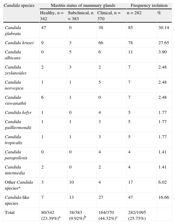
ResultsOut of the 1.095 milk samples analyzed, 282 (25.75%) were positive to yeast isolation (Table 1), obtaining in Queretaro 48/216, Hidalgo 119/697, Puebla 23/52 and Distrito Federal 92/130. Yeast isolates were obtained in 23.39% (80/342) out of milk samples from healthy mammary glands), 9.92% (38/383) from the subclinical mastitis group and 43.27% (164/370) from mammary glands with clinical mastitis. Significant differences (Chi square = 121.7, df = 2.1092, P<0.01) were found among the three groups for the yeast isolation frequencies. Only the genus Candida was identified in the samples that were positive to yeast isolation.
Milk samples and Candida species isolated from mammary glands of healthy cows and cows with subclinical and clinical mastitis.
| Candida species | Mastitis status of mammary glands | Frequency isolation | |||
| Healthy, n = 342 | Subclinical, n = 383 | Clinical, n = 370 | n=282 | % | |
| Candida glabrata | 47 | 0 | 38 | 85 | 30.14 |
| Candida krusei | 9 | 3 | 66 | 78 | 27.65 |
| Candida albicans | 0 | 5 | 6 | 11 | 3.90 |
| Candida zeylanoides | 2 | 3 | 2 | 7 | 2.48 |
| Candida norvegica | 1 | 1 | 5 | 7 | 2.48 |
| Candida viswanathii | 6 | 1 | 0 | 7 | 2.48 |
| Candida kefyr | 1 | 0 | 4 | 5 | 1.77 |
| Candida guilliermondii | 1 | 1 | 3 | 5 | 1.77 |
| Candida tropicalis | 1 | 1 | 3 | 5 | 1.77 |
| Candida parapsilosis | 0 | 0 | 4 | 4 | 1.41 |
| Candida intermedia | 2 | 0 | 2 | 4 | 1.41 |
| Other Candida species* | 3 | 10 | 4 | 17 | 6.02 |
| Candida-like species | 7 | 13 | 27 | 47 | 16.66 |
| Total | 80/342 (23.39%)a | 38/383 (9.92%)b | 164/370 (44.32%)c | 282/1095 (25.75%) | |
*Candida species isolated in < 1.41%.
a,b,cDifferent superscripts indicates statistical differences (p<0.01) among groups for isolates frequencies.
Twenty Candida species were identified, being the most frequently found Candida glabrata 30.14% (85/282), and C. krusei 27.65% (78/282) (Table 1).
Out of the 342 milk samples analyzed from healthy mammary glands, Candida isolates were obtained in 23.39% (80/342) from which 12 different species were identified. The species most frequently found was C. glabrata with 58.75% (47/80) isolates, followed by C. krusei with 11.25% (9/80) and Candida viswanathii with 7.5% (6/80). From the total isolates obtained, 8.75% (7/80) isolates could not been identified at to the species level, being considered as «Candida-like» according their growth characteristics, morphology and microscopy characteristics.
From the 383 samples analyzed from mammary glands with subclinical mastitis, only 9.92% (38/383) Candida isolates were obtained, and identified 15 different species. In this group, the most frequent species found were C. albicans 13.15% (5/38), C. krusei 7.89% (3/38), and Candida zeylanoides 7.89% (3/38), being 34.21% (13/38) of the isolates considered as Candida-like species.
In the 370 samples analyzed from mammary glands with clinical mastitis, 44.32% (164/370) Candida isolates were obtained, from which 13 different species were identified, being C. krusei the most frequently found with 40.24% (66/164) isolates, and C. glabrata with 23.17% (38/164). From these isolates, 16.46% (27/164) were classified as Candida-like species.
Other Candida species, such as C. zeylanoides, Candida norvegica and C. viswanathii were also found in 2.48% and Candida kefyr, C. guilliermondii, Candida tropicalis, Candida parapsilosis, Candida intermedia, Candida claussenii, Candida lusitaniae, Candida macedoniensis, Candida brumptii, Candida sloofii, Candida lambica, Candida cantarelli, Candida lipolytica and Candida incommunis, were also found at very low frequency (≤ 1.7%).
DiscussionIn this study, yeasts were isolated in the 25.75% of all samples analyzed, being Candida the only genus identified. C. glabrata was the predominant species, isolated in 58.75% of all positive samples (47/80) from the mammary glands from healthy cows, and in 23.17% of the Candida isolates (38/164) from those with clinical mastitis. The isolation of this Candida species from cattle affected with mastitis has also recently been reported in New Zealand by Williamson and di Menna29. However, a previous report from Lagneau18, also showed the presence of C. glabrata in the mammary glands of healthy animals, in a rather small frequency (0.2%) when compared with our findings. In contrast, no C. glabrata was obtained from milk samples of our group of cows with subclinical mastitis. This could be partially explained by the fact that in subclinical mastitis it is common to find a greater number of bacteria competing for substrates; in addition, some bacterial metabolites are known as yeasts antagonists, restricting their development20. It seems necessary to put more our attention to this species in order to establish its possible role in bovine mastitis.
The second most frequent species of Candida found in this study was C. krusei. This species was isolated in 11.25% of the healthy glands positive milk samples (9/80); in 7.89% of those with subclinical mastitis (3/38); and in 40.24% of those diagnosed with clinical mastitis (66/164). The presence of C. krusei in dairy cattle with mastitis has been recognized since the 1970s14,22, although it has been more frequently reported since the late 1990s4,11,16,19. There is, however, some discrepancy among the rates in which this yeast has been found. Whereas Casia4, reported in 2005 a 44.5% of C. krusei isolates from cows with mastitis, similarly to ours findings; Krukowski in 200016, and Langoni in 199519, found this species in 15.5% of mastitis samples and in less percentage than 2% in glands with subclinical and clinical mastitis, respectively. Both findings are lower than our results. This might be a consequence effect of several factors, such as the abuse of intramammary antibiotic treatment or the use of home made antibiotic infusions for mastitis therapy16; the natural resistance of C. krusei to antimycotics, like fluconazol11; yeast contaminated food or environment7,27; and inadequate milking procedures27, in addition to the presence of pathogenic strains of C. krusei27. Recently, Williamson and di Menna reported a self-limitating clinical mastitis infection due to C. krusei in cows at calving and at up to 5weeks post-partum29. The isolation of C. krusei from healthy mammary glands might indicate the yeast as part of their normal microbiota and therefore, acting as an opportunistic pathogen in mastitis cases.
Regardless the health status of the mammary glands analyzed, C. zeylanoides, C. norvegica, C. guilliermondii, and C. tropicalis were always isolated. All these species have been shown to be involved in clinical mastitis. C. zeylanoides and C. norvegica are considered opportunistic pathogens, while C. guilliermondii and C. tropicalis are recognized as pathogens4,16. However, our findings suggest that C. norvegica might act as a pathogen similar to C. guilliermondii and C. tropicalis, since the three species were found predominantly in milk from glands with clinical mastitis. In addition, our results on the isolation of Candida parapsilosis, only from animals with clinical mastitis, might support the probably pathogenicity of this Candida species16,18.
The isolation of C. albicans only from glands from the groups of cows with clinical and subclinical mastitis (but not from healthy mammary glands), is consistent with the recognized pathogenicity of this species. The percentages in which this yeast was isolated in this study were similar to those reported in cows with mastitis by Langoni19, Krukowski16, and Casia4.
Our findings in relation to C. viswanathii, which was identified in 7.5% (6/80) of the healthy glands group, and in 2.6% (1/38) of the subclinical mastitis group, suggest the opportunistic pathogen role that this Candida species could be playing in this disease.
C. kefyr, on the other hand, was identified in 2.43% (4/164) isolates in the clinical mastitis group, in partial agreement with previous reports in Europe that found C. kefir in milk samples at frequencies as high as 24.1%16,25.
The isolation of numerous Candida non-albicans species from bovine milk is also in agreement with other reports4,16,25, recognizing among others, antibacterial therapy without previous susceptibility tests as the principal predisposing factor for yeast infections. Furthermore, the wide distribution of yeasts in nature, e.g. soil, plants and water, to which cows are in contact with, might have implications regarding the presence of fungi in mammary glands4,8,16. Similar to our findings, there are some reports where Candida species other than C. albicans, have been isolated from milk of cows with mastitis2,3,8.
Of the yeast isolates identified as Candida-like species, 16.66% (47/282) could not be further characterized by the biochemical methodology used in this study. At present, it is well known that the use of routine yeasts identification methods based on morphology and biochemical test are time-consuming, and they often fail to identify yeast species other than C. albicans. Alternative identification methods, such as PCR or RAPD-PCR might be necessary to support identification and further characterization of these mycotic agents17.
The presence of yeasts in udders of healthy cows, as well as in those of cows with mastitis, may be an important factor in the dissemination of disease. These agents might increase their presence due to handling of dairy cattle, bacterial mastitis treatment and prevention as well as inadequate milking practices. Furthermore, the presence of Candida non-albicans species that have gained ground as pathogens of the bovine mammary gland must be highlighted. For instance, the association of C. glabrata and C. krusei with clinical mastitis is clear, probably acting as opportunistic pathogens, considering that they can also be found in milk samples coming from healthy cows.
Although C. albicans has been reported as the most common species of yeast pathogen found, this study shows that C. krusei seems to be of interest as a probable cause of mastitis problems.
FinancingPrograma de Apoyo a Proyectos de Investigación e Innovación Tecnológica (PAPIIT) grant IN 209908, Universidad Nacional Autónoma de México (UNAM). Consejo Nacional de Ciencia y Tecnología (CONACYT) 151676.
Conflict of interestAuthors have no conflict of interests.
The authors like to thank Sarahi Luna-Castro who collaborated with the biochemical characterization of the yeasts. We are particularly grateful to Dr. Cristina Escalante-Ochoa for her valuable review of this manuscript.

