


This study evaluates the diagnostic importance of a new parameter consisting of the densitometric and metabolic properties of the lesion measured by 18F-FDG PET/CT in differentiating between benign and malignant adrenal lesions in cancer patients.
Material and methodsWe evaluated PET/CT parameters of adrenal lesions of patients between 2017 and 2019. A new parameter, “Adrenal Dansitometabolic index (ADMI),” was investigated. Additionally, SUVmax, tumor-to-liver ratio (T/LR), Hounsfield Units (HU), metabolic tumor volume (MTV), and total lesion glycolysis (TLG) of adrenal lesions were analyzed. Logistic regression analyses were conducted to identify the most predictive parameter for malignant lesions.
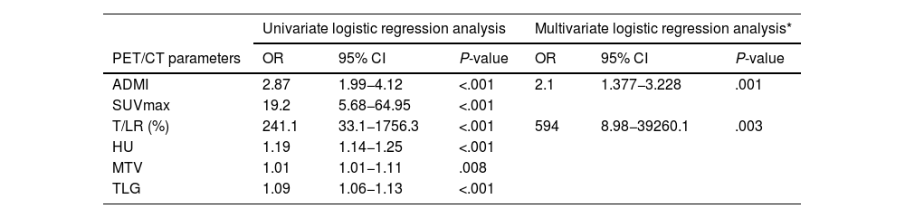
ResultsTwo hundred and four adrenal lesions underwent PET/CT for tumor evaluation. Based on ROC analysis, ADMI > 9.5, a SUVmax > 4.15, a T/L SUVmax ratio > 1.7, an attenuation value > 21.75 HU, a TLG > 14.61 and a MTV > 4.5 were chosen as the optimal cut-off values for differentiating malignant from benign lesions. ADMI, SUVmax, and T/L ratio demonstrated similarly high diagnostic performance (AUC values: 0.993, 0.992 and 0.988 (P = .000), respectively). ADMI exhibited the highest sensitivity and negative predictive value. A multivariate logistic regression analysis revealed that both ADMI and T/L ratio serve as independent prognostic factors for malignancy (P = .001 and P = .003, respectively).
ConclusionADMI, effectively distinguishes benign from malignant. This technique may facilitate investigations of the adrenal lesions to clinical outcomes but needs further large series studies.
Este estudio evalúa la importancia diagnóstica de un nuevo parámetro consistente en las propiedades densitométricas y metabólicas de la lesión, medidas mediante PET/TC 18F-FDG, para diferenciar entre lesiones suprarrenales benignas y malignas en pacientes con cáncer.
Materiales y métodosEvaluamos los parámetros PET/TC de las lesiones suprarrenales de pacientes entre 2017 y 2019. Se investigó un nuevo parámetro, el “índice densitometabólico suprarrenal (IDMS)”. Además, se analizaron el SUVmax, la relación tumor-hígado (T/H), las unidades Hounsfield (UH), el volumen metabólico del tumor (VMT) y la glucólisis total de la lesión (GTL) de las lesiones suprarrenales. Se realizaron análisis de regresión logística para identificar el parámetro más predictivo de las lesiones malignas.
ResultadosSe realizó PET/TC a 204 lesiones suprarrenales para la evaluación del tumor. Con base en el análisis ROC, se eligieron IDMS > 9,5, SUVmax > 4,15, una relación SUVmax T/H > 1,7, un valor de atenuación (UH) > 21,75 HU, una GTL > 14,61 y un VMT > 4,5 como los valores de corte óptimos para diferenciar lesiones malignas de benignas. IDMS, SUVmax y la relación T/H demostraron un rendimiento diagnóstico igualmente alto (valores de AUC: 0,993, 0,992 y 0,988 (P = ,000), respectivamente). IDMS exhibió la sensibilidad y el valor predictivo negativo más altos. Un análisis de regresión logística multivariante reveló que tanto IDMS como la relación T/H sirven como factores pronósticos independientes para la malignidad (P = ,001 y P = ,003, respectivamente).
ConclusionesEl IDMS distingue eficazmente las lesiones benignas de las malignas. Esta técnica puede facilitar la investigación de las lesiones suprarrenales para obtener resultados clínicos, pero se necesitan más estudios con series más amplias.
Article
If you experience access problems, you can contact the SEMNIM Technical Secretariat by email at secretaria.tecnica@semnim.es or by phone at +34 619 594 780.

Revista Española de Medicina Nuclear e Imagen Molecular (English Edition)
