metricas
covid
Revista Española de Medicina Nuclear e Imagen Molecular (English Edition) Dual time point 2-deoxy-2-[18F]fluoro-d-glucose PET/CT: Nodal staging in locally...
Journal Information
Vol. 33. Issue 1.
Pages 1-5 (January - February 2014)
Share
Download PDF
More article options
Visits
845
Vol. 33. Issue 1.
Pages 1-5 (January - February 2014)
Original article
Dual time point 2-deoxy-2-[18F]fluoro-d-glucose PET/CT: Nodal staging in locally advanced breast cancer
PET-TC con 2-deoxi-2-[18F]fluor-d-glucosa en doble fase: estadificación ganglionar en cáncer de mama localmente avanzado
Visits
845
A.M. García Vicentea,
Corresponding author
angarvice@yahoo.es

Corresponding author.
, Á. Soriano Castrejóna, M.Á. Cruz Morab, C. Ortega Ruiperezc, R. Espinosa Auniónd, A. León Martíne, A. González Ageitosf, O. Van Gómez Lópeza
a Nuclear Medicine Department, University General Hospital, Ciudad Real, Spain
b Oncology Department, Virgen de la Salud Hospital, Toledo, Spain
c Oncology Department, Virgen de la Luz Hospital, Cuenca, Spain
d Oncology Department, La Mancha Centro Hospital, Alcázar de San Juan, Ciudad Real, Spain
e Investigation Unit, University General Hospital, Ciudad Real, Spain
f Oncology Department, Nuestra Sra. del Prado Hospital, Talavera de la Reina, Toledo, Spain
This item has received
Article information
Abstract
Full Text
Bibliography
Download PDF
Statistics
Figures (2)
Tables (3)
Table 1. Statistical diagnostic parameters in lymph node status of conventional diagnostic imaging, metabolic imaging and combined.
Tables
Table 2. Metabolic lymph node involvement: distribution of extra-axillary locations.
Tables
Table 3. Distribution of cN and mN.
Tables
Show moreShow less
Abstract
Aim

To assess dual time point 2-deoxy-2-[18F]fluoro-d-glucose 18FFDG PET-CT accuracy in nodal staging and in detection of extra-axillary involvement.
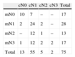
Material and methods

Dual time point [18F] FDG PET/CT scan was performed in 75 patients. Visual and semiquantitative assessment of lymph nodes was performed. Semiquantitative measurement of SUV and ROC-analysis were carried out to calculate SUVmax cut-off value with the best diagnostic performance. Axillary and extra-axillary lymph node chains were evaluated.
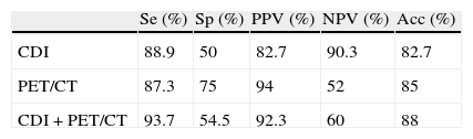
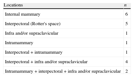
Results

Sensitivity and specificity of visual assessment was 87.3% and 75%, respectively. SUVmax values with the best sensitivity were 0.90 and 0.95 for early and delayed PET, respectively. SUVmax values with the best specificity were 1.95 and 2.75, respectively. Extra-axillary lymph node involvement was detected in 26.7%.

Conclusion

FDG PET/CT detected extra-axillary lymph node involvement in one-fourth of the patients. Semiquantitative lymph node analysis did not show any advantage over the visual evaluation.

Keywords:
18FFDG PET/CT
Locally advanced breast cancer
Dual time point PET
Lymph node staging
Resumen
Objetivo

Valorar la precision diagnóstica de la PET-CT con 2-deoxi-2-[18F]fluor-d-glucosa [18F] FDG en doble fase en la estadificación ganglionar y en la detección de afectación extra-axilar.

Material y métodos

Se realizó una [18F] FDG PET-TC en doble fase a 75 pacientes. Se valoraron los ganglios linfáticos de forma visual y semicuantitativa. Se realizaron medidas del SUV y análisis ROC para calcular el valor de SUV max con la mejor precisión diagnóstica. Se evaluaron los niveles axilares y extra-axilares.

Resultados

La sensibilidad y especificidad del análisis visual fue del 87.3% y 75% respectivamente. Los valores de SUVmax con la mejor sensibilidad fueron de 0.90 y 0.95 para el PET en fase precoz y tardía respectivamente. Los valores de SUV max con la mejor especificidad fueron de 1.95 y 2.75 respectivamente. Se detectó afectación ganglionar extra-axilar en el 26.7%.

Conclusión

La PET-TC con FDG detectó afectación ganglionar extra-axilar en una cuarta parte de las pacientes. El análisis semicuantitativo no pareció aportar ninguna ventaja sobre la valoración visual.

Palabras clave:
[18F] FDG PET-TC
Cáncer de mama localmente avanzado
PET en doble fase
Estadificación ganglionar

Article

These are the options to access the full texts of the publication Revista Española de Medicina Nuclear e Imagen Molecular (English Edition)
Member
If you are a member of the Spanish Society of Nuclear Medicine and Molecular Imaging (SEMNIM), you can access the full text of the contents of the Revista Española de Medicina Nuclear e Imagen Molecular through the journal links published on the SEMNIM website (link: http://socios.semnim.es/?ir-a=remnim), after logging in as a member.

If you experience access problems, you can contact the SEMNIM Technical Secretariat by email at secretaria.tecnica@semnim.es or by phone at +34 619 594 780.
Subscriber
Subscriber

If you already have your login data, please click here .

If you have forgotten your password you can you can recover it by clicking here and selecting the option “I have forgotten my password”
Subscribe
Subscribe to

Revista Española de Medicina Nuclear e Imagen Molecular (English Edition)

Purchase
Purchase article

Purchasing article the PDF version will be downloaded

Purchase now
Contact
Phone for subscriptions and reporting of errors
From Monday to Friday from 9 a.m. to 6 p.m. (GMT + 1) except for the months of July and August which will be from 9 a.m. to 3 p.m.
Calls from Spain
932 415 960
Calls from outside Spain
+34 932 415 960
E-mail
Article options
Tools