


Programmed death-ligand 1 (PD-L1) expression serves as a critical biomarker for selecting patients eligible for treatment with immune checkpoint inhibitors. Herein, we investigated the association between PD-L1 expression and various FDG PET/CT–derived metabolic parameters in patients with non-small cell lung cancer (NSCLC).
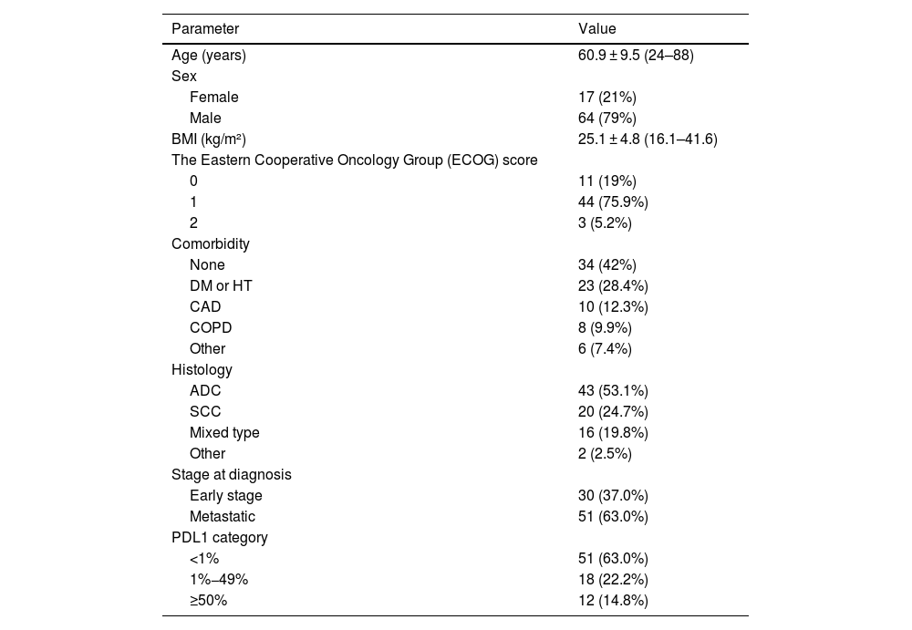
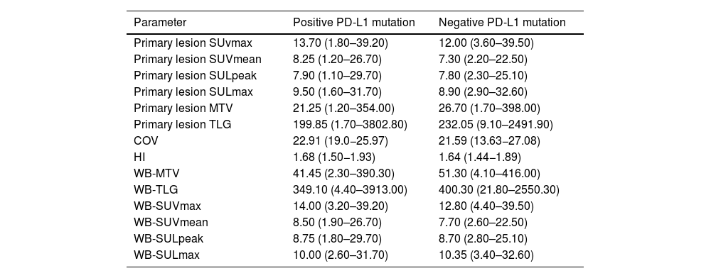
Materials and methodsThis retrospective study included 81 NSCLC patients who underwent pre-treatment F-18 FDG PET/CT imaging and histopathological evaluation of PD-L1 expression. PD-L1 tumour proportion score (TPS) was determined using the SP263 immunohistochemical assay. PD-L1 positivity was defined as TPS ≥ 1%. Quantitative PET/CT parameters—SUVmax, SUVmean, SULpeak, SULmax, metabolic tumor volume (MTV), total lesion glycolysis (TLG), and heterogeneity indices (coefficient of variation [COV] and SUV-based heterogeneity index [HI])—were analyzed in relation to PD-L1 TPS.
ResultsPD-L1 positivity was identified in 30 patients (37%). Although SUVmax, SUVmean, SULpeak, and SULmax values tended to be higher in PD-L1–positive patients, these differences were not statistically significant. Conversely, MTV and TLG were higher in the PD-L1–negative group. Among all parameters, HI was significantly elevated in the PD-L1–positive group (P = .031), and remained significant across PD-L1 expression strata (P = .037). In metastatic patients, HI and COV showed significant positive correlation with PD-L1 expression (r = 0.34 and 0.33, respectively). ROC analysis identified a HI cut-off of 1.59 to predict PD-L1 positivity with 90% sensitivity and 50% specificity (AUC = 0.674).
ConclusionsTumor heterogeneity indices, particularly HI and COV derived from FDG PET/CT, demonstrated stronger predictive value for PD-L1 expression than conventional metabolic parameters. These findings suggest that metabolic heterogeneity may serve as a useful noninvasive imaging biomarker for guiding immunotherapy in NSCLC.
La expresión del ligando de muerte programada 1 (PD-L1) sirve como un biomarcador crucial para seleccionar a los pacientes elegibles para el tratamiento con inhibidores de puntos de control inmunitarios. En este estudio, investigamos la asociación entre la expresión de PD-L1 y diversos parámetros metabólicos derivados de la tomografía por emisión de positrones/tomografía computarizada con FDG (PET/TC con FDG) en pacientes con cáncer de pulmón no microcítico (CPNM).
Materiales y métodosEste estudio retrospectivo incluyó a 81 pacientes con CPNM que se sometieron a una PET/TC con F-18 FDG antes del tratamiento y a una evaluación histopatológica de la expresión de PD-L1. La puntuación de proporción tumoral (TPS) de PD-L1 se determinó mediante el ensayo inmunohistoquímico SP263. Se definió positividad de PD-L1 como TPS ≥ 1%. Se analizaron los parámetros cuantitativos de la PET/TC —SUVmax, SUVmean, SULpeak, SULmax, volumen tumoral metabólico (MTV), glucólisis total de la lesión (TLG) e índices de heterogeneidad (coeficiente de variación [COV] e índice de heterogeneidad basado en SUV [HI])— en relación con el TPS de PD-L1.
ResultadosLa positividad de PD-L1 se identificó en 30 pacientes (37%). Aunque los valores de SUVmax, SUVmean, SULpeak y SULmax tendieron a ser más altos en los pacientes PD-L1 positivos, estas diferencias no fueron estadísticamente significativas. Por el contrario, los valores de MTV y TLG fueron mayores en el grupo PD-L1 negativo. Entre todos los parámetros, el HI fue significativamente más elevado en el grupo PD-L1 positivo (P = ,031) y permaneció significativo entre los diferentes niveles de expresión de PD-L1 (P = ,037). En pacientes con enfermedad metastásica, HI y COV mostraron una correlación positiva significativa con la expresión de PD-L1 (r = 0,34 y 0,33, respectivamente). El análisis ROC identificó un punto de corte para HI de 1,59 para predecir la positividad de PD-L1 con una sensibilidad del 90% y especificidad del 50% (AUC = 0,674).
ConclusionesLos índices de heterogeneidad tumoral, en particular HI y COV derivados de la PET/TC con FDG, demostraron un mayor valor predictivo para la expresión de PD-L1 que los parámetros metabólicos convencionales. Estos hallazgos sugieren que la heterogeneidad metabólica podría servir como un biomarcador de imagen no invasivo útil para orientar la inmunoterapia en el CPNM.
Article
If you experience access problems, you can contact the SEMNIM Technical Secretariat by email at secretaria.tecnica@semnim.es or by phone at +34 619 594 780.

Revista Española de Medicina Nuclear e Imagen Molecular (English Edition)
