


White matter lesions (WMLs), detected as hyperintensities in T2-weighted MRI, represent small vessel disease in the brain and are considered a potential risk factor for memory and cognitive impairment. It has not been sufficiently evidenced that cognitive impairment in patients with Alzheimer's disease is caused by WMLs as well as β-amyloid (Aβ) pathology. The aim of this study was to evaluate the relationship between WMLs and cerebral glucose metabolism in patients with cognitive impairment after adjustment of cerebral Aβ burden.
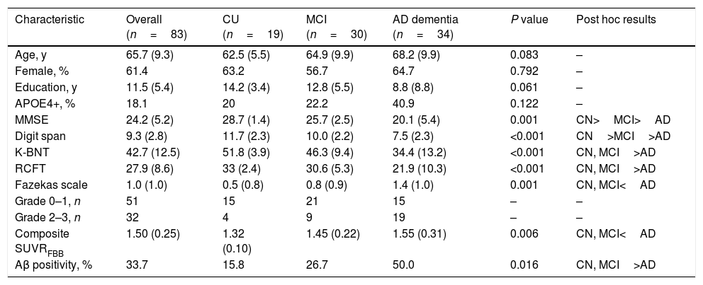
Materials and methodsEighty-three subjects with cognitive performance ranging from normal to dementia, who underwent brain MRI and 18F-florbetaben positron emission tomography (PET) and 18F-fluorodeoxyglucose PET, were included in this cross-sectional study. The Fazekas scale was used to quantify WMLs on brain T2-weighted MRI. The cerebral Aβ burden and cerebral glucose metabolism were quantitatively estimated using volume-of-interest analysis. Differences in the regional cerebral glucose metabolism were evaluated between low-WML (Fazekas scale<2) and high-WML (Fazekas scale≥2) groups. Multiple linear regression analysis adjusted for age, sex and cerebral Aβ burden was performed to evaluate the relationship between the Fazekas scale score and cerebral glucose metabolism.
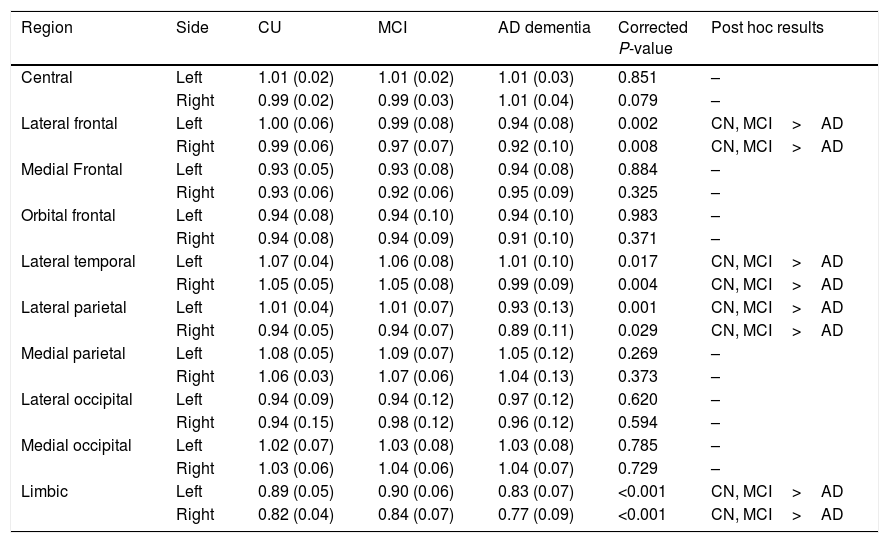
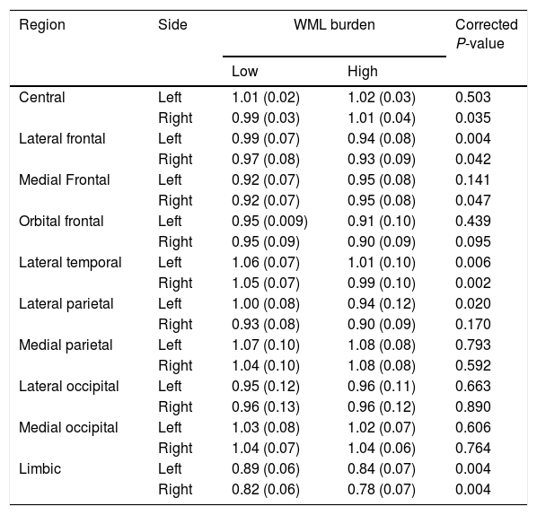
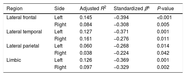
ResultsThe regional cerebral glucose metabolism for the bilateral frontal, temporal, and parietal cortices, and limbic lobes in the high-WML group were significantly lower than those in the low-WML group. There were significant negative correlations between the Fazekas scale score and regional cerebral glucose metabolism in the bilateral frontal, bilateral temporal and left parietal cortices, and bilateral limbic lobes. Multiple linear regression analysis revealed that the Fazekas scale score was an independent determinant of the glucose metabolism in the bilateral frontal and temporal cortices and limbic lobes.
ConclusionsWMLs are associated with decreased cerebral glucose metabolism. Our findings suggest that small vessel disease, as well as Aβ pathology, may contribute to cognitive impairment in patients with Alzheimer's disease.
Los daños en la sustancia blanca (DSB), detectados como hiperintensidades en las imágenes de RM ponderadas en T2, representan la enfermedad de pequeños vasos cerebrales, y están considerados como un factor de riesgo potencial de trastornos de la memoria y disfunción cognitiva. No se ha evidenciado suficientemente que la disfunción cognitiva en pacientes con enfermedad de Alzheimer esté causada por DSB y la patología β-amiloide (Aβ). El objetivo de estudio fue evaluar la relación entre los DSB y el metabolismo de la glucosa cerebral en pacientes con disfunción cognitiva, tras el ajuste de la carga cerebral de Aβ.
Materiales y métodosIncluimos en este estudio transversal a ochenta y tres sujetos con desempeño cognitivo que oscilaba entre normal y demencia, a quienes se realizó RM cerebral y PET con Florbetaben (18F) y PET con 18F-FDG. Utilizamos la escala Fazekas para cuantificar los DSB en la RM cerebral ponderada en T2. Estimamos cuantitativamente la carga cerebral de Aβ y el metabolismo de la glucosa cerebral utilizando el análisis del volumen de interés. Evaluamos las diferencias del metabolismo de la glucosa cerebral regional entre los grupos de bajo DSB (escala Fazekas<2) y alto DSB (escala Fazekas≥2). Realizamos un análisis de regresión lineal múltiple ajustado por edad, sexo y carga cerebral de Aβ, para evaluar la relación entre la puntuación de la escala Fazekas y el metabolismo de la glucosa cerebral.
ResultadosEl metabolismo de la glucosa cerebral regional para los lóbulos bilateral frontal, temporal, córtices parietales, y lóbulos límbicos en el grupo de alto DSB fueron significativamente menores que los del grupo de bajo DSB. Existieron correlaciones negativas significativas entre la puntuación de la escala Fazekas y el metabolismo de la glucosa cerebral regional en los córtices bilateral frontal, bilateral temporal y córtices parietales izquierdos, y lóbulos límbicos bilaterales. Los análisis de regresión lineal múltiple revelaron que la puntuación de la escala Fazekas era un determinante independiente del metabolismo de la glucosa en los córtices bilateral frontal y temporal y los lóbulos límbicos.
ConclusionesLos DSB están asociados a un descenso del metabolismo de la glucosa cerebral. Nuestros hallazgos sugieren que la enfermedad de pequeños vasos, así como lo patología de Aβ, pueden contribuir a la disfunción cognitiva en pacientes con enfermedad de Alzheimer.
Article
If you experience access problems, you can contact the SEMNIM Technical Secretariat by email at secretaria.tecnica@semnim.es or by phone at +34 619 594 780.

Revista Española de Medicina Nuclear e Imagen Molecular (English Edition)
