


We analyzed the morphology of colonies of endophytic bacteria and yeasts by fluorescence. Agrobacterium tumefaciens 6N2 (formerly 197MX) is a non-pathogenic isolate from sugarcane with features of relevance for microbial interactions1. Meyerozyma guilliermondii 6N was obtained from the same host3.
A. tumefaciens 6N2 was tagged with gfp.AAV-a, allowing the detection of cells actively expressing the fluorescence2. GFP-tagged 6N2 and M.guilliermondii 6N were cultured at 30°C in nutrient broth until late exponential phase1. Cell densities were adjusted to ∼0.1 OD600nm, and spot inoculated on nutrient agar (NA) and YPD agar plates5, as pure or mixed cultures (proportion 1:1). Plates were incubated at 30°C for 72h; colonies were imbibed with 10μl of 25μM Calcofluor White M2R (CW) that binds chitin and cellulose, and observed with a 4× objective lens under a Zeiss SteREO Lumar.V12 Stereomicroscope with GFP and DAPI filters.
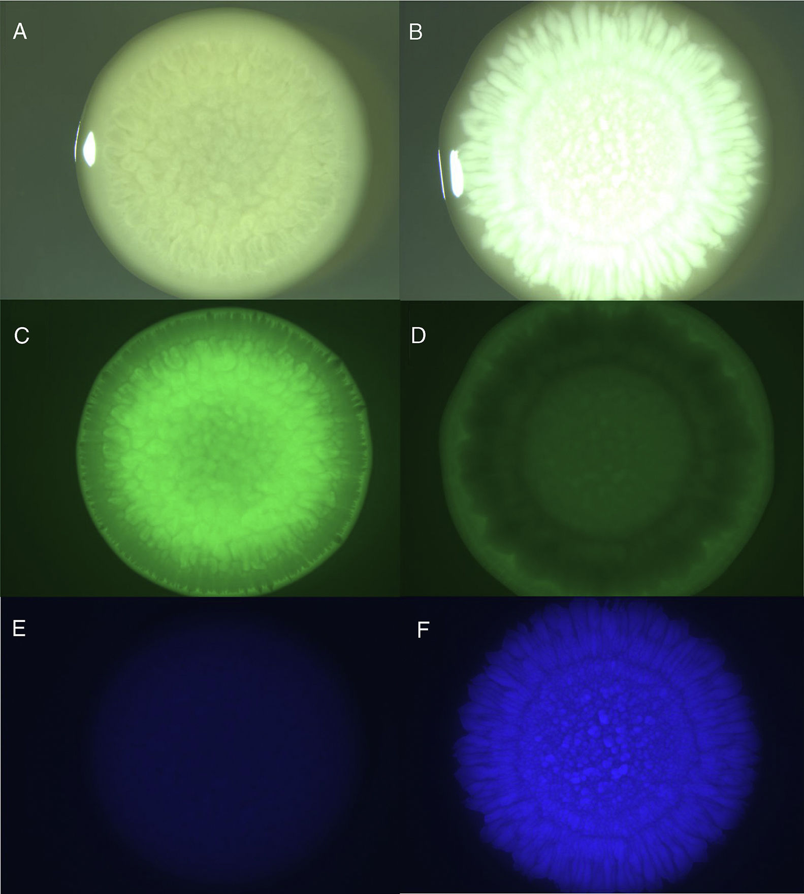
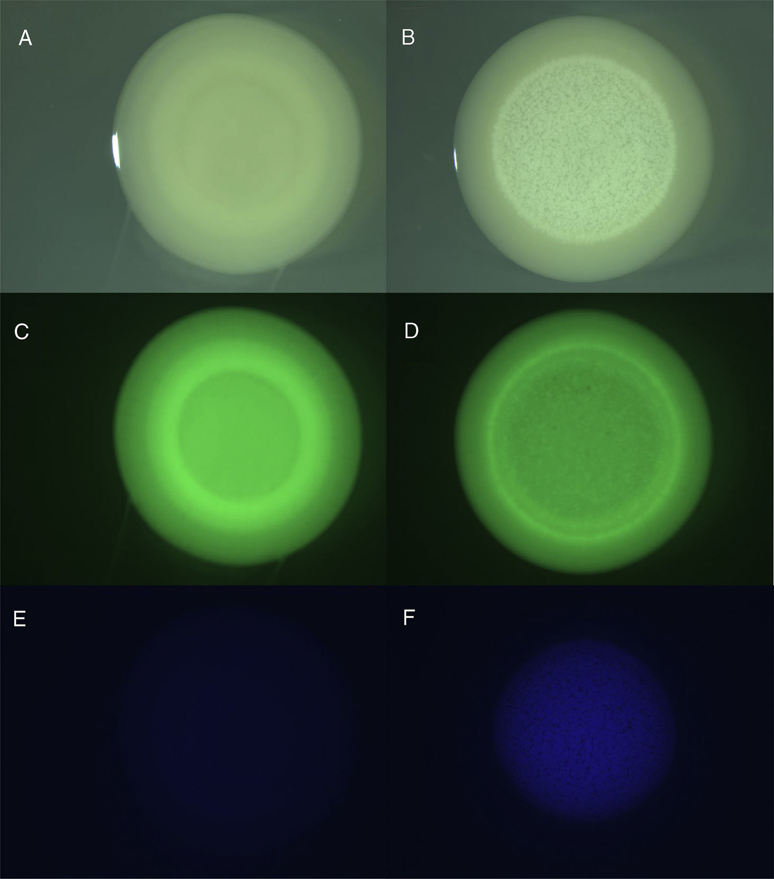
While 6N2 colonies exhibited a wrinkled surface on YPD agar (Fig. 1A and C), they were smooth and convex on NA (Fig. 2A and C). Under interaction conditions 6N2 showed singular patterns: 6N2 was located at the edges and the center on YPD agar (Fig. 1D), and was evenly distributed on NA (Fig. 2D). In both media 6N seemed to be located on top of the bacterial growth (Fig. 1B and B). 6N development was granular with lobate borders on YPD agar (Fig. 1B and F), and was smooth and dotted on NA (Fig. 2B and F). Noteworthy, 6N2 was not stained with CW (Fig. 1E and E). Beyond the effect of the interactions on the host plant, the images show the potential of the strains and the techniques for the study of interactions in a colony biofilm, even if the true location of each strain requires other techniques (e.g., confocal microscopy)4.

Colony biofilms of A. tumefaciens 6N2 and M. guilliermondii 6N growing on YPD agar. A, C and E: pure colony of 6N2 observed with visible illumination, GFP filter and DAPI filter, respectively. B, D and F: colony of mixed cultures of 6N2 and 6N observed with visible illumination, GFP filter and DAPI filter, respectively.

Colony biofilms of A. tumefaciens 6N2 and M. guilliermondii 6N growing on NA. A, C and E: pure colony of 6N2 observed with visible illumination, GFP filter and DAPI filter, respectively. B, D and F: colony of mixed cultures of 6N2 and 6N observed with visible illumination, GFP filter and DAPI filter, respectively.
This work was supported by the Consejo Nacional de Investigaciones Científicas y Técnicas (CONICET, PIP 946, PU-E 22920160100012CO), Agencia Nacional de Promoción Científica y Tecnológica (PICT 2016 No. 0532; PICT 2016 No. 2013), and Secretaría de Ciencia, Arte e Innovación Tecnológica from the Universidad Nacional de Tucumán (PIUNT D609).
Conflict of interestThe authors declare that they have no conflicts of interest.
We thank Jean-Michel Camadro (Institut Jacques Monod, UMR7592, CNRS Université Paris Diderot, Paris, France) for kindly providing access to the stereomicroscope facility.
