


La resonancia magnética cardíaca fetal es una nueva técnica para el estudio de las cardiopatías congénitas. El objetivo de este artículo es analizar los hallazgos adicionales que aporta respecto a la ecocardiografía y el impacto clínico en el diagnóstico y tratamiento en un grupo de pacientes.
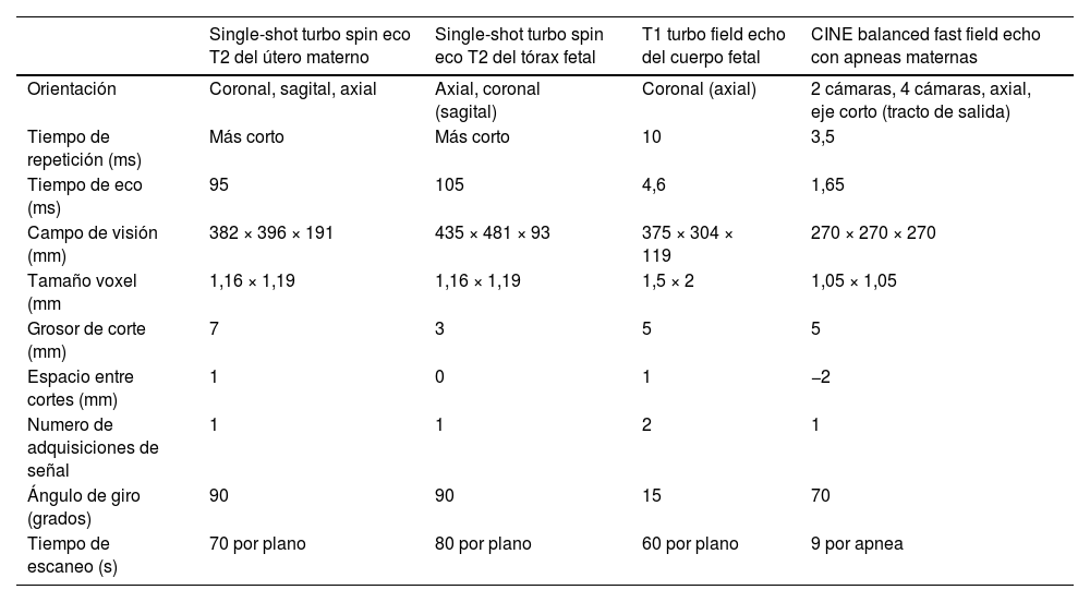
Material y métodoSe incluyeron las cardioRM fetales realizadas en 2023 y 2024. Se revisó retrospectivamente la historia clínica de las madres para analizar la edad de las gestantes, edad gestacional a la hora de las pruebas de imagen, hallazgos ecográficos, hallazgos de RM y tiempo entre ecografía y RM. Todas las ecocardiografías fueron indicadas, realizadas e informadas dentro del seguimiento clínico normal de estas pacientes por obstetras especializados en medicina fetal. Las RM fueron indicadas por los obstetras, realizadas en una RM 1,5 Tesla e informadas por un radiólogo y una pediatra especializados en cardiopatías congénitas. Se evaluó si la RM aportó nuevos hallazgos, si cambió la información pronóstica y/o el manejo clínico. El diagnóstico final se obtuvo analizando la historia clínica de las madres y el seguimiento de los niños durante la infancia.
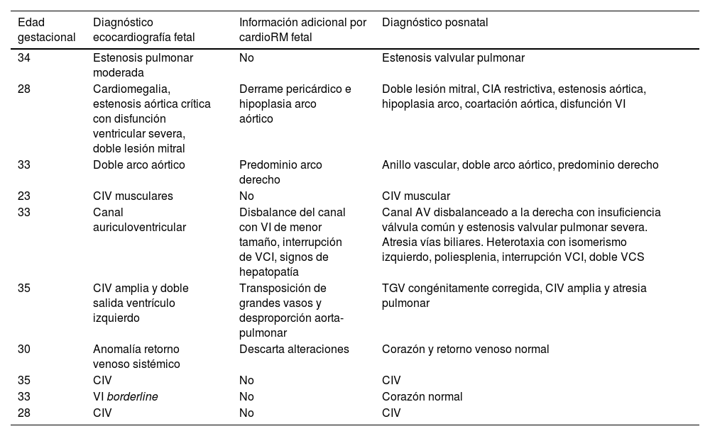
ResultadosDiez fetos entre la semana 23 y 35 de gestación fueron derivados para estudiar malformaciones cardiovasculares mediante RM (estenosis pulmonar, mitral, aórtica, transposición de grandes vasos, anillo vascular, comunicación interventricular, canal auriculoventricular, ventrículo izquierdo hipoplásico). La RM aportó información adicional a la ecocardiografía en la mitad de los casos, sin cambiar el manejo prenatal de ninguno.
ConclusiónLa RM fetal mostró ser una técnica útil para el diagnóstico prenatal, aportando información adicional relevante en muchos casos, sin cambiar el manejo.
.
Fetal cardiac magnetic resonance imaging (MRI) is a new technique for the study of congenital heart defects. The aim of this article is to analyse the additional findings it provides compared to echocardiography and its clinical impact on diagnosis and treatment in a group of patients.
Materials and methodsFetal cardiac MRIs performed in 2023 and 2024 were included. The maternal medical records were retrospectively reviewed to analyse the age of the pregnant women, gestational age at the time of imaging, ultrasound findings, MRI findings, and the time interval between ultrasound and MRI. All echocardiograms were indicated, performed and reported as part of the routine clinical follow-up of these patients by obstetricians specialised in fetal medicine. The MRIs were indicated by obstetricians, performed on a 1.5 Tesla scanner, and reported by a radiologist and a paediatrician specialised in congenital heart defects. MRIs were evaluated for new findings and for changes to prognostic information, and/or clinical management. The final diagnosis was obtained by analysing maternal medical records and follow-up during infancy.
ResultsTen fetuses between 23 and 35 weeks of gestation were referred for MRI assessment of cardiovascular malformations (pulmonary, mitral and aortic stenosis; transposition of the great vessels; vascular ring; ventricular septal defect; atrioventricular canal; hypoplastic left ventricle). MRI provided additional information to echocardiography in half of the cases, without altering prenatal management in any of them.
ConclusionFetal MRI has been shown to be a useful technique for prenatal diagnosis, providing relevant additional information in many cases without altering management.


