Buscar en
Medicina Clínica Práctica
Toda la web
Inicio Medicina Clínica Práctica Prosthetic valve endocarditis complicated with perivalvular pseudoaneurysm
Información de la revista
Vol. 4. Núm. 2.
(Abril - Junio 2021)
Compartir
Compartir
Descargar PDF
Más opciones de artículo
Vol. 4. Núm. 2.
(Abril - Junio 2021)
Image in medicine
Open Access
Prosthetic valve endocarditis complicated with perivalvular pseudoaneurysm
Endocarditis sobre válvula protésica complicada con pseudoaneurisma perivalvular
Visitas
729
Gonçalo Miranda
Autor para correspondencia
goncaloflmiranda@gmail.com

Corresponding author.
, Ana Sofia Rodrigues, Maria Inês Gonçalves, Paula Perestrelo, Isabel de La Cal Caballero, Pedro Lito, Anne Delgado Bohlen, Bruno Rodrigues
Centro Hospitalar Universitário Cova da Beira, Quinta do Alvito, 6200-251, Covilhã, Portugal
Este artículo ha recibido

Under a Creative Commons license
Información del artículo
Texto completo
Bibliografía
Descargar PDF
Estadísticas
Figuras (1)
Texto completo

Infective endocarditis is an infection of the endocardial surface of the heart. Patients with a prosthetic valve are at higher risk to develop infective endocarditis and more often develop complications of the disease. Pseudoaneurysms are severe complications associated with severe valvular and perivalvular damage.1

A seventy-two-year-old male with a past history of subacute mitral valve endocarditis with severe mitral regurgitation and moderate aortic regurgitation, who has undergone mitral valvuloplasty and placement of aortic biological prosthesis four years before, was admitted to our hospital with complaints of fever for one week. Urine and blood cultures were positive for Enterococcus faecalis and antibiotic therapy with ampicillin and gentamicin was initiated.

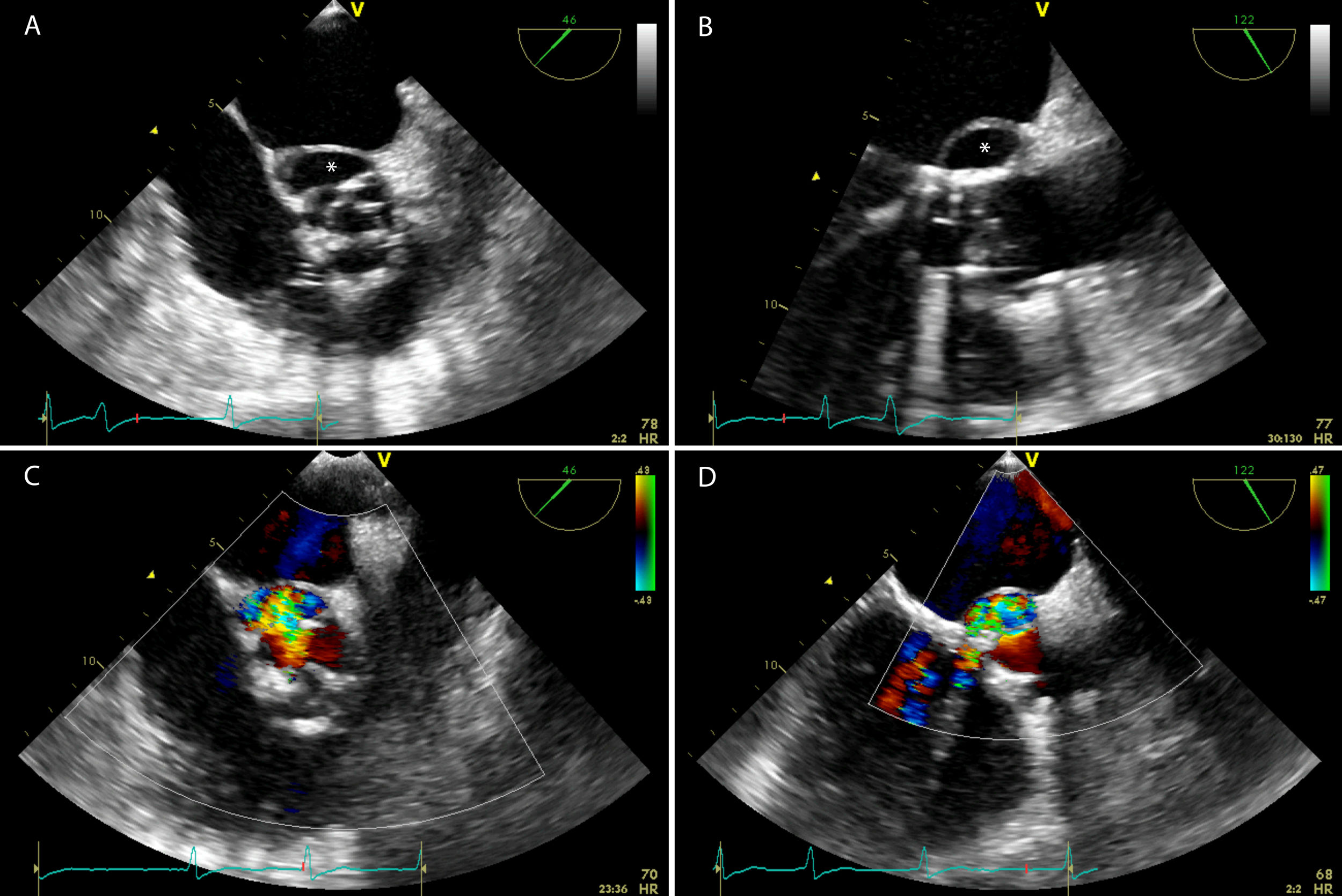
A transesophageal echocardiography (Fig. 1A and B) was performed and demonstrated a biological prosthesis in aortic position with reduced opening and a perivalvular echo-free saccular cavity along with the non-coronary and left coronary cusps, corresponding to a pseudoaneurysm. With Color-Flow Doppler (Fig. 1C and D) it was possible to observe a communication between the perivalvular cavity and the cardiovascular lumen with a high-velocity jet entering the left ventricle outflow tract.

Fig. 1
(0,39MB).

Antibiotic therapy was maintained and the patient was referred for cardiothoracic surgery.

Reference
[1]
G. Habib, P. Lancellotti, M.J. Antunes, M.G. Bongiorni, J.P. Casalta, F. Del Zotti, et al.
ESC guidelines for the management of infective endocarditis. The task force for the management of infective endocarditis of the European Society of Cardiology.
Eur Heart J, 36 (2015), pp. 3075-3128
Copyright © 2020. The Authors
Opciones de artículo
Herramientas
es en pt

¿Es usted profesional sanitario apto para prescribir o dispensar medicamentos?

Are you a health professional able to prescribe or dispense drugs?

Você é um profissional de saúde habilitado a prescrever ou dispensar medicamentos

Quizás le interese:
10.1016/j.mcpsp.2021.100245
No mostrar más