


This study investigates the concordance of bioelectrical impedance vector analysis (BIVA) measurements in both hemisomes in patients receiving fluid therapy, since clinical practice suggests performing them in the hemisoma contralateral to that of its administration to avoid interferences.
The primary objective of this study was to evaluate the concordance of phase angle (PhA), resistance and reactance, both standardized by height (Rz/h, Xc/h) between the hemisoma where the patient is receiving iv fluids and the other one.
MethodsClinical, prospective and descriptive study, which included 100 hospitalized patients with total parenteral nutrition (TPN) or other intravenous fluid therapy. BIVA measurements were taken in both hemisomes and analyzed by means of Bland–Altman plots, Passing–Block test and conditional method agreement trees (COAT).
Results100 patients were included (57 men and 43 women), with an average age of 67.2±15.1 years. Univariate analysis using the Bland–Altman plot showed no concordance of PhA, Rz/h and Xc/h between both hemisomes in patients who received fluid therapy, but Passing–Block test showed no systematic or proportional differences between hemisomas and multivariate COAT analysis did not show that the specified covariates affected concordance.
ConclusionsNo systematic or proportional differences between hemisomas in resistance and reactance has been demonstrated, suggesting the possibility of being able to perform the measurement independently of the side of fluid administration.
Este estudio investiga la concordancia de las mediciones del análisis de vector de impedancia bioeléctrica (BIVA) en ambos hemisomas en pacientes que reciben fluidoterapia, ya que la práctica clínica sugiere realizarlas en el hemisoma contralateral al de su administración para evitar interferencias.
El objetivo principal de este estudio fue evaluar la concordancia del ángulo de fase (PhA), resistencia y reactancia, ambas estandarizadas por altura (Rz/h, Xc/h) entre el hemisoma en el que el paciente está recibiendo fluidos intravenosos y el contralateral.
MétodosEstudio clínico, prospectivo y descriptivo, que incluyó 100 pacientes hospitalizados con nutrición parenteral total (NPT) u otra fluidoterapia intravenosa. Las mediciones de BIVA se realizaron en ambos hemisomas y se analizaron mediante gráficos de Bland-Altman, prueba de Passing Block y árboles de acuerdo de métodos condicionales (COAT).
ResultadosSe incluyeron 100 pacientes (57 hombres y 43 mujeres), con una edad media de 67,2±15,1 años. El análisis univariante mediante el gráfico de Bland-Altman no mostró concordancia de PhA, Rz/h y Xc/h entre ambos hemisomas en pacientes que recibieron fluidoterapia, el test Passing Block no mostró diferencias sistemáticas o proporcionales entre hemisomas y el análisis multivariante COAT no mostró que las covariables especificadas afectaran a la concordancia.
ConclusionesNo se han demostrado diferencias sistemáticas o proporcionales entre hemisomas en resistencia y reactancia, lo que sugiere la posibilidad de poder realizar la medición independientemente del lado de administración de fluidos.
Bioelectrical impedance vector analysis (BIVA) is a noninvasive, rapid, and relatively inexpensive technique that is widely used in the assessment of body composition and nutritional status nutritional. BIVA provides information on hydration and cell mass through the measurement of impedance (Z) and its components, resistance (Rz) and reactance (Xc). These electrical parameters are integrated into the phase angle (PhA), which has been shown to be a useful indicator of cellular integrity and nutritional status. PhA is related to the degree of hydration and tissue integrity, being a valuable prognostic marker in various pathologies, including infections, cancer and chronic diseases such as chronic renal failure.1
Resistance is inversely related to body water content, with higher resistance indicating lower hydration, while a decrease in resistance suggests overhydration. Reactance reflects cellular properties, especially the integrity of cell membranes and is directly proportional to it. A reduction in Xc may be associated with loss of mass or cell damage.2
The relevance of PhA in clinical practice has increased markedly in recent years due to its ability to reflect cellular health and its usefulness as a predictor of clinical outcomes. Recent research has shown that low PhA values are associated with worse prognosis and increased mortality in various clinical populations, including patients with cirrhosis, cancer, and those in intensive care units. In addition, numerous studies have suggested that PhA may be a marker of malnutrition and sarcopenia, providing valuable information for nutritional assessment and treatment monitoring.2–4
BIVA, and specifically PhA provides valuable information on body composition (body cell mass, i.e. nutritional status, and hydration). But in clinical practice, in patients receiving fluid therapy, it is usually recommended to perform BIVA measurements in the contralateral hemisome to which fluid therapy is administered, since the simultaneous passage of fluid could modify the hydration of that side of the body and thus alter resistance. However, there is insufficient literature addressing the influence of this aspect on the clinical significance of BIVA measurements. This knowledge gap represents an opportunity to investigate the concordance of BIVA measurements between both hemisomes in patients under fluid therapy.
Our hypothesis was that performing BIVA on the side of the body receiving intravenous fluid compared to the contralateral side has no clinical relevance. To test the hypothesis, we developed an experiment in which the agreement is analyzed with Bland–Altman plots and Passing–Block test.
The primary objective of this study was to evaluate the concordance of PhA, resistance and reactance, both standardized by height (Rz/h, Xc/h) in both hemisomes in patients receiving fluid therapy. As a secondary objective we set out to explore whether there were other covariates such as age, sex and side of the body where fluid therapy is administered, which may influence this aspect.
MethodsA prospective, descriptive, clinical study was carried out in which BIVA was performed in both hemispheres in a cohort of hospitalized patients receiving total parenteral nutrition (TPN) or other intravenous fluid therapy.
This project was approved by the Ethics Committee for Research with Medicines of the Gerencia de Atención Integrada de Albacete (Albacete Integrated Care Management) with internal code N°2023-099. The procedures and materials complied with the principles of the Declaration of Helsinki and with the regulations on data protection and research in Spain (European Union Regulation 2016/679).
Body composition analysis was performed using the 50kHz phase-sensitive bioimpendaciometer (Nutrilab Whole Body Bioimpedance Vector Analyser, AKERN, Florence, Italy) delivering 400μA via tetrapolar electrodes placed on the ipsilateral hand and foot.2,4,5
ParticipantsThis study included 100 patients over 18 years of age, hospitalized, under treatment with fluid therapy (TPN or other) at a rate at the time measurements were taken between 1500 and 3000ml of volume in 24h, all right-handed, who, after being informed, signed the consent for their participation. The measurement was performed in both body hemisomes.
To calculate the sample size, a pilot study was conducted in which Rz/h, the BIA parameter most susceptible to changes in hydration, was analyzed, and the average difference between the two hemispheres was 6.7Ω/m with a standard deviation of 10.8Ω/m. According to these data, following the method of Lu et al.,6 at α=0.05 and 80% power, it was concluded that the sample size requires 10 observations. Taking into account that if there was an influence on the concordance in the fluid therapy side variables, age and sex in the COAT method,7 8 graphs would be obtained (2×2×2), i.e. 80 observations would be necessary, which would have to be increased by 25%, up to 100, taking into account that each branch of the COAT decision tree will not divide exactly into two halves of equal size.
VariablesThe variables collected were age, sex, height, type of catheter through which fluid therapy (central catheter with peripheral insertion (PICC), another central catheter, midline or peripheral catheter), side of the body in which the measurement and type of fluid therapy (parenteral nutrition or cristaloid therapy) was administered. In addition, the resistance and reactance obtained by two sequential measurements in each hemisome.
In those cases, in which it was not possible to measure the patients, the height reported by them was used. In those cases, in which it was not known, the estimation was based on the mean height calculated with the ulnar distance and the heel-knee distance (calculator in the NutricAlba form at https://endoalba.org.es/nutricalba/).
The Rz/h and Xc/h of each hemisoma were calculated as the average of two measurements of Rz and Xc, respectively, taken without moving the electrodes, divided by the height in meters. With these values, PhA was determined by the arc-tangent of Xc/Rz, and bioimpedance, as the square root of the sum of the squares.
Data collectionClinical information with all data obtained per patient was collected in an electronic data notebook (case report form, (CRF)). For identification in the CRF, the patient's initials were used as a previously defined code known only to the research team. In a separate document, the initials were associated with the medical record number. The CRFs are under the custody of the principal investigator.
Statistical analysisA description of patient characteristics was performed.
Qualitative variables were expressed as frequencies and percentages. Quantitative variables were expressed as Huber's M-estimator (central tendency parameter, analogous to the arithmetic mean and median, but more robust than both, as it is not affected by outliers) and absolute deviation to the standardized median (scaling parameter, analogous to the standard deviation, but more robust than the latter, as it is not affected by outliers).8
Concordance between hemisomes was analyzed in two ways: by Bland–Altman plots9 and by Passing–Bablok tests.10
For the interpretation of the Bland–Altman plot, the acceptable difference between hemispheres is considered to be in the case of PhA±0.5, which was previously described by as the largest clinically acceptable difference.11
In the case of Rz/h and Xc/h, we do not know any previous publications on the maximum acceptable difference, so these were calculated by simulation, as the difference that will result in a PhA error<±0.5. To this, a random population of 1,000,000 people (50% male) was generated according the population data published by Piccoli.12 PhA was calculated for everyone in the population as arctg(Xc/Rz). These generated Rz/h Xc/h and PhA were considered “real values”. Then, several delta errors were generated, and we calculated what PhA each person would get for Rz/h±delta without Xc/h changes. Therefore, two vectors were generated, one for the PhA errors calculated with Rz/h−delta, and the other for the PhA errors calculated with Rz/h+delta. Then, these vectors joined in a third one with the absolute value of the PhA errors. The 95th percentile (p95) of the error's distribution was then calculated. We tested several delta errors and chose that one that p95 was very close to 0.5 but less than 0.5. In summary: the maximum admissible difference calculated was the delta that resulted in a clinically acceptable PhA in more than 95% of the measurements. An analogous algorithm was used to calculate the maximum admissible error of Xc. Therefore, the maximum admissible error for Rz/h is ±17.4, and the maximum admissible error for Xc/h is ±2.14.
Agreement was considered to be present when the confidence interval of the limits of agreement (ICLA) was within these differences, defining the ICLA as the one between the upper limit of the confidence interval of the upper limit of agreement and the lower limit of the confidence interval of the lower limit of agreement.13
To analyze whether agreement between measures of the variables of interest was affected by covariates (age, sex, side of the body on which fluid therapy is administered, and type of catheter), the conditional method agreement trees (COAT) proposed by Karapetyan et al.7 was used.
Statistical analysis was performed using the R programming language.14
The Huber M-estimator and the scale parameter were calculated using the hubers() function of the MASS package.15 The Bland–Altman plots and corresponding statistics were obtained using the bland.altman.plot() and bland.altman.stats() functions of the BlandAltmanLeh package.16 The Passing–Bablok test was performed using the mcreg() function of the mcr package.17
Multivariate concordance analysis was performed using the coat() function of the coat package.7
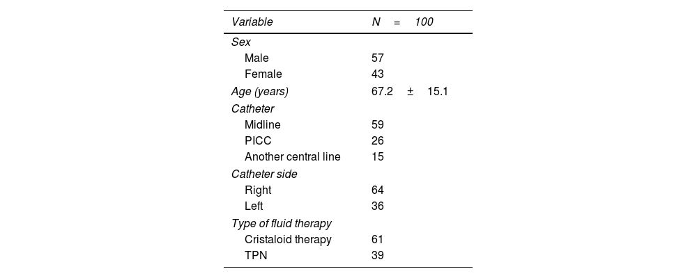
ResultsA total of 100 patients were included, of which, 57 were men and 43 were women, with an average age of 67.2±15.1 years (Huber's M-estimator±median absolute deviation).
Table 1 describes the characteristics of the patients, type of catheter and intravenous fluid therapy.
Characteristics of patients receiving intravenous fluids included in the study.
| Variable | N=100 |
|---|---|
| Sex | |
| Male | 57 |
| Female | 43 |
| Age (years) | 67.2±15.1 |
| Catheter | |
| Midline | 59 |
| PICC | 26 |
| Another central line | 15 |
| Catheter side | |
| Right | 64 |
| Left | 36 |
| Type of fluid therapy | |
| Cristaloid therapy | 61 |
| TPN | 39 |
PICC: central catheter with peripheral insertion (PICC); TPN: parenteral nutrition. Age is expressed as Huber's M-estimator±median absolute deviation's.
Table 2 shows the electrical values according to the hemisoma in which BIVA was performed and according to the presence or absence of the catheter (fluid administration side or contralateral side).
Electrical values as a function of hemisoma and the presence or absence of catheter (fluid).
| Left | Right | Catheter | No catheter | |
|---|---|---|---|---|
| Variable | Average H | Average H | Average H | Average H |
| Rz/h | 312 (70.6) | 308.5 (71.2) | 307.8 (73.5) | 312.8 (67.8) |
| Xc/h | 25.4 (7.9) | 25.8 (7.7) | 25.5 (7.9) | 25.7 (8.1) |
| PhA | 4.7 (0.9) | 4.8 (1) | 4.7 (1) | 4.7 (0.9) |
| Z/h | 313.1 (71.5) | 309.6 (71.2) | 308.9 (72.6) | 313.9 (69.1) |
Rz/h: standardized resistance; Xc/h: standardized reactance; PhA: phase angle; Z/h: standardized impedance; Average H: Huber's M-estimator (median absolute deviation).
Univariate analysis using the Bland–Altman plot showed the existence of no concordance of PhA, Rz/h and Xc/h in both hemisomes in patients who received intravenous fluid therapy.
It was found that the mean difference between one hemisoma and the contralateral hemisoma of the PhA was −0.04° with a confidence interval of the limits of agreement (ICLA) of (−0.60, +0.52°), which implies no concordance according to the pre-established criteria (Fig. S1).
Regarding Rz/h, the mean difference was 4.85Ω/m, ICLA (−25, +35Ω/m), being slightly lower on the side where fluid therapy was administered so that, again, the limits were out the range of acceptable difference of the agreement, thus demonstrating disagreement (Fig. S2).
In the case of Xc/h, the mean difference was 0.17Ω/m (−2.4, +2.8Ω/m), so that, again, the limits were out within the range of acceptable difference of the agreement, thus demonstrating disagreement (Fig. S3).
*Figs. S1–S3 correspond to the Bland–Altman plot graphs and are found in the supplementary material.
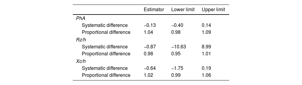
Passing–Block chartNext, the evaluation of univariate agreement was continued using the Passing–Block test, in which no significant systematic or proportional differences in PhA, Rz/h and Xc/h were demonstrated (Figs. 1–3; Table 3).
Statistical results of the Passing–Block test.
| Estimator | Lower limit | Upper limit | |
|---|---|---|---|
| PhA | |||
| Systematic difference | −0.13 | −0.40 | 0.14 |
| Proportional difference | 1.04 | 0.98 | 1.09 |
| Rz/h | |||
| Systematic difference | −0.87 | −10.63 | 8.99 |
| Proportional difference | 0.98 | 0.95 | 1.01 |
| Xc/h | |||
| Systematic difference | −0.64 | −1.75 | 0.19 |
| Proportional difference | 1.02 | 0.99 | 1.06 |
Lower and upper limits for every parameter show the 95% confidence interval. The confidence interval includes de 0 for all the systematics differences, and the 1 for all the proportional differences of parameters.
Rz/h: standardized resistance; Xc/h: standardized reactance; PhA: phase Angle; Z/h: standardized impedance.
Multivariate analysis using COAT showed that, when considering the covariates side on which the fluid was administered, sex, age and type of catheter, there was a significant difference in the concordance of the PhA depending on the side on which the fluid was administered, so that when fluid therapy was administered on the right side the mean difference between hemisomes was −0.1° with an ICLA (−0.67, +0.48), while when it was administered on the left side the mean difference between hemisomes was +0.07° with an ICLA (−0.48, +0.62), this difference in concordance between hemisomes being significant (p=0.015) in the COAT analysis. In any case, this difference were upper than the acceptable pre-established difference, so that there is no concordance in phase angle between hemisomes regardless of the side on which fluid therapy is administered.
In the case of Rz/h, multivariate analysis showed that sex can affect concordance. In men the mean Rz/h difference between hemisomes was +1.52Ω/m with an ICLA (−29, +32) and in women the mean Rz/h difference between hemisomes was +9.26Ω/m with an ICLA (−22, +40) (p=0.02). In any case the ICLAs were upper than the preestablished acceptable differences, so in both cases there is no concordance.
The concordance of Xc/h was not affected by any of the covariates studied.
DiscussionOur hypothesis that it is possible to perform BIVA on both sides of the body, regardless of the simultaneous passage of intravenous fluid therapy, was accepted. To this end, we evaluated the concordance of PhA, Rz/h, and Xc/h in both hemisomes of patients receiving fluid therapy. Our results show no systematic or proportional differences in raw BIVA parameters, regardless of whether the measurement is taken on the fluid-receiving hemisome or the contralateral one. However, Bland–Altman plots indicate a lack of concordance.
BIVA is a fundamental tool in the diagnosis of malnutrition, as it allows the assessment of body composition and hydration status. Since muscle mass loss is a key parameter in the Global Leadership Initiative on Malnutrition (GLIM) criteria, accurate methods for its evaluation are essential. Currently, bioelectrical impedance analysis (BIA), dual-energy X-ray absorptiometry (DXA), and computed tomography (CT) are available for this purpose.
The appendicular muscle mass index (ASMI) is a metric used to assess skeletal muscle mass, adjusted for the height of the individual. It is an essential tool to identify low muscle mass, especially relevant in cases of sarcopenia and malnutrition, and even more so in those patients in whom muscle loss is not clinically evident.18,19
Body weight is another relevant criterion in the diagnosis of malnutrition, but this can vary significantly depending on the patient's hydration status. BIA helps to identify whether weight changes are due to fluctuations in body water content (such as edema or dehydration) or to an actual loss of lean and/or fatty tissue. This improves diagnostic accuracy and avoids errors when classifying nutritional status.18
Oxidative stress and chronic inflammation can damage cell membranes, making the PhA a rapid and effective marker for assessing cellular integrity. Its utility lies in its ability to identify inflammatory processes and predict adverse health outcomes, as a reduced PhA can be associated with loss of muscle mass, increased systemic inflammation, and a poorer prognosis. In patients with malnutrition, a low PhA is associated with a higher risk of complications and increased morbidity and mortality.20 Therefore, monitoring changes in body composition is essential to adjust treatment on an individual basis. Although its use was initially focused on outpatient clinics, it has been shown to be crucial in hospitalization units as well, where it is key for the comprehensive assessment and management of the patient.21
Classically, it has been recommended to perform the measurement on the right side,22 to avoid artifacts in the measurement produced by the administered fluid and/or the catheter. Our study shows that it is possible to perform BIVA in one hemisoma or the other regardless of fluid therapy administration without getting systematic or proportional biased results. This simplifies the measurement process and offers greater flexibility in clinical practice and to be able to always perform the measurement on the same side in the same patient. Cofactors such as sex, age and side of administration have been shown not to influence the test result.
The literature on concordance and BIVA, especially in different body segments or hemisomes, is still scarce. In the work of Jacob Dellingere et al., they analyzed the concordance of PhA, Rz and Xc between different bioimpedance devices: supine bioimpedance spectroscopy, supine single frequency bioelectrical impedance analysis and standing multifrequency bioelectrical impedance analysis; and supine and standing positions. The results revealed greater concordance in the female sex. The resistance and phase angle were equivalent between the devices in the supine position but not with the standing analyzer, revealing notable differences in the measurements.23
On the other hand, several studies have evaluated the agreement between different BIVA devices. Ángeles Espinosa Cuevas et al. studied the degree of agreement of PhA, Rz, Xc, fat-free mass (FFM) and fat mass (FM) between different analyzers: spectroscopic (BIA-BIS), multifrequency (BIA-MF), single-frequency (BIA-SF) and segmental multifrequency (BIA-MS) techniques in hemodialysis patients. The agreement between the four devices was good for BIVA diagnosis. BIA-BIS and DEXA analyzers had the lowest bias for both FFM and FM, although with higher limits of agreement. The lowest limits of agreement were found with the BIA-MS analyzer.24
Fernanda Bernal et al. evaluated 406 ambulatory patients with chronic stable heart failure, the accuracy between BIA-MF and BIA-SF bioelectrical impedance devices in the measurement of PhA, Rz, Xc together with hydration and body cell mass. Strong correlation was observed between both methods in all raw variables (r≥0.90). Lin's concordance correlation coefficient (CCC) value was high for Rz 0.99 (95% CI 0.997–0.998), moderate for Xc, 0.93 (0.92–0.94) and poor for PhA, 0.88 (0.85–0.90). MF-BIA and SF-BIA demonstrated good agreement for the measurement of the Rz parameter; however, the Xc and PhA parameters should be used with caution due to reported variability.25
One of the main weaknesses of the study lies in the acceptability limits stipulated for the assessment of concordance. The acceptability limits for the PhA have been ±0.5 standard deviation of the distribution in the general population, recently published.11 The limits of Rz and Xc have been calculated from the PhA as explained above, since there is nothing published or validated in this regard to date.
On the other hand, although no systematic or proportional differences were found in the electrical parameters between the two hemisomes, in some patients there were important differences when the measurement was made in the hemisome in which fluid therapy was administered or in the other; but these differences in some patients were positive and in others negative. We believe that these variations may be due to the position of the electrodes, their adherence, or the patient's skin conditions, which underlines the importance of following a rigorous technique to obtain accurate measurements.
Another limitation of the study is that the maximum volume of fluid administered at the time of measurement was 3000ml in 24h, so the results cannot be generalized to patients in whom higher volumes are administered.
Among the strengths of the research we can highlight the measurement in real clinical practice, which increases the relevance and applicability of the results; the systematic way in which the BIVA was carried out, making two measurements in each hemisome to minimize variability; and a complete statistical analysis for the evaluation of the concordance, using several statistical techniques, including Bland–Altman graphs, Passing–Block test and conditional method agreement trees (COAT), providing a robust and detailed evaluation.
ConclusionAfter carrying out our study, we can confirm that we have not found systematic or proportional differences when performing bioimpedance on both sides of the body, which suggests the possibility of being able to perform the measurement independently of the side of administration of the fluid; this represents a novelty and advantage in clinical practice, as the measurement can always be performed on the same side for greater reproducibility, less bias in the comparison of successive measurements and greater accuracy in the results for making diagnostic/therapeutic decisions.
Future studies could focus on the evaluation of the concordance of the phase angle in different contexts of fluid therapy, considering variables such as volume and speed of infusion. In addition, it would be useful to investigate the influence of other factors such as the previous hydration status of the patients and the presence of comorbidities. The development of standardized protocols for the measurement of BIVA in hospitalized patients would also be a significant advance for clinical practice.
CRediT authorship contribution statementMarina Jara Vidal: investigation, writing – original draft, data curation; Andrés Ruiz de Assín Valverde: writing – review & editing; Marta Gallach Martínez: writing – review & editing; Noel Roig Marín: writing – review & editing; César Gonzalvo Díaz: writing – review & editing; Rosa Pilar Quílez Toboso: writing – review & editing; Lourdes García Blasco: writing – review & editing; Silvia Aznar Rodríguez: writing – review & editing; Jose Juan Lozano García: writing – review & editing; Jose Joaquín Alfaro Martínez: conceptualization, formal analysis, supervision, writing – review & editing.
Sources of fundingThis research has not received any specific support from public sector agencies, the commercial sector or non-profit entities.
Conflicts of interestNone.
To our colleagues from the Endocrinology and Nutrition Service, including endocrinologists, nutritionists and nurses, and to the rest of colleagues who have contributed to the implementation of morphofunctional assessment in clinical practice.


