


metricas
Clínica e Investigación en Arteriosclerosis Levels of sCD163 in women rheumatoid arthritis: Relationship with cardiovascular...
Información de la revista
Vol. 37. Núm. 1.
(Enero - Febrero 2025)
Cita
Cita
Compartir
Descargar PDF
Más opciones de artículo
Visitas
573
Vol. 37. Núm. 1.
(Enero - Febrero 2025)
Original article
Acceso a texto completo

Levels of sCD163 in women rheumatoid arthritis: Relationship with cardiovascular risk markers

Niveles de sCD163 en mujeres con artritis reumatoide: relación con los marcadores de riesgo cardiovascular
Visitas
573
Oscar Zaragoza-Garcíaa, Olivia Briceñob, José Rafael Villafan-Bernalc, Ilse Adriana Gutiérrez-Péreza, Héctor Ugo Rojas-Delgadod, Gustavo Adolfo Alonso-Silverioa, Antonio Alarcón-Paredesa, José Eduardo Navarro-Zarzae, Cristina Morales-Martínezd, Rubén Rodríguez-Garcíaf, Iris Paola Guzmán-Guzmána,
Autor para correspondencia
pao_nkiller@yahoo.com.mx

Corresponding author.
a Laboratory of Multidisciplinary Research and Biomedical Innovation, Universidad Autónoma de Guerrero, Chilpancingo, Guerrero, Mexico
b Infectious Diseases Research Center, Instituto Nacional de Enfermedades Respiratorias Ismael Cosío Villegas, Mexico City, Mexico
c Laboratory of Immunogenomics and Metabolic Diseases, Instituto Nacional de Medicina Genomica, Mexico City, Mexico
d Hospital General Dr. José G. Parres, Cuernavaca, Morelos, Mexico
e Hospital General Dr. Raymundo Abarca Alarcón, Chilpancingo, Guerrero, Mexico
f Laboratorio de Clínico, Instituto Mexicano del Seguro Social, Hospital General Regional, Cuernavaca, Morelos, Mexico
Este artículo ha recibido
Información del artículo
Resumen
Texto completo
Bibliografía
Descargar PDF
Estadísticas
Figuras (4)
fig0005
fig0010
fig0015
fig0020
Tablas (2)
Table 1. Cardiovascular risk factors in women with RA.
Tablas
Table 2. Effect of cardiovascular risk factors on levels of sCD163 in women with RA.
Tablas
Abstract
Aim

The soluble scavenger receptor differentiation antigen 163 (sCD163), a monocyte/macrophage activation marker, is related to cardiovascular mortality in the general population. This study aimed to evaluate their relationship between serum levels of sCD163 with cardiovascular risk indicators in rheumatoid arthritis (RA).

Methods

A cross-sectional study was performed on 80 women diagnosed with RA. The cardiovascular risks were determined using the lipid profile, metabolic syndrome, and QRISK3 calculator. For the assessment of RA activity, we evaluated the DAS28 with erythrocyte sedimentation rate (DAS28-ESR). The serum levels of sCD163 were determined by the ELISA method. Logistic regression models and receiver operating characteristics (ROC) curve were used to assess the association and predictive value of sCD163 with cardiovascular risk in RA patients.

Results

Levels of sCD163 were significantly higher in RA patients with high sensitivity protein C-reactive to HDL-c ratio (CHR)0.121 (p=0.003), total cholesterol/HDL-c ratio>7% (p=0.004), LDL-c/HDL-c ratio>3% (p=0.035), atherogenic index of plasma>0.21 (p=0.004), cardiometabolic index (CMI)1.70 (p=0.005), and high DAS28-ESR (p=0.004). In multivariate analysis, levels of sCD1631107.3ng/mL were associated with CHR0.121 (OR=3.43, p=0.020), CMI1.70 (OR=4.25, p=0.005), total cholesterol/HDL-c ratio>7% (OR=6.63, p=0.044), as well as with DAS28-ESR>3.2 (OR=8.10, p=0.008). Moreover, levels of sCD163 predicted CHR0.121 (AUC=0.701), cholesterol total/HDL ratio>7% (AUC=0.764), and DAS28-ESR>3.2 (AUC=0.720).

Conclusion

Serum levels of sCD163 could be considered a surrogate of cardiovascular risk and clinical activity in RA.

Keywords:
sCD163
Cardiovascular risk
Atherogenic risk
Clinical activity
Rheumatoid arthritis
Resumen
Objetivo

El antígeno de diferenciación del receptor carroñero soluble 163 (sCD163), un marcador de activación de monocitos/macrófagos, está relacionado con la mortalidad cardiovascular en la población general. Este estudio tuvo como objetivo evaluar su relación entre los niveles séricos de sCD163 con indicadores de riesgo cardiovascular en la artritis reumatoide (AR).

Métodos

Se realizó un estudio transversal en 80 mujeres diagnosticadas con AR. El riesgo cardiovascular se determinó mediante el perfil lipídico, el síndrome metabólico y la calculadora QRISK3. Para la evaluación de la actividad de la AR, evaluamos el DAS28 con velocidad de sedimentación globular (DAS28-VSG). Los niveles séricos de sCD163 se determinaron mediante el método ELISA. Se utilizaron modelos de regresión logística y la curva de características operativas del receptor (ROC) para evaluar la asociación y el valor predictivo de sCD163 con el riesgo cardiovascular en pacientes con AR.

Resultados

Los niveles de sCD163 fueron significativamente mayores en pacientes con AR con elevado índice proteína C reactiva de alta sensibilidad/HDL-c (CHR)0,121 (p=0,003), cociente colesterol total/HDL-c>7% (p=0,004), cociente c-LDL/c-HDL>3% (p=0,035), índice aterogénico del plasma >0,21 (p=0,004), índice cardiometabólico (CMI)1,70 (p=0,005) y DAS28-VSG elevado (p=0,004). En el análisis multivariado, los niveles de sCD1631.107,3ng/ml se asociaron con CHR0,121 (OR=3,43; p=0,020), CMI1,70 (OR=4,25; p=0,005), cociente colesterol total/cHDL>7% (OR=6,63; p=0,044), así como con DAS28-ESR>3,2 (OR=8,10; p=0,008). Además, los niveles de sCD163 predijeron CHR0,121 (AUC=0,701), cociente colesterol total/HDL>7% (AUC=0,764) y DAS28-ESR>3,2 (AUC=0,720).

Conclusión

Los niveles séricos de sCD163 podrían considerarse un marcador subrogado del riesgo cardiovascular y la actividad clínica en la AR.

Palabras clave:
sCD163
Riesgo cardiovascular
Riesgo aterogénico
Actividad clínica
Artritis reumatoide
Texto completo
Introduction

CD163 is a 130-kDa transmembrane protein expressed on monocytes and macrophages whose primary function is as a scavenger receptor that recognizes and mediates endocytosis of the haptoglobin–hemoglobin complex.1 Its expression is regulated by acute phase reactants, hemoglobin, lipopolysaccharide, chemokines, granulocyte, and monocyte colony-stimulating factor, proinflammatory and anti-inflammatory cytokines, as well as corticosteroid use.2–5

Activation of monocytes and macrophages, as well as the release of the soluble form of CD163 (sCD163), are increased in the serum and plasma of patients with atherosclerosis,6,7 acute coronary syndrome,8 and acute ischemic stroke (AIS).9 Elevated sCD163 levels are associated with cardiovascular disease (CVD)-related and all-cause mortality.10 Thus, elevated concentrations of sCD163 are a biomarker of CVD risk in the general population.9

In rheumatoid arthritis (RA) have about 1.5-fold higher risk of CVD, death from myocardial infarction, coronary revascularization, and stroke.11–13 Currently, biomarkers such as adiponectin, osteoprotegerin, asymmetric dimethylarginine,14 endocan, paraoxonase 1, lipoproteins, antibodies against HDL-c,15 and monocyte activation molecules such as CD163 could be are emergent predictors of CVD in RA. However, its role as a biomarker of CVD risk in RA remains unelucilated.

In RA, the time of monocyte circulation in the blood decreases, and their turnover and activation are more prominent in joints.16 High sCD163 concentrations have been related to macrophage activation during synovitis in RA.17 Moreover, sCD163 serum levels were associated with autoantibody positivity, clinical activity, radiographic progression, acute phase reactants, and proinflammatory cytokines in inflammatory arthritis such as psoriatic arthritis, juvenile idiopathic arthritis, and RA.17–23 While in patients with systemic lupus erythematosus (SLE), sCD163 levels are predictors of carotid plaque progression and risk of cardiovascular events.24,25 This study analyzes the association between serum sCD163 levels and cardiovascular risk markers in women RA patients.

Materials and methodsSubjects

Eighty Mexican women diagnosed with RA, according to the 2010 American College of Rheumatology (ACR) and the European League Against Rheumatism (EULAR) criteria26 were enrolled in this study between February 2019 and February 2020 in the General Hospital. The sample size estimated through Cohen's f2 effect size for an F-test formula yields a minimal sample of 79 subjects, considering an estimated anticipated effect size of 0.32, a power level of 0.80, and a probability level of 0.05. In our population, the ratio of women: men attending care associated with RA is 9:1. Therefore, in this study focused on evaluating the relationship of sCD163 with cardiovascular risk (CVR) markers in women with RA. Subjects were selected according to the following exclusion criteria: (a) overlapping autoimmune diseases, (b) bacterial or viral infections at enrollment, (c) therapy with lipid-lowering, and (d) hormonal treatment, e.g., oral contraceptive pills. All participants signed written informed consent, and the research was conducted under the principles established by the Declaration of Helsinki. The study was approved by the Ethics Research Committee.

Clinical and anthropometric data

The clinical and treatment characteristics were evaluated during the consultation and from the clinical records. The standard treatments were mainly methotrexate, received orally, in combination with chloroquine and prednisone; this study does not include patients treated with biological agents because this is not a therapy available at our hospital. The presence of hypertension, type 2 diabetes (T2D), smoking, and duration of RA was recorded. Ten RA patients had pharmacological treatment based on angiotensin-converting enzyme inhibitors, and two patients had a combination of diuretics. For T2D, three patients reported treatment with metformin, one with insulin, and two with a combination of metformin and glibenclamide.

During the medical consultation, the rheumatologist performed the clinical examination of the participants. The health assessment questionnaire disability index (HAQ-DI) and disease activity score 28 with erythrocyte sedimentation rate (DAS28-ESR) were assessed. Most of the participants had functional capacity, and the HAQ-DI cut-off for considering disability presence was ≥1. The DAS28-ESR cut-off points that defined the clinical activity for the disease were <2.6 for remission, >2.6–3.2 for low activity, >3.2 to ≤5.1 for moderate activity, and >5.1 for high activity.

The weight and body mass index (BMI) were evaluated by bioimpedance (Omron, IL, USA), and the height was determined using a portable stadiometer (Seca 240, Hamburg, Germany). We classified BMI according to OMS criteria,27 considering the underweight range<18.5kg/m2, normal weight range 18.5–24.9kg/m2, overweight range 25–29.9kg/m2, and obese range ≥30kg/m2. The waist circumference (WC) was measured using a measuring tape with an accuracy of ±0.1cm (Seca 201, Hamburg, Germany). The systolic blood pressure (SBP) and diastolic blood pressure (DBP) were measured on the left arm in duplicate at intervals of 5min and after a minimum rest state of 5min using an automated sphygmomanometer HEM-712C (Omron, IL, USA).

Measurement of inflammation and autoantibodies

Blood samples were collected after 8h of overnight fasting via venipuncture and transferred to tubes with EDTA and without clot activator. An erythrocyte sedimentation rate (ESR) test was performed, as well as the quantification of serum levels of high sensitivity protein C reactive (hsCRP) and rheumatoid factor (RF) were assessed according to the manufacturer instructions [COBAS C311; Roche Diagnostics GmbH, Germany]. IgG isotype antibodies against citrullinated peptides (anti-CCPs) [DIASTAT anti-CCP Axis-Shield, Dundee, United Kingdom] and antibodies against mutated citrullinated vimentin (anti-MCV) [ORGENTEC Diagnostika GmbH, Mainz, Germany] were measured following manufacturer's specifications. Serum levels of RF, anti-MCV, and anti-CCPs were considered positive when values supered 20IU/mL, 20U/mL, and 5U/mL, respectively.

Lipid profile, surrogate cardiovascular risk markers, and metabolic syndrome

Total cholesterol, triglycerides, and high-density lipoprotein cholesterol (HDL-c) were analyzed by colorimetric assays at a semi-automated equipment Cobas Miras [Roche Diagnostics GmbH, Germany] using reagents of Wiener lab brand. The Friedewald formula was employed to determine the levels of low-density lipoprotein cholesterol (LDL-c), as follows: LDL-c (mg/dL)=[total cholesterol][HDL-c+triglycerides/5].28 The cut-off points to define impaired lipid parameters were total cholesterol200mg/dL, triglycerides150mg/dL, low HDL-c<50mg/dL, LDL-c100mg/dL.29

The high-sensitivity C-reactive protein to high-density lipoprotein cholesterol ratio (CHR) was also determinate, and the cut-off point to define high CVR was ≥0.121, according to values above the second tertile. For the atherogenic indices, the cut-offs to define risk were: for total cholesterol/HDL-c ratio<4.5% low risk, 4.5–7% moderate risk, and >7% high risk; for the LDL-c/HDL-c and triglycerides/HDL-c ratios, scores>3% were considered high risk.30 The atherogenic index of plasma (AIP) was calculated using the logarithm of the triglyceride to HDL-c ratio. The AIP was categorized into low risk (AIP<0.11), moderate risk (AIP 0.11–0.21), and high risk (AIP>0.21).31

We estimated the following cardiometabolic markers, lipid accumulation product index (LAP) estimated from WC (cm) and triglycerides (mmol/L) using the equation for women [(WC-58) (triglycerides)],32 and the cardiometabolic index (CMI) using the equation [(triglycerides/HDL-c) (waist to height ratio)].33 We considered values for LAP35.91cmmmol/L and CMI1.70 as criteria for high cardiovascular risk.30

QRISK3 index was calculated online considering age, gender, ethnicity, deprivation, SBP, standard deviation of repeated measures of SBP, BMI, total cholesterol/HDL-c ratio, smoking, family history of coronary heart disease in a first-degree relative aged less than 60 years old, type 1 diabetes, T2D, treated hypertension, RA, atrial fibrillation, chronic kidney disease (stage 3, 4, or 5), migraine, corticosteroids, LES, atypical antipsychotics, severe mental illness, and HIV/AIDs. Through the QRISK3 index, we evaluated the risk of heart attack or stroke within the next ten years, according to a score>10% that defines high risk.34

The metabolic syndrome (MetS) was defined by the presence of three or more components using the modified NCEP-ATPIII criteria: abdominal obesity (WC80cm for women), elevated SBP (≥130mmHg) and/or DBP (≥85mmHg), elevated triglycerides (≥150mg/dL), low HDL-c (<50mg/dL in women), and high glucose fasting (≥100mg/dL).29

Measurement of sCD163 serum levels

The determination of sCD163 was measured by a specific sandwich ELISA method (R&D Systems, Minneapolis, MN, USA), following the manufacturer's instructions in an automatized equipment Multiskan Go [Thermo Scientific, Finland]. The absorbance was measured at 450nm based on a standard curve, and the levels of sCD163 were quantified. The sensitivity of sCD163 ranged from 0.058 to 0.613ng/mL. In this study, the median value for sCD1631107.30ng/mL was considered for the statistical models to establish an association with cardiovascular risk surrogates.

Statistical analysis

Statistical analysis was performed using STATA v.13.0 (StataCorp, College Station, TX, USA), GraphPad Prism v.8.4 (GraphPad Software, San Diego, CA, USA), and Origin 2023b [OriginLab Corp., Northampton, MA, USA] for Windows. Categorical variables were expressed as frequencies and inferentially tested using the Chi-square test. Quantitative data of cardiovascular risk indicators were compared using Mann–Whitney U tests after confirming the non-parametrical distribution of the data by the Shapiro–Wilk test. Spearman's correlation coefficient assessed the linear relationship between quantitative variables. The association between sCD613 and cardiovascular risk indicators was analyzed using logistic regression and ROC curves. For estimating the area under the curve (AUC), we use the following criteria: CHR0.121, total cholesterol/HDL-c ratio>7%, LDL-c/HDL-c ratio>3%, AIP>0.21, CMI1.70, and DAS28-ESR>3.2. p values<0.05 were considered statistically significant.

ResultsPrevalence of CVR factors in the participants

This study included a total of 80 women diagnosed with RA. The mean age was 44.98±12.48 years. 100% of the participants were positive for anti-CCPs and anti-MCV autoantibodies. Regarding treatment, participants had anti-rheumatic treatment with DMARDs, mostly in combination with prednisone. One-third of the population had a DAS28-ESR score>3.2 despite treatment.

A 36.25% of the participants had obesity, and 76.25% had abdominal obesity. The frequency of T2D (7.5%), hypertension (15%), and smoking (15%) were lower in comparison with the frequency of MetS (50%) and the frequency of indicators for high cardiovascular risk. The most common CVR factors was low HDL-c levels (83.75%), followed by CHR (67.5%), high LAP index (65%), AIP (48.7%), and QRISK3 (16.25%) (Table 1).

Table 1.

Cardiovascular risk factors in women with RA.

Variables  n=80 (%) 
Age40 years, n (%)  52 (65) 
BMI30kg/m2, n (%)  29 (36.25) 
Abdominal obesity80cm, n (%)  61 (76.25) 
Smoking, n (%)  12 (15) 
Type 2 diabetes, n (%)  6 (7.5) 
Hypertension, n (%)  12 (15) 
Total cholesterol200mg/dL, n (%)  12 (15) 
Triglycerides150mg/dL, n (%)  30 (37.5) 
Glucose100mg/dL, n (%)  22 (27.5) 
LDL-c100mg/dL, n (%)  37 (46.25) 
HDL-c<50mg/dL, n (%)  67 (83.75) 
CHR0.121, n (%)  54 (67.5) 
Total cholesterol/HDL-c ratio 4.5–7%, n (%)  25 (31.25) 
Total cholesterol/HDL-c ratio>7%, n (%)  12 (15) 
LDL-c/HDL-c ratio>3%, n (%)  30 (37.5) 
Triglycerides/HDL-c ratio>3%, n (%)  44 (55) 
AIP 0.11–0.21, n (%)  5 (6.25) 
AIP>0.21, n (%)  39 (48.75) 
LAP35.91cm mmol/L, n (%)  52 (65) 
CMI1.70, n (%)  46 (57.5) 
MetS, n (%)  40 (50) 
QRISK310%, n (%)  13 (16.25) 
Duration of illness>5 years, n (%)  45 (56.96) 
hsCRP10mg/L, n (%)  27 (33.75) 
ESR20mm/h, n (%)  60 (75) 
DAS28-ESR>3.2, n (%)  33 (41.25) 
HAQ-DI1, n (%)  7 (8.75) 
Rheumatoid factor>20UI/mL, n (%)  59 (89.39) 
Anti-CCPs>5U/mL, n (%)  80 (100) 
Anti-MCV>20U/mL, n (%)  80 (100) 
Prednisone, n (%)  63 (78.75) 
Methrotexate, n (%)  79 (98.75) 
Chloroquine, n (%)  61 (76.25) 

AIP, atherogenic index of plasma; Anti-CCPs, antibodies anti-cyclic citrullinated peptide antibodies; Anti-MVC, antibodies against mutated citrullinate vimentin; BMI, body mass index; CD163, cluster of differentiation 163; CHR, high-sensitivity C-reactive protein to high density lipoprotein cholesterol ratio; CMI, cardiometabolic index; DAS28, disease activity score 28; ESR, erythrocyte sedimentation rate; HAQ-DI, health assessment questionnaire disability index; HDL-c, high-density lipoprotein cholesterol; hsCRP, high sensibility protein C reactive; LAP, lipid accumulation products; LDL-c, low-density lipoprotein cholesterol; MetS, metabolic syndrome; RA, rheumatoid arthritis.

Categorical data are presented as numbers (n) and percentages (%).

Serum levels of sCD163 was associated with CVR indicators

Serum sCD163 levels were elevated, with a median value of 1107.30ng/mL [807.28–1651.04ng/mL (5th and 95th percentiles)]. Moreover, we found that serum sCD163 levels were positively correlated with age (r=0.29, p=0.008), CHR (r=0.30, p=0.005), total cholesterol/HDL-c ratio (r=0.28, p=0.012), LDL-c/HDL-c ratio (r=0.28, p=0.011), triglycerides/HDL-c ratio (r=0.26, p=0.020), CMI (r=0.30, p=0.007), and QRISK3 (r=0.32, p=0.004), as well as with the markers of RA activity, hsCRP (r=0.28, p=0.010), DAS28-ESR (r=0.31, p=0.004) and HAQ-DI score (r=0.35, p=0.001) and negatively correlated with HDL-c levels (r=−0.298, p=0.007) (Fig. 1).

Figure 1.

Correlation between levels of sCD163 with CVR and clinical parameters. A Spearman's correlation analysis was used. p-Value<0.05 was considered statistically significant.

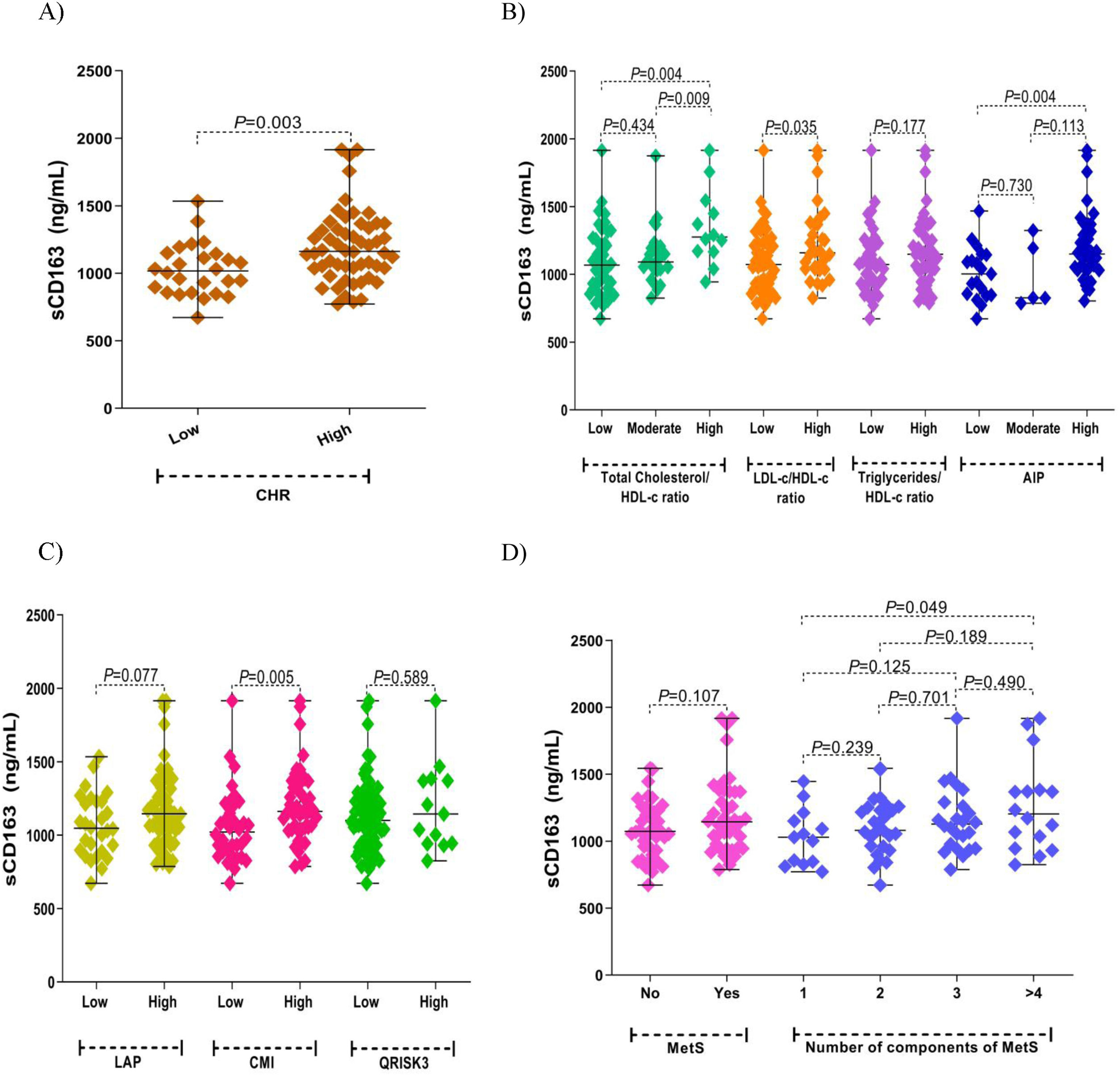
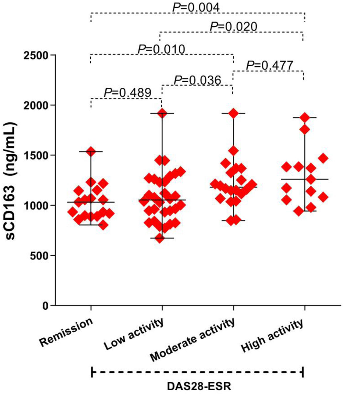
The analysis of CVR indicators demonstrated increased serum levels of sCD163 in patients with CHR0.121 (p=0.003) (Fig. 2A), total cholesterol/HDL-c ratio>7% (p=0.004), with an LDL/HDL-c ratio>3% (p=0.035) and AIP>0.21 (p=0.004) (Fig. 2B), CMI score1.70 (p=0.005) (Fig. 2C), as well as in those with >4 MetS components (p=0.049) (Fig. 2D), and those with moderate (p=0.010) and high (p=0.004) clinical RA activity (Fig. 3).

Figure 2.

Association between levels of sCD163 with CVR factors in women with RA. (A) CHR. (B) Atherogenic indices. (C) Cardiometabolic markers. (D) MetS. Mann–Whitney test. p-Value<0.05 was considered statistically significant.

Figure 3.

Association between levels of sCD163 with clinical activity in women with RA. Mann–Whitney test. p-Value<0.05 was considered statistically significant.

Serum sCD163 levels as predictor of CVR

In a multivariate logistic regression model, we found that the presence of CHR0.121 (β=172.53ng/mL, p=0.003), total cholesterol/HDL-c ratio>7% (β=258.0ng/mL, p=0.006), AIP (β=116.13ng/mL, p=0.046), CMI1.70 (β=123.49ng/mL, p=0.033) and high clinical RA activity (β=210.45ng/mL, p=0.024) were associated with an increase in sCD163 levels (Table 2).

Table 2.

Effect of cardiovascular risk factors on levels of sCD163 in women with RA.

Variables  Multivariate analysis
  sCD163 ng/mLβ (95% CI), p value  sCD1631107.3ng/mLOR (95% CI), p value 
BMI 25–29.9kg/m2  9.77 (−134.42 to 153.97) 0.893  0.78 (0.22–2.70) 0.702 
BMI30kg/m2  128.62 (−12.23 to 269.47) 0.073  3.38 (0.99–11.45) 0.050 
Abdominal obesity80cm  82.76 (−54.41 to 219.94) 0.233  1.37 (0.44–4.22) 0.581 
Total cholesterol200mg/dL  −83.80 (−248.85 to 81.24) 0.315  0.39 (0.10–1.56) 0.187 
Triglycerides150mg/dL  21.22 (−97.58 to 140.03) 0.723  1.08 (0.41–2.81) 0.870 
Glucose100mg/dL  29.04 (−106.31 to 164.40) 0.670  0.99 (0.33–2.96) 0.991 
HDL-c<50mg/dL  190 (16.70 to 364.92) 0.032  5.14 (1.07–24.65) 0.041 
LDL-c100mg/dL  −1.32 (−121.27 to 118.63) 0.983  0.91 (0.35–2.41) 0.865 
CHR0.121  172.53 (58.75 to 286.31) 0.003  3.43 (1.21–9.69) 0.020 
Total cholesterol/HDL-c ratio 4.5–7%  59.51 (−65.93 to 184.95) 0.348  1.44 (0.49–4.19) 0.497 
Total cholesterol/HDL-c ratio>7%  258.00 (76.21 to 439.79) 0.006  6.63 (1.05–41.84) 0.044 
LDL-c/HDL-c ratio>3%  117.18 (−7.15 to 241.53) 0.064  1.61 (0.57–4.52) 0.363 
Triglycerides/HDL-c ratio>3%  81.53 (−33.16 to 196.23) 0.161  2.40 (0.91–6.30) 0.075 
AIP>0.21  116.13 (2.08 to 230.19) 0.046  2.59 (0.98–6.86) 0.054 
LAP35.91cmmmol/L  94.88 (−25.27 to 215.04) 0.120  1.82 (0.67–4.90) 0.236 
CMI1.70  123.49 (9.91 to 237.08) 0.033  4.25 (1.56–11.60) 0.005 
MetS  88.07 (−28.05 to 204.20) 0.135  1.63 (0.63–4.19) 0.310 
2 components of MetS  45.94 (−124.74 to 216.63) 0.593  1.70 (0.40–7.24) 0.471 
3 components of MetS  86.33 (−89.90 to 262.56) 0.332  2.19 (0.49–9.73) 0.299 
≥4 components of MetS  177.16 (−19.45 to 373.78) 0.077  2.72 (0.52–14.24) 0.235 
hsCRP10mg/L  121.45 (−2.19 to 245.10) 0.054  2.87 (0.99–8.30) 0.052 
QRISK310%  −53.60 (−235.06 to 127.85) 0.558  0.55 (0.12–2.42) 0.431 
DAS28-ESR>2.6–3.2  31.89 (−113.56 to 177.35) 0.663  1.18 (0.31–4.50) 0.802 
DAS28-ESR>3.2 to ≤5.1  186.24 (26.29 to 346.18) 0.023  8.10 (1.74–37.60) 0.008 
DAS28-ESR>5.1  210.45 (28.01 to 392.88) 0.024  3.77 (0.70–20.12) 0.120 
HAQ-DI107.34 (−106.24 to 320.92) 0.320  2.14 (0.34–13.40) 0.415 

AIP, atherogenic index of plasma; BMI, body mass index; CD163, cluster of differentiation 163; CHR, high-sensitivity C-reactive protein to high density lipoprotein cholesterol ratio; CMI, cardiometabolic index; DAS28, disease activity score 28; ESR, erythrocyte sedimentation rate; HAQ-DI, health assessment questionnaire disability index; HDL-c, high-density lipoprotein cholesterol; hsCRP, high sensibility protein C reactive; LAP, lipid accumulation products; LDL-c, low-density lipoprotein cholesterol; MetS, metabolic syndrome; RA, rheumatoid arthritis.

Reference category: 18.24.9kg/m2 for BMI; <80cm for abdominal obesity; total cholesterol<200mg/dL; triglycerides<150mg/dL; glucose<100mg/dL; HDL-c<50mg/dL; LDL-c<100mg/dL; CHR<0.121; total cholesterol/HDL-c<4.5%; LDL-c/HDL-c<3%; triglycerides/HDL-c<3%; AIP0.21; LAP<35.91cmmmol/L; CMI<1.70; MetS was not; components of MetS was 1; hsCRP<10mg/L; QRISK3<1%; DAS28-ESR<2.6%; HAQ-DI<1.

β=coefficient of regression. p-Value<0.05 was considered statistically significant.

OR=odds ratio. p-Value<0.05 was considered statistically significant.

Model adjusted by age, duration of illness, and treated with prednisone.

Here, we considered elevated sCD163 levels of ≥1107.30ng/mL for the logistic regression analysis after adjusting for age, years of RA disease progression, and prednisone usage. The levels of sCD163 were not associated to obesity and obesity-related parameters. We found an association of sCD163 levels ≥1107.30ng/mL with the presence of HDL-c<50mg/dL (OR=5.14, p=0.041), CHR0.121 (OR=3.43, p=0.020), total cholesterol/HDL-c ratio>7% (OR=6.63, p=0.044), CMI1.70 (OR=4.25, p=0.005), and with a DAS28-ESR>3.2 (OR=8.10, p=0.008) (Table 2).

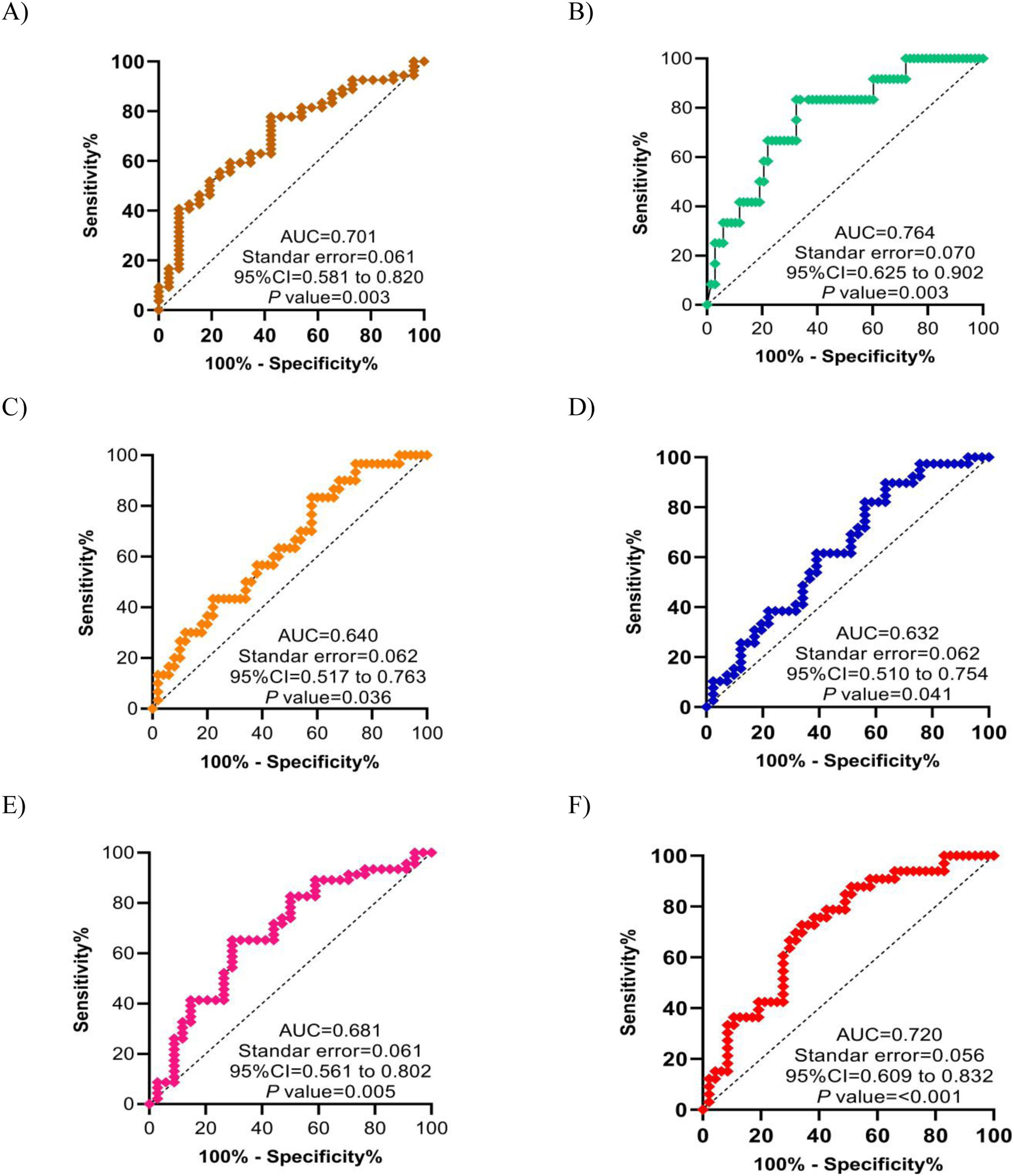
Furthermore, we determined the capacity of sCD163 levels to predict CHR0.121 (AUC=0.701, p=0.003) (Fig. 4A), total cholesterol/HDL-c ratio>7% (AUC=0.764, p=0.003) (Fig. 4B), and CMI1.70 (AUC=0.681, p=0.005) (Fig. 4E). Additionally, we found that serum levels of sCD163 significantly predicted LDL-c/HDL-c ratio>3% (AUC=0.640, p=0.036) (Fig. 4C), AIP>0.21 (AUC=0.632, p=0.041) (Fig. 4D), as well as high clinical activity in RA (AUC=0.720, p<0.001) (Fig. 4F).

Figure 4.

ROC curve of sCD163 on CVR factors and clinical activity in women with RA. (A) CHR0.121. (B) Total cholesterol/HDL-c ratio>7%. (C) LDL-c/HDL-c ratio>3%. (D) AIP>0.21. (E) CMI1.70. (F) DAS28-ESR>3.2. p-Value0.05 was considered statistically significant.

Discussion

This study establishes the utility of serum sCD163 levels as a surrogate marker of CVR in women with RA. We found that serum sCD163 levels of our cohort are higher compared to levels reported in RA population from Denmark,18,20 Romania,19 and Japan.17 However, in RA population from Finland, sCD163 levels are even higher than in our patients.21 The variability in sCD163 levels in RA patients may be related to different degrees of monocytes and synovial macrophage activation16,17 since such activation might contribute to the release of sCD163 into circulation.35

Serum sCD163 levels in our patients were up to 20-fold higher compared to levels reported in patients with SLE,25 to long-standing RA,19 cerebrovascular events25 and peripheral arterial disease.7 Although the exact reason for such elevated serum levels of sCD163 remains unknown, some studies found previously that sCD163 serum levels are positively correlated to CRP and TNF-α serum levels.17–20,36,37 The macrophage TNF-α-converting enzyme (TACE), also called ADAM17, separates the ectodomain of CD163, contributing to the generation of the soluble form of CD163,38 so that inflammation induced by TNF-α in RA contribute to the elevated serum levels of sCD163 and possibly is linked to metabolic alterations and risk of atherosclerosis.39–41 In this sense, there is evidence that some synovial macrophage subpopulations act locally at synovial fibroblast as mediators of inflammation and modulators of lipid metabolism.42 In RA, several factors contribute to the development of inflammation, and although the described mechanisms do not show a direct relationship between CVR and sCD163, the evidence suggests it could be a cardiovascular risk marker.

We found the relationship between sCD163 levels with novel CVR markers such as CHR and CMI, as well as with the atherogenic indices and the number of MetS components. Similarly, sCD163 levels have been related to serum triglycerides43 and LDL-c36 levels, as well as other MetS components.37,43–45 In Taiwanese adults, elevated sCD163 levels are associated with a 5.35 times higher risk of MetS.46 Similarly, our study observed that four or more MetS components are related to increased sCD163 levels. Furthermore, it has been described that the common comorbidities related to obesity (e.g., MetS) could affect the modulation of sCD163 levels through direct or indirect mechanisms.46 In our population, 36.25% of women with RA are obesity and have high CVR.

We analyzed the 10-year risk of cardiovascular events in women with RA, and we find a positive relationship between the sCD163 levels and the score QRISK3 calculator. In our cohort, 16.25% of the participants had an elevated 10-year risk of cardiovascular events compared to more than 70% in an Argentine population with RA.47 The QRISK3 calculator and the combination of QRISK3+2015/2016 EULAR mSCORE exhibit a high sensitivity for identifying the presence of atherosclerotic plaque in RA.48 So, this study show our patients’ subclinical atherosclerosis risk and the importance of assessing traditional CV factors and new soluble biomarkers as sCD163.

The sCD163 levels could be considered as a surrogate marker of CVR in RA. Furthermore, we found that a total cholesterol/HDL-c ratio of >7% was related to a 7-fold higher probability of increasing serum sCD163. It coincides with the study of Sun et al.9 who found that hypercholesterolemia was associated with sCD163 levels in patients with AIS. Therefore, we suggest that the inflammation in RA could by associated to a mechanism of altered the lipid metabolism, proinflammatory cytokines, and acute phase reactants that subsequently becomes related to sCD163 levels. Finally, the monitoring surrogate markers of CVR may help predict and prevent major vascular events in clinical course of RA.49,50 The cardiovascular prevention in RA is based on controlling the inflammation and reducing the modifiable traditional risk factors.

Strengths and limitations

A limitation of this study was including patients from only one hospital, and non-characterization of other CVR markers (for example, the use of advanced visualization techniques for the detection and monitoring of cardiovascular affections). The inclusion men with RA and a control group that was comparable. However, external validation studies and cohort studies should be performed based on the present findings. By other hand, although the evaluation of a modest sample size could be considered a limitation, is strength the characterization of the patient's enrollment. Another strength of the study was the sCD163 levels was related to clinical activity and CVR markers. Therefore, sCD163 could by a surrogate biomarker of disease activity and CVR in women with RA.

Conclusion

Elevated sCD163 levels are associated with the presence of CVR markers, as well as with clinical activity in women with RA. External validation and replication of our findings should be executed in the future to consider sCD163 as a clinically helpful CVR marker.

CRediT authorship contributions statement

IPGG: Conceptualization, Supervision, Methodology, Visualization, Formal analysis, Validation, Investigation, Writing-original draft preparation, Writing-review & editing. OZG: Investigation, Methodology, Formal analysis, Writing-original draft preparation. OB: Methodology, Writing-review & editing. JRVB: Methodology, Writing-review & editing. IAGP: Methodology, Investigation. HURD: Methodology. CMM: Methodology. GAAS: Methodology. AAP: Methodology. JENZ: Methodology. RRG: Methodology. All authors contributed to the manuscript revision, and read, and approved the submitted version.

Ethical standards

The study was approved by the Ethics and Research Committee of the University Autonomous of Guerrero (project identification code CB-004/2017). Informed consent was obtained from all participants included in the study. All experiments conformed to the Declaration of Helsinki.

Funding

None.

Conflict of interest

The authors declare that the research was conducted in the absence of any commercial or financial relationships that could be construed as a potential conflict of interest.

References
[1]
M. Kristiansen, J.H. Graversen, C. Jacobsen, O. Sonne, H.J. Hoffman, S.K. Law, et al.
Identification of the haemoglobin scavenger receptor.
Nature, 409 (2001), pp. 198-201
[2]
M.M. Van den Heuvel, C.P. Tensen, J.H. van As, T.K. Van den Berg, D.M. Fluitsma, C.D. Dijkstra, et al.
Regulation of CD163 on human macrophages: cross-linking of CD163 induces signaling and activation.
J Leukoc Biol, 66 (1999), pp. 858-866
[3]
C. Buechler, M. Ritter, E. Orsó, T. Langmann, J. Klucken, G. Schmitz.
Regulation of scavenger receptor CD163 expression in human monocytes and macrophages by pro- and antiinflammatory stimuli.
J Leukoc Biol, 67 (2000), pp. 97-103
[4]
C.A. Gleissner, I. Shaked, C. Erbel, D. Böckler, H.A. Katus, K. Ley.
CXCL4 downregulates the atheroprotective hemoglobin receptor CD163 in human macrophages.
Cir Res, 106 (2010), pp. 203-211
[5]
T. Kaempfer, E. Duerst, P. Gehrig, B. Roschitzki, D. Rutishauser, J. Grossmann, et al.
Extracellular hemoglobin polarizes the macrophage proteome toward Hb-clearance, enhanced antioxidant capacity and suppressed HLA class 2 expression.
J Proteome Res, 10 (2011), pp. 2397-2408
[6]
L.P. Aristoteli, H.J. Møller, B. Bailey, S.K. Moestrup, L. Kritharides.
The monocytic lineage specific soluble CD163 is a plasma marker of coronary atherosclerosis.
Atherosclerosis, 184 (2006), pp. 342-347
[7]
J.A. Moreno, T. Dejouvencel, J. Labreuche, D.M. Smadja, M. Dussiot, J.L. Martin-Ventura, et al.
Peripheral artery disease is associated with a high CD163/TWEAK plasma ratio.
Arterioscler Thromb Vas Biol, 30 (2010), pp. 1253-1262
[8]
J.A. Moreno, A. Ortega-Gómez, S. Delbosc, N. Beaufort, E. Sorbets, L. Louedec, et al.
In vitro and in vivo evidence for the role of elastase shedding of CD163 in human atherothrombosis.
Eur Heart J, 33 (2012), pp. 252-263
[9]
H. Sun, X. Zhang, J. Ma, Z. Liu, Y. Qi, L. Fang, et al.
Circulating soluble CD163: a potential predictor for the functional outcome of acute ischemic stroke.
Front Neurol, 12 (2021), pp. 740420
[10]
P. Durda, L.M. Raffield, E.M. Lange, N.C. Olson, N.S. Jenny, M. Cushman, et al.
Circulating soluble CD163, association with cardiovascular outcomes and mortality, and identification of genetic variants in older individuals: the cardiovascular health study.
J Am Heart Assoc, 11 (2022), pp. e024374
[11]
B.B. Løgstrup, T. Ellingsen, A.B. Pedersen, B. Darvalics, K.K.W. Olesen, H.E. Bøtker, et al.
Cardiovascular risk and mortality in rheumatoid arthritis compared with diabetes mellitus and the general population.
Rheumatology (Oxford), 60 (2021), pp. 1400-1409
[12]
V. Restivo, S. Candiloro, M. Daidone, R. Norrito, M. Cataldi, G. Minutolo, et al.
Systematic review and meta-analysis of cardiovascular risk in rheumatological disease: symptomatic and non-symptomatic events in rheumatoid arthritis and systemic lupus erythematous.
Autoimmun Rev, 21 (2022), pp. 102925
[13]
J.A. Avina-Zubieta, J. Thomas, M. Sadatsafavi, A.J. Lehman, D. Lacaille.
Risk of incident cardiovascular events in patients with rheumatoid arthritis: a meta-analysis of observational studies.
Ann Rheum Dis, 71 (2012), pp. 1524-1529
[14]
R. López-Mejías, S. Castañeda, C. González-Juanatey, A. Corrales, I. Ferraz-Amaro, F. Genre, et al.
Cardiovascular risk assessment in patients with rheumatoid arthritis: the relevance of clinical, genetic and serological markers.
Autoimmun Rev, 15 (2016), pp. 1013-1030
[15]
A. Mandel, A. Schwarting, L. Cavagna, K. Triantafyllias.
Novel surrogate markers of cardiovascular risk the setting of autoimmune rheumatic diseases: current data and implications for the future.
Front Med (Lausanne), 9 (2022), pp. 820263
[16]
B. Smiljanovic, A. Radzikowska, E. Kuca-Warnawin, W. Kurowska, J.R. Grün, B. Stuhlmüller, et al.
Monocyte alterations in rheumatoid arthritis are dominated by preterm release from bone marrow and prominent triggering in the joint.
Ann Rheum Dis, 77 (2018), pp. 300-308
[17]
N. Matsushita, M. Kashiwagi, R. Wait, R. Nagayoshi, M. Nakamura, T. Matsuda, et al.
Elevated levels of soluble CD163 in sera and fluids from rheumatoid arthritis patients and inhibition of the shedding of CD163 by TIMP-3.
Clin Exp Immunol, 130 (2002), pp. 156-161
[18]
S.R. Greisen, H.J. Moller, K. Stengaard-Pedersen, M.L. Hetland, K. Horslev-Petersen, A. Jorgensen, et al.
Soluble macrophage-derived CD163 is a marker of disease activity and progression in early rheumatoid arthritis.
Clin Exp Rheumatol, 29 (2011), pp. 689-692
[19]
C. Jude, D. Dejica, G. Samasca, L. Balacescu, O. Balacescu.
Soluble CD163 serum levels are elevated and correlated with IL-12 and CXCL10 in patients with long-standing rheumatoid arthritis.
Rheumatol Int, 33 (2013), pp. 1031-1037
[20]
S.R. Greisen, H.J. Møller, K. Stengaard-Pedersen, M.L. Hetland, K. Hørslev-Petersen, P. Junker, et al.
Macrophage activity assessed by soluble CD163 in early rheumatoid arthritis: association with disease activity but different response patterns to synthetic and biologic DMARDs.
Clin Exp Rheumatol, 33 (2015), pp. 498-502
[21]
J. Parantainen, G. Barreto, R. Koivuniemi, H. Kautiainen, D. Nordström, E. Moilanen, et al.
The biological activity of serum bacterial lipopolysaccharides associates with disease activity and likelihood of achieving remission in patients with rheumatoid arthritis.
Arthritis Res Ther, 24 (2022), pp. 256
[22]
N. Barbarroja Puerto, I. Arias de la Rosa, C. Román-Rodriguez, I. Gómez garcía, C. Perez-Sanchez, C. López Medina, et al.
POS1001 Clinical and surrogate cardiovascular risk assessment and its relationship with psoriatic arthritis pathogenesis.
Ann Rheum Dis, 80 (2021), pp. 769
[23]
N. Sakumura, M. Shimizu, M. Mizuta, N. Inoue, Y. Nakagishi, A. Yachie.
Soluble CD163, a unique biomarker to evaluate the disease activity, exhibits macrophage activation in systemic juvenile idiopathic arthritis.
Cytokine, 110 (2018), pp. 459-465
[24]
C. David, G. Divard, R. Abbas, B. escoubet, J. Chezel, M.P. Chauveheid, et al.
Soluble CD163 is a biomarker for accelerated atherosclerosis in systemic lupus erythematosus patients at apparent low risk for cardiovascular disease.
Scand J Rheumatol, 49 (2020), pp. 33-37
[25]
C. David, N. Costedoat-Chalumeau, D. Belhadi, C. Laouénan, A. Boutten, J. Chezel, et al.
Soluble CD163 and incident cardiovascular events in patients with systemic lupus erythematosus: an observational cohort study.
J Intern Med, 292 (2022), pp. 536-539
[26]
D. Aletaha, T. Neogi, A.J. Silman, J. Funovits, D.T. Felson, C.O. Bingham 3rd, et al.
2010 Rheumatoid arthritis classification criteria: an American College of Rheumatology/European League Against Rheumatism collaborative initiative.
Arthritis Rheum, 62 (2010), pp. 2569-2581
[27]
Organization, World Health. Obesity: preventing and managing the global epidemic: World Health Organization; 2000.
[28]
W.T. Friedewald, R.I. Levy, D.S. Fredrickson.
Estimation of the concentration of low-density lipoprotein cholesterol in plasma, without use of the preparative ultracentrifuge.
Clin Chem, 18 (1972), pp. 499-502
[29]
S.M. Grundy, J.I. Cleeman, S.R. Daniels, K.A. Donato, R.H. Eckel, B.A. Franklin, et al.
Diagnosis and management of the metabolic syndrome: an American Heart Association/National Heart, Lung, and Blood Institute scientific statement: executive summary.
Crit Pathw Cardiol, 4 (2005), pp. 198-203
[30]
O. Zaragoza-García, I.P. Guzmán-Guzmán, M.E. Moreno-Godínez, J.E. Navarro-Zarza, V. Antonio-Vejar, M. Ramírez, et al.
PON-1 haplotype (−108C>T, L55M, and Q192R) modulates the serum levels and activity PONase promoting an atherogenic lipid profile in rheumatoid arthritis patients.
Clin Rheumatol, 40 (2021), pp. 741-752
[31]
M. Dobiásová, J. Frohlich.
The plasma parameter log (TG/HDL-C) as an atherogenic index: correlation with lipoprotein particle size and esterification rate in apoB-lipoprotein-depleted plasma (FER(HDL)).
Clin Biochem, 34 (2001), pp. 583-588
[32]
A.G. Ioachimescu, D.M. Brennan, B.M. Hoar, B.J. Hoogwerf.
The lipid accumulation product and all-cause mortality in patients at high cardiovascular risk: a PreCIS database study.
Obesity (Silver Spring), 18 (2010), pp. 1836-1844
[33]
I. Wakabayashi, T. Daimon.
The “cardiometabolic index” as a new marker determined by adiposity and blood lipids for discrimination of diabetes mellitus.
Clin Chim Acta, 438 (2015), pp. 274-278
[34]
J. Hippisley-Cox, C. Coupland, P. Brindle.
Development and validation of QRISK3 risk prediction algorithms to estimate future risk of cardiovascular disease: prospective cohort study.
BMJ, 357 (2017), pp. j2099
[35]
H.N. Daghestani, C.F. Pieper, V.B. Kraus.
Soluble macrophage biomarkers indicate inflammatory phenotypes in patients with knee osteoarthritis.
Arthritis Rheumatol, 67 (2015), pp. 956-965
[36]
N.M. Al-Daghri, O.S. Al-Attas, L.S. Bindahman, M.S. Alokail, K.M. Alkharfy, H.M. Draz, et al.
Soluble CD163 is associated with body mass index and blood pressure in hypertensive obese Saudi patients.
Eur J Clin Invest, 42 (2012), pp. 1221-1226
[37]
M.V. Zanni, T.H. Burdo, H. Makimura, K.C. Williams, S.K. Grinspoon.
Relationship between monocyte/macrophage activation marker soluble CD163 and insulin resistance in obese and normal-weight subjects.
Clin Endocrinol (Oxf), 77 (2012), pp. 385-390
[38]
A. Etzerodt, M.B. Maniecki, K. Møller, H.J. Møller, S.K. Moestrup.
Tumor necrosis factor α-converting enzyme (TACE/ADAM17) mediates ectodomain shedding of the scavenger receptor CD163.
J Leukoc Biol, 88 (2010), pp. 1201-1205
[39]
H. Bruunsgaard, P. Skinhøj, A.N. Pedersen, M. Schroll, B.K. Pedersen.
Ageing, tumour necrosis factor-alpha (TNF-alpha) and atherosclerosis.
Clin Exp Immunol, 121 (2000), pp. 255-260
[40]
S. Parveen, R. Jacob, L. Rajasekhar, C. Srinivasa, I.K. Mohan.
Serum lipid alterations in early rheumatoid arthritis patients on disease modifying anti rheumatoid therapy.
Indian J Clin Biochem, 32 (2017), pp. 26-32
[41]
C. Charles-Schoeman, X. Wang, Y.Y. Lee, A. Shahbazian, I. Navarro-Millán, S. Yang, et al.
Association of triple therapy with improvement in cholesterol profiles over two years follow-up in the Treatment of Early Aggressive Rheumatoid Trial.
Arthritis Rheumatol, 68 (2016), pp. 577-586
[42]
S. Alivernini, L. MacDonald, A. Elmesmari, S. Finlay, B. Tolusso, M.R. Gigante, et al.
Distinct synovial tissue macrophage subsets regulate inflammation and remission in rheumatoid arthritis.
Nat Med, 26 (2020), pp. 1295-1306
[43]
H.J. Møller, R. Frikke-Schmidt, S.K. Moestrup, B.G. Nordestgaard, A. Tybjaerg-Hansen.
Serum soluble CD163 predicts risk of type 2 diabetes in the general population.
Clin Chem, 57 (2011), pp. 291-297
[44]
T. Parkner, L.P. Sørenser, A.R. Nielsen, C.P. Fischer, B.M. Bibby, S. Nielsen, et al.
Soluble CD163: a biomarker linking macrophages and insulin resistance.
Diabetologia, 55 (2012), pp. 1856-1862
[45]
K. Fjeldborg, T. Christiansen, M. Bennetzen, H.J. Møller, S.B. Pedersen.
The macrophage-specific serum marker, soluble CD163, is increased in obesity and reduced after dietary-induced weight loss.
Obesity (Silver Spring), 21 (2013), pp. 2437-2443
[46]
T.Y. Hu, S.Y. Lee, C.K. Shih, M.J. Chou, M.C. Wu, I.C. Teng, et al.
Soluble CD163-associated dietary patterns and the risk of metabolic syndrome.
Nutrients, 11 (2019), pp. 940
[47]
W. Masson, E. Rossi, R.N. Alvarado, G. Cornejo-Peña, J.I. Damonte, N. Fiorini, et al.
Rheumatoid arthritis, statin indication and lipid goals: analysis according to different recommendations.
Rheumatol Clin (Engl Ed), 18 (2022), pp. 266-272
[48]
A. Corrales, N. Vegas-Revenga, B. Atienza-Mateo, C. Corrales-Selaya, D. Prieto-Peña, J. Rueda-Gotor, et al.
Combined use of QRISK3 and SCORE as predictors of carotid plaques in patients with rheumatoid arthritis.
Rheumatology (Oxford), 60 (2021), pp. 2801-2807
[49]
L.D. Heftdal, K. Stengaard-Pedersen, L.M. Ornbjerg, M.L. Hetland, K. Horslev-Petersen, P. Junker, et al.
Soluble CD206 plasma levels in rheumatoid arthritis reflect decreased in disease activity.
Scand J Clin Lab Invest, 77 (2017), pp. 385-389
[50]
S. Rodríguez-Muguruza, A. Altuna-Coy, V. Arreaza-Gil, M. Mendieta-Homs, S. Castro-Oreiro, M.J. Poveda-Elices, et al.
A serum metabolic biomarker panel for early rheumatoid arthritis.
Front Immunol, 14 (2023), pp. 1253913
Copyright © 2024. Sociedad Española de Arteriosclerosis
asdasdasd
Opciones de artículo
Herramientas