


The main objective is to determine if there are differences in visual function and retinal structure in patients with bipolar disorder compared to healthy subjects.
Material and methodsCross-sectional observational study of cases and controls, adjusted for age and sex. A total of 43 controls (86 eyes) and 82 cases (163 eyes) were included. Visual function was assessed by measuring best corrected visual acuity (BCVA) using high contrast and low contrast visual charts. Optical coherence tomography (OCT) model modelo DRI OCT Triton-Swept Source (Topcon, Tokyo, Japón) was used for retinal structural analysis.
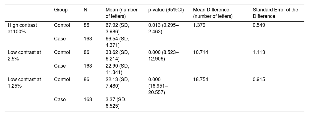
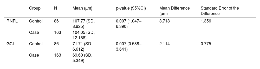
ResultsBCVA with high contrast, as well as with reduced contrast at 2.5% and 1.25%, showed significant differences (p < 0.05) between both groups, being the mean of each variable lower in the case group. OCT analysis also showed significant differences in the mean thickness of the nerve fiber layer (RNFL) and ganglion cell layer (GCL) between the two groups, with the mean of each variable lower in the case group (p-value = 0.007 in both). No significant differences were observed in the mean thickness of these retinal layers between type I and type II bipolar patients (p-value = 0.556 and 0.871 respectively).
Conclusionsthere are significant differences in visual function and in the mean thickness of retinal layers between bipolar patients and healthy controls.
El objetivo principal es determinar si existen diferencias en la función visual y en la estructura de la retina en pacientes con trastorno bipolar, en comparación con sujetos sanos.
Material y métodosEstudio observacional transversal de tipo casos y controles, ajustados por edad y sexo. Se han incluido un total de 43 controles (86 ojos) y 82 casos (163 ojos). La función visual se ha evaluado midiendo la mejor agudeza visual corregida (MAVC) con láminas de alto contraste y bajo contraste visual. Para llevar a cabo el análisis estructural de la retina se ha utilizado la tomografía de coherencia óptica (OCT) modelo DRI OCT Triton-Swept Source (Topcon, Tokyo, Japón).
ResultadosLa MAVC con alto contraste, así como con contraste reducido al 2,5% y 1,25% muestran diferencias significativas (p < 0,05) entre ambos grupos, siendo la media de cada una de estas variables menor en el grupo de casos. En el análisis mediante OCT también se detectan diferencias significativas entre grupos en el espesor medio de la capa de fibras nerviosas (RNFL) y de células ganglionares (GCL) (p-valor = 0,007 en ambas variables), siendo la media de cada variable menor en el grupo de casos. No se han observado diferencias significativas en el espesor medio de estas capas de la retina entre pacientes bipolares tipo I y tipo II (p-valor = 0,556 y 0,871 respectivamente).
ConclusionesExisten diferencias significativas en la función visual y en el espesor medio de las capas estructurales de la retina entre pacientes bipolares y controles sanos.
