


The effects of acute continuous positive airway pressure therapy on left ventricular diastolic function and functional capacity in patients with compensated systolic heart failure remain unclear.
METHODS:This randomized, double-blind, placebo-controlled clinical trial included 43 patients with heart failure and a left ventricular ejection fraction <0.50 who were in functional classes I-III according to the New York Heart Association criteria. Twenty-three patients were assigned to continuous positive airway pressure therapy (10 cmH2O), while 20 patients received placebo with null pressure for 30 minutes. All patients underwent a 6-minute walk test (6MWT) and Doppler echocardiography before and immediately after intervention. Clinicaltrials.gov: NCT01088854.
RESULTS:The groups had similar clinical and echocardiographic baseline variables. Variation in the diastolic function index (e′) after intervention was associated with differences in the distance walked in both groups. However, in the continuous positive airway pressure group, this difference was greater (continuous positive airway pressure group: Δ6MWT = 9.44+16.05×Δe′, p = 0.002; sham group: Δ6MWT = 7.49+5.38×Δe′; p = 0.015). There was a statistically significant interaction between e′ index variation and continuous positive airway pressure for the improvement of functional capacity (p = 0.020).
CONCLUSIONS:Continuous positive airway pressure does not acurately change the echocardiographic indexes of left ventricle systolic or diastolic function in patients with compensated systolic heart failure. However, 30-minute continuous positive airway pressure therapy appears to have an effect on left ventricular diastolic function by increasing functional capacity.
Noninvasive ventilation with continuous positive airway pressure (CPAP) has been described as a non-pharmacological approach in patients with compensated systolic heart failure (HF). Previous studies suggested that the benefits of CPAP included improved oxygenation, reduced myocardial oxygen demand, decreased respiratory work and decreased left ventricular (LV) preload and afterload (1-4). However, the effects of CPAP on the LV systolic function indexes of HF patients are controversial. The increases in left ventricular ejection fraction (LVEF) and cardiac output have been shown to persist for over 1 hour of rest following a single session of noninvasive therapy (bilevel positive airway pressure) (5). However, other authors did not observe any CPAP-related changes in these indexes, regardless of whether the evaluation was performed during or after the intervention (6,7).
Notably, most of the studies evaluating the hemodynamic effects of CPAP in HF patients included subjects with obstructive sleep apnea. Therefore, it is difficult to establish whether the cardiac improvement is associated with the apnea treatment or is due to a direct effect on the heart (8).
Patients with compensated HF and reduced LVEF present decreased functional capacity related to increased LV diastolic filling pressure rather than a decrease in the ejection fraction itself. Diastolic dysfunction causes dyspnea, which is the most important limiting symptom and can be used as a predictor of major outcomes such as decompensated HF and death (9,10).
The effects of CPAP on myocardial oxygenation and metabolic demand may increase diastolic function by improving relaxation. Therefore, it is reasonable to hypothesize that the CPAP-induced increase in diastolic function indexes would have a beneficial effect on functional capacity in patients with compensated systolic HF.
Thus, the aim of this study was to investigate the acute effects of first-time CPAP application on LV diastolic function and its influence on functional capacity in asymptomatic HF patients with reduced LVEF.
METHODSStudy PopulationInformed consent was provided by each patient before study participation, and the protocol was approved by the Institutional Committee of Research Ethics (OF.405/2009-CEP).
The study was a prospective, unicenter, randomized, double-blind trial. Recruitment began in January 2008 and ended in January 2010, and 43 consecutive patients with compensated HF and reduced ejection fraction were enrolled from the outpatient clinics of our hospital.
The inclusion criteria were as follows: men and women who were previously diagnosed with systolic HF; NYHA functional class I-III; LVEF<0.50; receiving optimal pharmacological treatment (11,12); and a stable clinical condition, as shown by the absence of dyspnea exacerbation for at least 3 months and the ability to cooperate with the procedures.
The exclusion criteria included unstable angina or acute coronary syndrome in the last 6 months, arterial blood pressure>140/90 mmHg, atrial fibrillation, complex ventricular arrhythmia, chronic obstructive pulmonary disease, obstructive sleep apnea, more than mild cardiac valve disease, active infection, or physical impairment that would cause a walking limitation.
This study was registered at ClinicalTrials.gov (NCT01088854).
Study ProtocolAll patients underwent baseline 2-dimensional and Doppler echocardiographic examinations and were randomized using a random-figure computerized program in a proportion of 1:1. Twenty patients were included in the sham group (simulated CPAP), and 23 patients were included in the CPAP group. The patients, the echocardiographer, and the physiotherapist who conducted the 6MWT were blinded to the randomization.
After clinical evaluation, the patients underwent echocardiography followed by the initial 6MWT in an indoor 30-meter-long corridor according to the American Thoracic Society guidelines (13). In addition to the walked distance, the heart rate, respiratory rate, pulse oximetry, blood pressure, and modified Borg Scale score were recorded before and after the test. All patients had already experienced at least one 6MWT in the last month; therefore, they were already familiar with the procedure. At the conclusion of the initial 6MWT, patients allocated to the CPAP group were positioned comfortably for the CPAP procedure with 10 cmH2O positive pressure for 30 minutes. Subsequently, the CPAP was withdrawn, and a second echocardiogram was performed. Patients allocated to the sham group underwent the same procedure except for the null pressure. Because the patients had never experienced this intervention, we assumed that they were unaware of whether they were receiving CPAP. Finally, a second 6MWT was performed by the same therapist who was blinded to the CPAP procedure.
EchocardiographyStandard transthoracic echocardiograms were performed by the same examiner (SGZB) using an HDI 5000 SONOCT ultrasound (Phillips, Andover, MA, USA) according to American Society of Echocardiography and Canadian Consensus recommendations (14,15). In our laboratory, the intra-observer variability is below 5%. Left atrial (LA) and LVEF volumes were obtained using Simpson's rule. The LVEF was calculated as the normalized difference between the diastolic and systolic volumes. Spectral Doppler recordings from the mitral valve as well as the Doppler velocities of the septal and lateral annular tissue were obtained from the apical 4-chamber view. Peak early (E wave) and late (A wave) transmitral flow velocities, E/A ratio, isovolumetric relaxation time (IRT), and deceleration time (DTE) were measured. Longitudinal systolic (Sm), early diastolic (e′), and late diastolic (a′) mitral annular velocities were measured, and average velocities were calculated from three consecutive cardiac cycles.
Statistical AnalysisSummary data are expressed as the mean±standard deviation. Etiology, comorbidity, and medication frequencies were compared between groups using the chi-square (χ2) test or Fisher's exact test. Student's t test was used for other comparisons between groups either before or after intervention. Linear regression analysis was used to assess the association between changes in walked distance and variations in diastolic index (e′) immediately after CPAP. A general linear model was used to analyze the interaction between e′ index variation and CPAP to improve functional capacity. A 2-tailed significance level of p<0.05 was considered statistically significant, with a power equal to 80%. All analyses were performed using the commercially available statistical package SAS for Windows, version 9.2 (Cary, NC, USA).
RESULTSDespite the overt heart failure, all patients were oligosymptomatic. A total of 151 of the 195 patients who were observed in the outpatient clinics of our hospital from January 2008 to January 2010 presented with one or more of the exclusion criteria; therefore, 44 patients (27 men and 17 women) were included in the study protocol. Of these patients, 20 were randomly assigned to the sham group, while 23 were assigned to the CPAP group.
Table1 shows the anthropometric and cardiac morphometric data before CPAP intervention. There were no differences between the groups.
Baseline anthropometric and morphometric characteristics of the patients.
| Sham group(n = 20) | CPAP group(n = 23) | p | |
|---|---|---|---|
| Sex (men/women) | 13/7 | 13/10 | 0.490 |
| Age (years) | 55.0±11.11 | 54.3±9.66 | 0.827 |
| BMI (kg/m2) | 25.7±5.07 | 26.7±4.95 | 0.512 |
| LVM (g) | 280±56.7 | 260±78.4 | 0.123 |
| LAV (mL/m2) | 53.81±15.19 | 54.7±16.80 | 0.854 |
| LVDd (cm) | 6.4±0.67 | 6.4±1.01 | 0.764 |
| LVDs (cm) | 5.2±0.77 | 5.2±1.10 | 0.868 |
| IVSd (cm) | 1.00±0.144 | 0.96±0.145 | 0.275 |
| PWd (cm) | 0.99±0.112 | 0.94±0.106 | 0.105 |
Values are presented as the mean±SD. BMI: body mass index; IVSd: interventricular septum diameter at end-diastole; LAV: left atrial volume; LVDd: left ventricular end-diastolic dimension; LVDs: left ventricular systolic dimension; LVM: left ventricular mass; PWd: posterior wall diameter at end-diastole.
The etiologies of cardiomyopathy were ischemia, arterial hypertension, alcoholism, and Chagas disease. HF induced by chemotherapy was observed in one patient in the sham group. The comorbidities were current smoking, hypothyroidism, hyperlipidemia, diabetes mellitus, and arterial hypertension. The two groups had similar causes of HF and comorbidities. Also, there were no differences between the groups as medications were concerned (Table2).
Etiologies and comorbidities.
| Sham group(n = 20) | CPAP group (n = 23) | p*) | |
|---|---|---|---|
| Etiology | |||
| Ischemic | 6 | 5 | |
| Hypertensive | 5 | 7 | |
| Idiopathic | 2 | 5 | |
| Alcoholism | 1 | 1 | |
| Chagas disease | 5 | 5 | |
| Chemotherapy | 1 | 0 | |
| 0.534 | |||
| Comorbidities | |||
| Hypertension | 11 | 15 | |
| Current smoking | 7 | 2 | |
| Hypothyroidism | 1 | 1 | |
| Hyperlipidemia | 5 | 6 | |
| Diabetes mellitus | 6 | 9 | |
| 0.220 | |||
| Medications | |||
| Diuretics | 16 | 22 | |
| ACE/BRA | 18 | 23 | |
| Beta-blockers | 15 | 23 | |
| Spirolactone | 11 | 14 | |
| Digoxin | 12 | 12 | |
| Aspirin | 7 | 11 | |
| Statin | 6 | 7 | |
| Warfarin | 3 | 1 | |
| 0.927 |
ACE: Angiotensin-converting enzyme; ARB: angiotensin II receptor blocker.
The parameters recorded in the first and second 6MWT were similar between the groups. Before CPAP, the mean distances walked by patients in the sham and CPAP groups were 451±55.8 meters and 437±70.7 meters, respectively (p = 0.480). After the intervention, patients in the sham group walked 452±62 meters, while those in the CPAP group walked 443±67.7 meters (p = 0.656). Table3 shows the echocardiography data. There was no difference between the groups before or after CPAP.
Systolic and diastolic function indices before and after intervention.
| Pre-intervention | Post-intervention | ||||||
|---|---|---|---|---|---|---|---|
| Indexes | Sham group (n = 20) | CPAP group (n = 23) | p*) | Sham group (n = 20) | CPAP group (n = 23) | p*) | |
| LVEF | 0.36±0.07 | 0.37±0.09 | 0.551 | 0.38±0.07 | 0.39±0.09 | 0.477 | |
| CO (L/min) | 3.56±0.52 | 3.62±0.42 | 0.662 | 3.59±0,50 | 3.65±0.46 | 0.680 | |
| SV (mL) | 51.5±9.5 | 53±9.5 | 0.647 | 52.7±9.6 | 54.5±10.3 | 0.567 | |
| E | 69±28.5 | 81±24.5 | 0.162 | 73±28.6 | 81±20.0 | 0.291 | |
| A | 70±21.9 | 69±25.5 | 0.820 | 68±21.5 | 69±25.5 | 0.863 | |
| E/A (cm/s) | 1.19±0.87 | 1.51±1.06 | 0.286 | 1.27±0.82 | 1.45±0.92 | 0.498 | |
| DTE (ms) | 241±81.2 | 205±65.2 | 0.117 | 231±84.6 | 208±60.8 | 0.306 | |
| IRT (ms) | 123±27.6 | 108±25.4 | 0.061 | 124±31.8 | 111±24.8 | 0.135 | |
| s′ (cm/s) | 7.5±1.1 | 7.6±1.4 | 0.359 | 7.5±1.0 | 7.9±1.3 | 0.834 | |
| e′ (cm/s) | 9.8±1.7 | 10.5±1.7 | 0.211 | 9.3±1.7 | 10.3±1.6 | 0.075 | |
| a′ (cm/s) | 11.8±2.4 | 12.9±2.5 | 0.682 | 12.7±2.1 | 13.0±2.7 | 0.652 | |
| E/e′ | 7.22±3.35 | 7.86±2.65 | 0.491 | 7.91±2.96 | 8.11±2.48 | 0.815 |
Data are expressed as the mean±SD. A: peak late transmitral flow velocity; a′: peak late diastolic mitral annular velocity (average of lateral and septal sites); CO: cardiac output; DTE: deceleration time of E; E: peak early transmitral flow velocity; e′: peak early diastolic mitral annular velocity (average of lateral and septal sites); IRT: isovolumetric relaxation time; LVEF: left ventricular ejection fraction (Simpson's rule); s′: peak systolic mitral annular velocity (average of lateral and septal sites); SV: stroke volume.
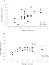
Before- and after-intervention differences in ventricular function analyzed using linear regression indicated a statistically significant association between e′ index variation and walked distance variation in the CPAP group (Figure1). That is, for each centimeter increase in annular mitral diastolic velocity, there was a mean increase of 16.05 meters in the 6MWT (Δ6MWT = 9.44+16.05×Δe′; R = 0.61; p = 0.002). This association, although statistically significant in the sham group, had less of an influence on physical capacity (Δ6MWT = 7.49+5.38×Δe′; R = 0.53; p = 0.015). Furthermore, there was a statistically significant interaction between e′ index variation and CPAP for the improvement of functional capacity (p = 0.020). There was no association between the systolic function indexes and 6MWT in either group.
DISCUSSION
The present study revealed the acute effects of 30-minute CPAP application in patients with compensated HF and systolic ventricular dysfunction through the randomization of patients with similar baseline characteristics.
Echocardiograms revealed cardiac enlargement in all individuals as well as myocardial hypertrophy and reduced ejection fraction. These results establish that the studied sample showed significantly impaired left ventricular function although they showed no signs of congestion.
After the intervention, there was no difference between the groups with respect to the variables obtained during the 6MWT. These results differ from those described by Chermont et al. (16) and Lima et al. (17). These authors evaluated the effects of 30-minute CPAP treatment in 12 patients with HF in a randomized study. The CPAP group showed increases in the distance covered and peak heart rate. Improved exercise tolerance was also found in a study by Steiner et al. (18), who examined a series of 11 patients with compensated HF without sleep apnea (only 6 patients completed the study). Overnight 6-hour CPAP applications were performed every night for approximately 8 months. Cardiopulmonary exercise testing showed improved peak oxygen consumption, exercise duration, and workload. However, in these studies, fewer patients were included and the protocols used were distinctly different from ours. Bradley et al. (4) conducted a study that included 258 HF patients who were randomly assigned to receive nocturnal CPAP or no treatment for 2 months. CPAP was associated with improved physical capacity in the 6MWT.
In the present study, the lack of improved exercise capacity after CPAP could be explained by the short-term administration; however, other researchers obtained similar results even with more prolonged use (6,7,19). Therefore, it is reasonable to state that the question of whether CPAP actually exerts effects on exercise tolerance in patients with compensated systolic HF must be definitively answered before more in-depth studies can be performed.
No significant changes in LV systolic function were observed in either group in the current study. In the normal heart, the initiation of CPAP therapy acutely increases intrathoracic pressure and decreases venous return and LV filling, resulting in decreased cardiac output (3,20). However, studies have shown that the simultaneous reduction of preload (caused by decreased filling pressure) and afterload (due to decreased transmural pressure) would improve systolic function in patients with HF (3),.
Despite the potential beneficial effects of CPAP on systolic function, the literature examining this issue is contradictory. Few studies have found improved hemodynamics in HF patients after the acute use of continuous or bilevel positive airway pressure (3,5,16,22). Long-term CPAP therapy has also been found to increase ejection fraction and cardiac output (4,23,24). Yoshinaga et al. (2) and Johnson et al. (20) compared the two application periods and observed improved ejection fraction and stroke volume only after chronic therapy.
On the contrary, in a randomized placebo-controlled trial performed by Smith et al. (6), no difference was found in ejection fraction after 6 weeks of nocturnal auto-titrating CPAP.
Interestingly, most patients in these studies presented with obstructive sleep apnea in addition to chronic HF, which would influence the effect of long-term CPAP on cardiac function and exercise tolerance. For example, Sin et al. (8) described an improvement in LVEF after 3 months of nightly CPAP only in HF patients with Cheyne-Stokes respiration and central sleep apnea. CPAP had no significant effect on LVEF in patients without these respiratory conditions.
There is a paucity of data on the effects of CPAP on LV diastolic function, despite its major impact on the symptoms and prognosis of patients with HF. Johnson et al. (20) investigated the effects of acute and long-term CPAP therapy on LV systole and diastole. In that study, 7 patients with HF and obstructive sleep apnea who used CPAP were compared with 5 patients with HF without obstructive sleep apnea who were not using CPAP. Only chronic CPAP therapy improved systolic function, and diastolic function was not affected by either chronic or acute CPAP use.
Conversely, we previously described a series of 11 patients with compensated HF who received acute CPAP therapy. Echocardiograms were performed at baseline and 30 minutes later while the patients were still actively receiving CPAP. CPAP therapy did not alter LV systolic function but did significantly improve diastolic function (25).
In the present study, LV diastolic function indices were evaluated at baseline and after CPAP withdrawal, and we found that the use of CPAP had no benefit. Despite this apparent lack of effect, our findings regarding the association between intervention-related variations in the diastolic function index (e′) and the walked distance were interesting. In both groups, improved diastolic function was associated with a proportional improvement in physical performance. This result was expected because it is well known that diastolic function influences exercise capacity (26-28). However, whereas in the sham group, variations in the e′ index were associated with an increase of only 5.38 meters in walked distance, in the CPAP group, each increment in the e′ index was associated with a mean increase of 16.05 meters in walked distance. This result indicated an interaction between e′ index variation and CPAP for the improvement of functional capacity. Therefore, in a general sense, although acute CPAP therapy did not affect ventricular function, it may cause an incremental increase in functional capacity in those patients who respond to acute CPAP with improved diastolic function. Our results also indicate that the effect of CPAP may involve factors other than ventricular function, including, for instance, pulmonary gas exchange, which was not analyzed in this study.
The main limitation of our study was the small number of included patients, which prevented the demonstration of further effects of CPAP on diastolic function indexes. Despite this limitation, our data raised an important question about the relevance of LV diastolic function in the functional capacity of patients with compensated systolic HF as well as the beneficial effect of CPAP. Further studies are needed to confirm these results.
CPAP does not acutely alter echocardiographic indexes of LV systolic or diastolic function in patients with compensated systolic HF. However, 30-minute CPAP therapy appears to influence LV diastolic function by increasing functional capacity.
ACKNOWLEDGMENTSWe would like to thank Fundação de Amparo à Pesquisa do Estado de São Paulo - FAPESP (Grant 2009/50249-0), which provided financial support for this study.
AUTHOR CONTRIBUTIONSMatsubara BB coordinated the study, performed the statistical analysis and reviewed the manuscript. Zanati SG coordinated the study and performed echocardiography. Matsubara LS and Minamoto ST performed the statistical analysis. Guirado GN collected data. Polegato BF and Roscani MG collected data and enrolled patients. Bussoni MF collected data, enrolled patients, and wrote the manuscript.
No potential conflict of interest was reported.