This study reports on the experience of one hospital regarding the surgical aspects, anatomic investigation and outcomes of the management of 21 conjoined twin pairs over the past 20 years.
METHODSAll cases of conjoined twins who were treated during this period were reviewed. A careful imaging evaluation was performed to detail the abdominal anatomy (particularly the liver), inferior vena cava, spleen and pancreas, either to identify the number of organs or to evaluate the degree of organ sharing.
RESULTSThere were eight sets of ischiopagus twins, seven sets of thoracopagus twins, three sets of omphalopagus twins, two sets of thoraco-omphalo-ischiopagus twins and one set of craniopagus twins. Nine pairs of conjoined twins could not be separated due to the complexity of the organs (mainly the liver and heart) that were shared by both twins; these pairs included one set of ischiopagus twins, six sets of thoracopagus twins and one set of thoraco-omphalo-ischiopagus twins. Twelve sets were separated, including seven sets of ischiopagus twins, three sets of omphalopagus twins, one set of thoracopagus twins and one set of craniopagus conjoined twins. The abdominal wall was closed in the majority of patients with the use of mesh instead of the earlier method of using tissue expanders. The surgical survival rate was 66.7%, and one pair of twins who did not undergo separation is currently alive.
CONCLUSIONA detailed anatomic study of the twins and surgical planning must precede separation. A well-prepared pediatric surgery team is sufficient to surgically manage conjoined twins.
Conjoined twins have fascinated mankind throughout the centuries because of the rarity of this type of birth; however, conjoined twins have always been a challenge for physicians. Eng and Chang Bunker are likely the most famous pair of conjoined twins. These twins were born in Siam in 1811, taken to the United States by a circus company and exhibited as the curious “Siamese Twins”, thus providing the origin of the colloquial term (1). The brothers lived for 63 years without considering separation, and they married sisters and fathered 21 children.
As a rare outcome of a monoamniotic and monochorionic gestation, conjoined twins occur when two identical individuals are joined by part of their anatomy and share one or more organs. The incidence of conjoined twins ranges from 1:50,000 to 1:100,000 live births. This number could be higher, but most of these pregnancies result in miscarriages and still births; only 18% of all conjoined infants survive (2), and approximately 35% of live births die within the first 24 hours, and only 18% of all conjoined twins survive longer than 24 hours. In Brazil, where abortion is not legal, we believe that the incidence of conjoined twins is likely higher than in most developed countries.
Conjoined twins are classified based on the terminology proposed by Spencer and colleagues (3). Based on this terminology, we use the most prominent site of union plus the suffix “pagus,” which is a Greek word meaning “that which is fixed.” Spencer et al. also divided the twins into three major groups: twins with a ventral union, twins with a dorsal union and twins with a lateral union. The first major group includes four types: cephalopagus (head), thoracopagus (chest), omphalopagus (umbilicus) and ischiopagus (hip). The dorsal union includes three types: pygopagus (sacrum), rachipagus (spine) and craniopagus (cranium). The last major group includes just one type of twins that is referred to as parapagus (side) twins.
The surgical separation of conjoined twins is now the principal aim of all medical teams who treat this uncommon condition. However, separation presents both surgical and anesthetic challenges. In addition, this surgery is sometimes not possible because the anomalies are rare and difficult to manage, even for experienced surgeons. If we share our experiences and learn from others, we can enhance our knowledge and skills for treating conjoined twins.
In Brazil, when a diagnosis of conjoined twinning is made, the pregnant mother is usually referred to a tertiary hospital that specializes in obstetric and perinatal care. Although there are several reports in the medical literature about conjoined twinning, only one Brazilian study has been published on this issue; that study was performed in a tertiary perinatology referral university center over a period of 25 years (4). The authors reported the occurrence of 14 pairs of conjoined twins and the successful separation of only one pair of omphalopagus twins. The present study aims to report the experience of one Brazilian hospital over a period of 20 years, focusing on surgical aspects, anatomic investigations and outcomes.
PATIENTS AND METHODSThis is a retrospective review of all cases of conjoined twins treated between January 1992 and July 2012 at the Pediatric Surgery Division and Liver Transplantation Unit of the Child Institute of the Faculdade de Medicina da Universidade de São Paulo. The study was approved by the ethics committee of the institution, and we obtained parental approval to publish the children's pictures.
Patient information was obtained by reviewing medical records and the intranet database of the hospital. Perinatal data included prenatal ultrasound diagnosis, gender, birth weight and the anatomy of the twins. The patients were classified based on the most prominent site of fusion, based on the embryological classification proposed by Spencer (3). Data regarding stillbirths, miscarriages and conjoined twin gestations were excluded. Cases of fetus-in-fetus, considered by some authors to be“incomplete conjoined twins,” were also excluded from this study.
A careful imaging evaluation was performed for all conjoined twins, as described in Table 1. Angiographic studies of the liver circulation were performed when the computed angiotomography or magnetic resonance imaging studies did not provide consistent information about these important anatomic details.
Conjoined twin type and anatomical evaluation.
| Type | Evaluation |
|---|---|
| Ischiopagus | Ultrasonography of the abdomen, skull and pelvis |
| Echocardiography | |
| Radiography | |
| Doppler ultrasound | |
| Contrast meal and enema | |
| Computed tomography | |
| Computed angiotomography | |
| Magnetic resonance imaging | |
| Micturating uretrocystography | |
| Endoscopy | |
| Cavography | |
| Hepatic venography | |
| Thoracopagus | Ultrasonography of the abdomen and skull |
| Echocardiography | |
| Radiography | |
| Fetal echocardiography | |
| Computed angiotomography | |
| Doppler ultrasound of the abdomen | |
| Magnetic resonance imaging | |
| Omphalopagus | Ultrasonography of the abdomen and skull |
| Echocardiography | |
| Radiography | |
| Doppler ultrasound of the abdomen | |
| Craniopagus | Computed tomography of the brain and skull |
| Computed angiotomography of the brain | |
| Complete ultrasonography examination of the abdomen | |
| Echocardiography |
After a careful imaging evaluation, the possibility of separation surgery was determined, and the twins were divided into two groups as follows:
- 1
Conjoined twins who were not candidates for surgical separation for the reasons described above.
- 2
Conjoined twins who underwent surgical separation. In this group, the following data were collected and analyzed: the age and weight at the time of the separation surgery, the length of surgery, the duration of anesthesia during the separation surgery, a detailed description of the separation surgery, the type of abdominal wall closure, postoperative complications and death.
Numerical data are presented as the mean±standard deviation. Statistical analyses were performed using the Student's t-test.
RESULTSTwenty-one sets of conjoined twins were analyzed. Most of the pairs were female, with 13 female sets and 8 male sets. The mean birth weight of the twin pairs was 2,921.17±1,078.05 g. A prenatal diagnosis was made in 19 pregnancies (90.5%). The mean age at separation surgery, excluding the two conjoined twins who underwent emergency separations during the newborn period, was 9 mo 24 d±4 mo 25 d. The mean weight of the twin sets at the time of the separation surgery, excluding the emergency separations, was 9,656.08±4,594.02 g.
The 21 sets included eight sets of ischiopagus twins, seven sets of thoracopagus twins, three sets of omphalopagus twins, two sets of thoraco-omphalo-ischiopagus twins and one set of craniopagus twins. The data collected for the groups are described above.
Non-operative managementNine pairs of twins were not candidates for separation based on imaging evaluations. The separation procedure was not possible due to the complexity of organs that were shared by both twins, mainly the liver and heart. The decision was made after consulting with the parents and the Ethical Committee of the Institution.
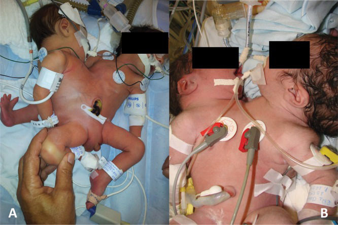
IschiopagusOnly one set of ischiopagus twins was not separated due to severe perinatal asphyxia that evolved to death within one day of life. This set had a diaphragmatic hernia with the stomach and spleen occupying one twin's thorax and the stomach and hepatic lobe occupying the other twin's thorax; this placement may have caused a pneumothorax in both twins. The twins shared one liver and pelvis; in addition, they had three legs (classified as “ischiopagus tripus”) and severe cardiovascular defects. The other seven sets of ischiopagus twins were separated (Figure 1A).
ThoracopagusMost sets of thoracopagus twins were not separated (6 of 7) because of complex cardiac anomalies, including two hearts sharing a ventricular wall and one shared heart containing four fused atria and two ventricles. The liver was also shared in all sets. Three sets also presented with duodenal sharing. Five of six sets died during the neonatal period (Figure 1B), and the remaining set died within four months.
Thoraco-omphalo-ischiopagusTwo sets of complex thoraco-omphalo-ischiopagus twins did not undergo separation. One of these sets of twins died three days after birth due to serious cardiac defects.
The second set of thoraco-omphalo-ischiopagus twins presented with four arms, two legs, one bladder, one pelvis, fused small intestines from the terminal portion to the anus and one set of female genitalia. Therefore, the two infants had individual stomachs, duodenums, jejunums and most of the ilea. The twins had horseshoe kidneys and a bicornuate uterus. The livers were fused and drained to the inferior vena cava of just one infant. Furthermore, the portal veins were crossed. For this case, the pediatric surgery group considered separation. However, in addition to liver sharing, one of the infants had complex cardiac anomalies (aorta and pulmonary artery emerging from the right ventricle and aortic coarctation) that led the parents to refuse surgical separation. This decision was supported by the ethical committee of our institution, as previously described, and the ethical committee of the Regional Medical Council of São Paulo. At the time of publication, these twins were still alive and growing.
Operative managementWhen separation was pursued after exhaustive imaging examinations, the technical aspects were always discussed with the anesthesiologists and rehearsed. The twins were comfortably positioned on the surgical table, and the procedure was started. Four anesthesiologists were required (two for each twin). Following endotracheal intubation, each twin underwent central vein and radial arterial cannulation for complete monitoring during the operation. A Foley bladder catheter was also inserted. The separation procedure was always performed by a single surgical team. After the separation, the second twin was moved with the corresponding anesthetic equipment and a second team of surgeons to another surgical room for the final reconstruction procedures. The reconstruction of the first twin was completed by the surgical team that performed the separation. After the separation procedure, all of the infants were transferred to the intensive care unit for strict monitoring.
Among the 21 sets of conjoined twins, 12 underwent separation surgery (seven ischiopagus, three omphalopagus, one thoracopagus and one craniopagus). The mean anesthesia time for the separation surgery was 8 h 45 min±4 h 49 min, and the mean surgery time was 6 h 52 min±4 h 10 min.
IschiopagusSix ischiopagus tripus and one ischiopagus tetrapus (with four normal legs) twin pairs underwent separation. The mean anesthesia time was 11 h 35 min±3 h 57 min, and the mean surgery time was 8 h 29 min±3 h 09 min.
The separation procedure began by making a large longitudinal incision across the anterior abdominal wall. The fused livers were separated along the anterior midline, after verifying that each liver had its own hilum and hepatic veins. Each infant had a normal stomach, duodenum, gallbladder, pancreas and spleen. All sets had shared intestines. In twins with a single anus, a colostomy or ileostomy was performed in one infant, depending on whether the small or large intestine was shared, and an intestinal anastomosis was performed on the second infant who kept the anus. In twins with one anus per twin, the intestines were separated according to the origin of intestinal sharing irrigation. A colostomy or ileostomy was performed when the anus was considered nonviable. For the urinary tract, crossed ureters were a common finding (5 of 6 sets); in these cases the ureter of one twin implanted in the bladder of the other twin. In these cases, the ureters were divided close to the bladder of the other twin and then reimplanted in the bladder of the corresponding twin. The separation was completed by adequate and anatomical division of internal and external genital organs and the bones of the pelvis using appropriate orthopedic instruments.
The reconstruction phase for each separated twin was performed by a reconstruction of the digestive system with intestinal anastomoses, followed by reimplantation of the divided ureters.
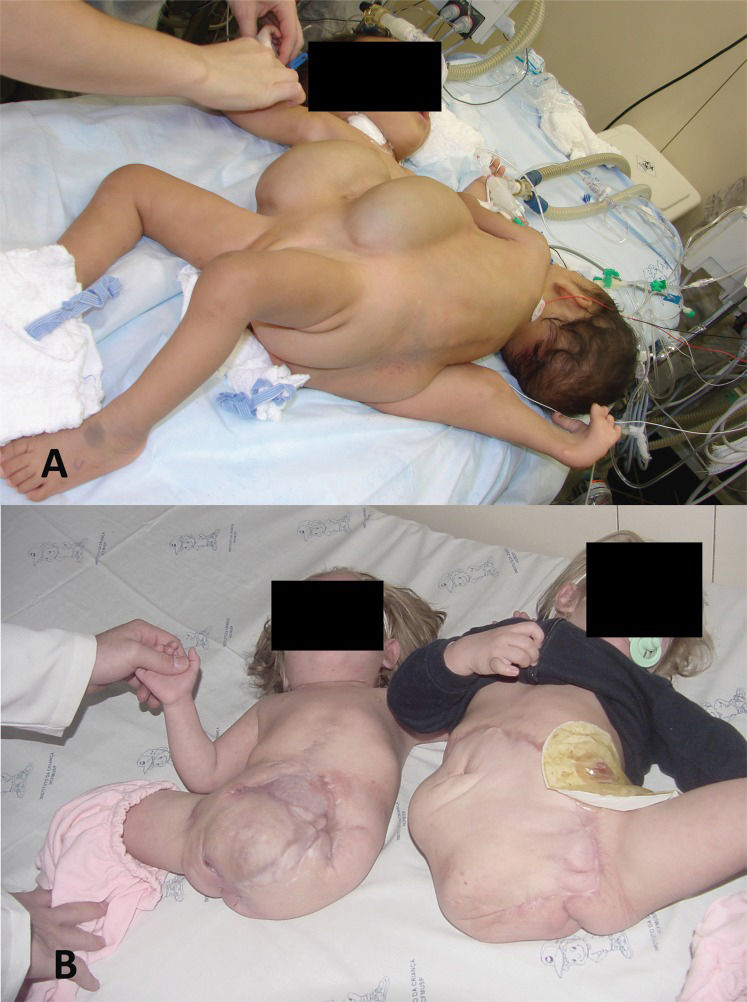
The last phase of reconstruction was the closure of the abdominal wall. For all cases of ischiopagus tripus twins, we preferred to use the third abnormal leg for soft-tissue coverage. The bones were excised after careful dissection, and a large flap containing skin and skeletal muscles was obtained for complete closure of the abdominal cavity of one of the twins. The abdominal wall of the other infant was closed with mesh. In the first two pairs of conjoined twins, tissue expanders were used prior to the separation procedure; however, no advantage was noted because mesh was still required to close the abdomen and to cover the viscera. In one of these twin pairs, the abdominal skin became necrotic one week after the separation procedure, most likely due to ischemia caused by the previous placement of the tissue expander. The necrotic skin was excised, and the child completely recovered due to the formation of granulation tissue and wound healing by secondary intention (Figure 2. Based on the experience of these two cases, we abandoned the use of tissue expanders in subsequent cases. However, even in the infant for whom the third leg was used, mesh was needed to complete the abdominal wall closure.
Of the seven sets of twins who underwent separation, one set developed sepsis and did not survive. These infants had serious complications during the preparation phase, including pulmonary infection and respiratory insufficiency. In addition, both infants had serious renal dysplasia and renal insufficiency. Despite these complications, the separation procedure was performed after obtaining consent from the family. Among the 12 infants who survived, 10 developed a wound infection (83.3%) or a urinary tract infection. Two patients developed late enteric fistulas due to the exposure of the intestine, despite the presence of the mesh. These infants underwent another operation during which the fistulas were successfully closed and a new mesh was inserted. One infant developed evisceration and needed a second operation to close the abdomen and place a new mesh. One twin developed late sepsis and died.
ThoracopagusOnly one set of thoracopagus twins underwent separation surgery. This set had a single pericardial sac, and a small part of the anterior ventricular wall was shared between both hearts. However, the two hearts also had serious anomalies, interventricular communication in one heart and a hypoplastic right ventricle in the other heart. The thoracic cavities were shared from the nipple level to the inferior abdomen. These twins shared one liver with two hila, two gallbladders, two stomachs and two duodenums. The intestines were separate. During the investigation period, one infant developed a pulmonary hemorrhage and died, leading to an emergency separation. The live infant survived the separation procedure and died of sepsis after 11 months.
OmphalopagusThe omphalopagus twins required a shorter duration of anesthesia (mean time of 5 h 12 min±2 h 3 min) and a shorter duration of surgery (mean time of 3 h 13 min±1 h 27 min compared with the ischiopagus twins (p = 0.02 and p = 0.005 for anesthesia and surgery time, respectively).
Although all sets of twins had a shared liver, none had a complex or shared biliary tract. There were two independent hepatic circulations, and each twin had an inferior vena cava. In addition, we did not encounter any congenital heart defects, and separation was possible in all sets.
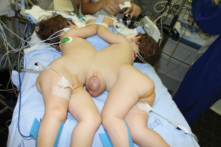
In all patients, the abdominal wall was closed with mesh, without the use of previously placed tissue expanders. One pair of twins was separated at 11 months of life and had an omphalocele that spontaneously epithelialized; complete skin coverage was achieved without difficulty (Figure 3.
In one pair of twins, one of the twins died within one hour of birth. An emergency separation procedure was performed, and the live infant survived the separation procedure. After five days, she presented with a gastric rupture that was repaired with gastric suture. This child recovered very well.
CraniopagusThere was one pair of craniopagus twins with serious associated defects. One of the twins had a skull and brain with normal volumes, anorectal agenesis and a recto-vesical fistula. The other twin presented with microcephaly and sirenomelia (“mermaid syndrome”). Both twins underwent an emergency colostomy during the neonatal period and were referred to us when they were 10 months old. Tomographic and angiotomographic studies of the brain revealed that complete separation would be impossible, and the twin with microcephaly and sirenomelia would have to be sacrificed during separation. After obtaining consent from the family, the separation procedure was performed by a team of neurosurgeons. The twin who survived underwent surgery for his anorectal agenesis six months after the separation. The colostomy was finally closed after two months and the infant recovered very well.
Regarding the mortality rate of our series, among the 42 infants (21 pairs of twins), 24 died and 18 were alive at the time this manuscript was written. Because one pair of twins is alive and did not undergo separation and 16 children survived the separation procedure, we conclude that the final survival rate for the procedure was 66.7% (16 infants alive among 24 infants [12 sets of twins] who underwent surgery). The most recent follow-up of these infants indicated that they are living and experiencing normal quality of life.
Table 2 summarizes the anatomical details and outcomes of the twins.
Conjoined twins treated at the Child Institute of the Faculdade de Medicina da Universidade de São Paulo.
| Case | Gender | Type | Main organs shared | Outcome |
|---|---|---|---|---|
| 1 | F | Thoracopagus | Heart, liver and small intestine | Cardiac insufficiency and set death with 11 days of life |
| 2 | F | Thoracopagus | Heart and liver | Perinatal asphyxia and progressive bradycardia due to congenital cardiac defects; set died with 1 day of life |
| 3 | F | Thoracopagus | Heart, liver, spleen, and small intestine | Perinatal asphyxia, submitted to ventilation; set died on the 27th day of life |
| 4 | F | Thoracopagus | Heart and liver | Cardiogenic shock and death on the 11th day of life |
| 5 | F | Thoracopagus | Heart | Cardiac insufficiency, pulmonary edema and sepsis by Enterobacter cloacae; died with 28 daysof life |
| 6 | M | Thoracopagus | Heart and liver | Bronchopneumonia and hypoxia leading to death at 4 months of life |
| 7 | F | Thoracopagus | Pericardium, liver and small intestine | Twin 1 died of sepsis and pulmonary hemorrhage leading to emergency separation, twin 2 died with 11 months of life |
| 8 | F | Ischiopagus | Four legs, liver, large intestine and crossed ureters | Successful separation at 9 months of life |
| 9 | M | Ischiopagus | Third malformed leg, large intestine, anus, scrotum, bladder and liver | Set died of perinatal asphyxia within 1 day of life |
| 10 | F | Ischiopagus | Third malformed leg, vulva, urethra, bladder, liver, large intestine and crossed ureters | Successful separation at 10 months of life |
| 11 | M | Ischiopagus | Third malformed leg, liver, large intestine and urethra. Crossed ureters | Set submitted to separation; one survived |
| 12 | F | Ischiopagus | Third malformed leg, anus, large intestine, liver and crossed ureters | Successful separation with 16 months of life |
| 13 | F | Ischiopagus | Four normal legs, sigmoid and rectum, anus, liver, | Successful separation with 16 months of life |
| 14 | M | Ischiopagus | Third malformed leg, ileum and large intestine, liver and crossed ureters | Successful separation with 10 months of life |
| 15 | M | Ischiopagus | Third malformed leg, liver, small intestine and anus. Crossed ureters | Both infants evolved, after separation, with sepsis and bradycardia leading to death |
| 16 | M | Omphalopagus | Large intestine and bladder | Twin 1 died leading to separation surgery. Twin 2 survived |
| 17 | F | Omphalopagus | Liver and duodenum | Separated with 3 days of life. |
| 18 | F | Omphalopagus | Liver | Successful separation with 11 months of life |
| 19 | F | Thoracoomphaloischiopagus | 2 legs, bladder and pelvis, colon, genitalia and anus, just one inferior vena cava.Portal vein of twin 1 drains to portal vein of twin 2. | Parents denied the separation due to high risks. The twins are alive. |
| 20 | M | Thoracoomphaloischiopagus | 3 legs, complex cardiopathy | Twins died three days after birth |
| 21 | M | Craniopagus | Brain vascular connections. Twin 1 with microcephaly and sirenomelia. Twin 2 with anorectal agenesis. | Separation with 10 months of life. Twin 1 was sacrificed and twin 2 is alive and well. |
The present series of patients is impressive because the largest sample of such malformations ever studied included 383 reviewed sets of conjoined twins. That study was published in 2011, and the most important findings included a marked variation in pregnancy outcomes, similarity in the proportion of types of twins among hospitals, significant female predominance and apparently increasing prevalence in South American countries. Additionally, no significant genetic, environmental or demographic associated factors were detected (5).
Although the literature advises the use of a multidisciplinary approach for surgical separation to improve survival rates (2,6,7), the separations in our institution were performed by a team composed only of trained pediatric surgeons. Despite this difference, our results did not differ significantly from those of other studies in terms of the final outcomes of the patients and mortality rates.
The problems encountered in our series and the lessons learned from our experience during the treatment of these 21 conjoined twins enrich our knowledge regarding surgery in complex pediatric patients. The first difficulty encountered was the anesthesia required for imaging investigations during the pre-separation phase. These imaging investigations included interventional radiological investigations and angiotomographic examinations. These procedures provided invaluable insight into potential or actual problems that may arise during the separation, such as the difficulties encountered in the ischiopagus tripus twins who died one week after surgery (patient 15).
As with every surgery, surgical separation of conjoined twins carries its own risks, which may be avoided by reducing the surgery and anesthesia times. Therefore, a meticulous investigation of the twins' anatomy is as crucial as the improvement of appropriate surgical techniques by training and experience. However, we also learned that, despite carefully studying the twins' anatomy, unexpected anatomical variations are frequently identified during the surgery; the surgical team must be prepared for these variations. Therefore, the separation must be performed by a team of trained general pediatric surgeons. In our experience, the inclusion of several specialties (orthopedic, plastic, urologic and cardiovascular surgeons) during the separation procedure was often confusing and did not lead to better results. In only one case was a team of neurosurgeons involved because the surgery required neurosurgical expertise that we did not have.
There are various controversies regarding the ideal age at which the separation procedure should be performed. Spitz et al. (8) preferred to operate at approximately three months of age, which allows time for detailed investigations to be conducted and enables separation to take place when the body wall can still rapidly expandto close substantial defects. However, a high incidence of postoperative wound infection can occur, and separation is very harmful to the body's functional reserves. Therefore, we preferred to operate at approximately 10 months of age, despite some psychosocial issues that may occur during the waiting period. Consequently, the mean age of our patients at the time of surgery was 10 months and 9 days.
Skin closure always presents a challenge and should be carefully considered before the separation procedure has begun. Many surgeons tend to use tissue expanders (1), and sometimes mesh (8,11). We opted for mesh instead of tissue expanders because of our personal experience, and data from the literature show that tissue expanders lead to complications in as many as 57.2% of twins in whom they are used (1). These complications include insufficient skin expansion, tissue expander infection, skin necrosis over the expander, exposure of the device and seroma formation. These complications require additional operations and general anesthesia. Moreover, the surgery to place the expanders represents a risk itself and involves unnecessary delays and costs. In a recent review of 12 separated sets of conjoined twins, the author concluded that tissue expanders are not required in most patients (12). In our series, we did not use other reported techniques that are available for providing adequate coverage of the abdominal contents and viscera. The utilization of skin grafts, although skin grafts were recently reported for use in separating conjoined twins, they should not be utilized for covering the abdominal viscera (1). Skin grafts can be appropriately used to cover a granulating surface that develops in a surviving twin. However, we have noted that during the late postoperative period of these separated twins, skin grafts are unnecessary; spontaneous epithelialization occurs if adequate nutritional support is provided to the infant. The other recently reported technique involves creating a pneumoperitoneum during the preoperative period by injecting 500 to 1,500 mL of air every 3 days to increase the abdominal circumference and promote soft tissue and skin expansion (1). Because the literature does not report positive results for this technique (13,14), we think that it may be abandoned.
Our results show a high incidence of ischiopagus (38.1%) and thoracopagus (42.8%) twins. Interestingly, the incidence of thoracopagus twins at our facility is similar to that reported in the collaborative study cited above (4). We believe that the low incidence of omphalopagus twins (14.3%) is due to the easier separation of this type at other centers, which leads to fewer transfers of these twins to a reference center. In contrast, there is a high incidence of stillbirth and miscarriage among thoraco-omphalo-ischiopagus twins because of the associated complex cardiac anomalies. However, our incidence of ischiopagus twins is quite different from the 1.8% reported in the collaborative study (5). We have no explanation for this difference. Finally, the least common and perhaps the most difficult type of twin to separate is the craniopagus type because the cranial union often involves a variety of neural and vascular connections.
In Brazil, legally allowed abortion should be considered for sets of conjoined twins with poor prognoses, particularly for thoracopagus twins, who are unlikely to be successfully separated and have a low survival rate. In thoracopagus sets, fetal echocardiography plays an important role in determining the anatomy of the hearts and helps the pediatric and surgical teams prepare. Fortunately, a prenatal diagnosis was made in 90.5% of our cases. This diagnosis is very useful for preparing the obstetric and pediatric surgical teams for a successful delivery and for researching the possibility of a surgical separation.
The surgical mortality rate of our series was 33.3%, which is in accordance with other published series. In a recent publication from the Philippines, the mortality rate of nine sets of twins was 17.7% (10). Spitz in the United Kingdom reported a mortality rate of 50% in 12 separation procedures, seven of which were performed emergently (6). Based on several publications and the present experience, emergency separations always have dismal outcomes. Therefore, careful deliberation and complete evaluation of the twins before any surgical intervention are important to ensuring that the surgery proceeds smoothly and with good results.
The Pediatric Surgery Division of the Child Institute of the University of São Paulo Medical School has treated 21 sets of conjoined twins over 20 years, with surgical experience in 12 pairs of patients. Considering the surgical outcomes, it is concluded that a well-prepared pediatric surgery team is sufficient for the surgical management of conjoined twins. Occasionally, emergency separation is needed; low survival rates are expected in this circumstance because of the poor condition of the patients and the limited availability of imaging exams. A good anatomical survey and proper surgical planning must precede the separation, and the twins must be as healthy as possible. Recent advances in imaging techniques for preoperative investigations provide adequate anatomic diagnosis and predict the possibility of separation. Moreover, advances in anesthetic care and postoperative critical care have improved outcomes and survival rates.
AUTHOR CONTRIBUTIONSTannuri AC and Batatinha JA reviewed the medical records and prepared the manuscript. Velhote MC and Tannuri U are the surgeons who reviewed the final version of manuscript.
This study was initiated by the second author under the supervision of the first author, and it was financed by Fundação de Amparo a Pesquisa do Estado de São Paulo (project number 2011/08273-0).
No potential conflict of interest was reported.


