






Edited by: Dr. Calos Cerdán
Cirugía General y Aparato Digestivo. Unidad de Cirugía Digestiva. Hospital Universitario de la Princesa. Madrid
Dr. Matteo Frasson
Cirugía general. Hospital Universitario y Politécnico La Fe. Valencia
Last update: January 2026
More infoPelvic exenteration (PE) is a potentially curative surgical option for patients with locally advanced rectal cancer (LARC) orlocallyrecurrent rectal cancer (LRRC) involvingmultivisceralstructures. Originally conceived as a palliative procedure, advancements in surgical techniques and multidisciplinary management have broadened its indications, enabling R0 resections in over 60% of cases. Appropriate patient selection, based on advanced imaging and comprehensive functional assessment, is essential to optimize oncologic outcomes. Currently, the surgical indication is primarily determined by the reasonable possibility of achieving an R0 resection with acceptable morbidity in a suitable candidate, which may translate into high survival rates. Surgical planning should follow a compartmental approach and require close collaboration among colorectal, urologic, gynecologic, vascular, orthopedic, plastic, and reconstructive surgeons. Urinary, gynecologic, vascular, osseous, and soft tissue reconstructions are tailored to the tumour’s extent and the patient's individual needs. In selected cases, PE may be performed with palliative intent. Intraoperative radiotherapy (IORT) and minimally invasive approaches have emerged as valuable adjuncts. Long-term oncologic and functional outcomes are closely linked to the achievement of negative margins. While postoperative morbidity remains significant, mortality rates have declined in high-volume centres. Postoperative quality of life has become a key outcome, with increasing emphasis on patient-reported outcome measures (PROMs) to guide clinical decision-making. PE continues to challenge traditional limits of resectability, offering renewed hope to carefully selected patients.
La exenteración pélvica (EP) representa una opción terapéutica potencialmente curativa para pacientes con cáncer de recto localmente avanzado (LARC) o recurrencia local (LRRC) con afectación multivisceral. Inicialmente concebida como un procedimiento paliativo, los avances técnicos y el enfoque multidisciplinar han ampliado sus indicaciones, permitiendo resecciones R0 en más del 60% de los casos. La correcta selección de pacientes, basada en estudios de imagen avanzados y una valoración funcional integral, es esencial para maximizar los beneficios oncológicos. En la actualidad, el límite de la indicación quirúrgica se establece en la posibilidad razonable de lograr una resección oncológica completa (R0) con una morbilidad aceptable en el paciente adecuado, lo que puede traducirse en tasas elevadas de supervivencia. La cirugía debe abordarse de forma compartimental y con la colaboración de múltiples especialidades. La reconstrucción urinaria, ginecológica, vascular, ósea y de tejidos blandos se adapta a la extensión tumoural y las necesidades del paciente. En casos seleccionados, la EP puede utilizarse con intención paliativa. La radioterapia intraoperatoria (IORT) y la cirugía mínimamente invasiva han demostrado ser estrategias complementarias prometedoras. Los resultados oncológicos y funcionales dependen en gran medida de la obtención de márgenes negativos. Aunque la morbilidad puede ser elevada, las tasas de mortalidad han disminuido en centros especializados. La calidad de vida posoperatoria se ha convertido en un parámetro clave, destacando el papel de las medidas reportadas por los pacientes (PROMs) en la toma de decisiones. La EP continúa desafiando los límites tradicionales de la resecabilidad, ofreciendo esperanza a pacientes seleccionados.
Pelvic exenteration (PE) is a radical surgical procedure consisting of the en bloc multivisceral resection of at least two contiguous pelvic organs, with the goal of obtaining oncologically negative margins in cases of locally advanced or recurrent pelvic neoplasia.1,2 This technique has transformed the approach to patients previously considered inoperable, offering a curative possibility in complex settings with extensive tumour invasion.3,4
Initially described in 1948 by Alexander Brunschwig for the palliative management of recurrent cervical carcinoma, PE has evolved from a palliative approach to a viable curative option, particularly in the context of locally advanced primary rectal cancer (LARC) or locally recurrent primary rectal cancer (LRRC).1,5 Although high mortality rates and poor survival outcomes initially limited its adoption, advances in anaesthesia, transfusion, radiology, and surgical techniques have significantly improved the safety and efficacy of PE.3,5
Achieving malignancy-free surgical margins (R0) is considered the most determining factor in predicting both long-term survival and postoperative quality of life.3,6–9 Currently, both LARC and LRRC achieve R0 resection rates greater than 60%.10–13
Surgical planning by multidisciplinary committees, based on tumour topography and the prediction of invaded structures, optimises surgical outcomes.14,15 For tumour staging, the most commonly used techniques are magnetic resonance imaging (MRI), computed tomography (CT), and positron emission tomography (PET/CT), occasionally supported by 3D reconstructions.15
Given the radical nature of the procedure, there is a general consensus that PE should be reserved for cases of resectable disease with curative intent.7
Athough PE is mostly performed by laparotomy, laparoscopic and robotic approaches are becoming increasingly common.16 Perioperative management of PE requires a specialised and multidisciplinary approach, which has driven centralisation in high-volume units, favouring the standardisation of procedures and improving both oncological outcomes and morbidity, and thereby expanding the limits of resectability.2,7,8,17–19
In addition to surgical advances, the role of total neoadjuvant therapy (TNT) is being investigated, which enables improved adherence, early treatment of micrometastases, and disease downstaging, with higher rates of pathological complete response.20–23
With the safety and oncological outcomes of PE now established, the current focus is on quality of life, promoting better patient selection and more accurate preoperative counseling. PROMs (Patient Reported Outcomes Measures) have gained relevance as a key tool to assess the true impact of surgery from the patient's perspective, reinforcing the need for a comprehensive approach to cancer.24,25
Indications, contraindications, and evolving surgical limitsLARC and LRRC, which affect pelvic organs or adjacent bony or neurovascular structures, are the most common indications for PE.3,26
PE is more common in recurrent disease, due to the earlier diagnosis of the primary cancer.27 A multidisciplinary approach, multivisceral resection, and resection of important neurovascular and bone structures, along with reconstructive and rehabilitative techniques, have allowed for larger resections, reducing the impact on patient function and quality of life,26,28,29 and also improving long-term survival.26,28 Currently, the limit of surgical indication can be considered the reasonable possibility of achieving complete oncological resection (R0) with acceptable morbidity in the appropriate patient, achieving high survival rates,1,3,7,30,31 (Table 1).
Indications and contraindications of pelvic exenteration.
| Indications |
|---|
| Possibility of achieving R0 with acceptable morbidity:Locally advanced rectal cancer (LARC) or recurrent rectal cancer (LRRC) unresectable with standard TMEOther advanced pelvic tumoursPalliative PE (PPE)a for relief of intractable symptoms: pain, bleeding, sepsis, obstruction, or fistulas |
| Relative contraindicationsb |
| Resectable metastasesSacral S1 infiltrationExternal iliac vessel infiltrationGreater sciatic notch extension |
| Absolute contraindications |
| Bilateral sciatic nerve involvementCircular pelvic bone involvementUnresectable disseminated metastatic diseasePoor performance status or severe comorbidities (ASA > 3) |
Additionally, in selected cases, PE may be considered palliative care (PPE) with the intention of relieving debilitating symptoms, especially in patients with limited options after prior radiation therapy.3
PE is limited to patients with good general health,3,35 excluding those with poor functional status, defined as a score greater than 3 on the American Society of Anaesthesiologists (ASA) score.26
The role of PE in patients with metastatic disease is uncertain. The presence of distant metastases unsuitable for curative resection contraindicates PE, but if they are resectable, they can be considered a relative contraindication, and simultaneous liver resection is feasible. The decision should be individualisd, especially given the prolonged recovery time that may limit the administration of adjuvant chemotherapy.7,26,36–38
Historically, pelvic wall involvement was considered an absolute contraindication, particularly in patients with unilateral hydronephrosis and sciatic pain. However, technical advances and high-volume units have made it increasingly possible to perform extra-anatomical R0 resections, even in complex cases involving bone or vascular resections.7,26
Infiltration of the external iliac vessels, sacral infiltration above S2, and extension through the greater sciatic notch may be contraindications, although these are increasingly relative, as surgical complexity and the risk of poor outcomes increase.7,30,34,36
Absolute contraindications include bilateral sciatic nerve involvement or circumferential bone involvement, which are associated with adverse outcomes and technical limitations in achieving an R0 resection.34
Although the indications for this procedure have expanded significantly, careful patient selection remains essential to optimise outcomes.
Diagnosis and preoperative evaluation for pelvic exenteration in rectal cancerA thorough multidisciplinary preoperative evaluation, including a concise perineal physical examination, including vaginal examination, colonoscopy, pelvic MRI, and even cystoscopy, is essential to confirm the diagnosis, assess for pelvic visceral involvement, and consider the possibility of achieving an R0 resection.39 Histological confirmation of malignancy is imperative for patients considered for PE for cancer.
In addition, the presence of metastatic disease should be assessed, and the patient's fitness to endure such a radical surgical procedure should be evaluated.39
MRI, CT, and PET/CT are essential for preoperative planning, assessing cancer spread and enabling accurate surgical mapping. These tests should be performed as close to the time of surgery as possible.9 High-resolution MRI is the test of choice for defining local spread to adjacent pelvic structures,7,15 since sensitivity and specificity are both very high.40 It also enables three-dimensional multiplanar volumetric reconstructions that aid in the assessment of tumour boundaries.18 CT of the chest, abdomen, and pelvis is the first-line modality for the detection of potential metastatic lesions. PET/CT complements this assessment by providing information on metabolic activity and tumour spread in the pelvis, which can modify therapeutic decisions in up to 40% of cases.15,41,42 In addition, CT arteriography and venography are useful for planning pelvic resections.15 Radiological reports should be standardised, based on recent clinical guidelines.43,44
Currently, mathematical methods are being applied to MRI that make it possible to generate three-dimensional images of the pelvis, which define the optimal surgical plane for achieving R0 resections45 (Fig. 1). Furthermore, advances in artificial intelligence, three-dimensional bioprinting, radiomics, and radiogenomics promise to expand the limits of resections and improve diagnostic accuracy in oncologic surgery.11,46

Magnetic resonance images (coronal and sagittal sections) showing a tumour corresponding to a second pelvic recurrence of a rectal neoplasm with involvement of neighboring pelvic structures, including the vaginal vault, bladder, bowel loops, and left levator ani muscle. Below, a three-dimensional anatomical reconstruction (Cella Medical Solutions® proprietary software) allows precise spatial visualisation of tumour extension and its relationship to organs and vascular structures, facilitating surgical planning in complex cases of pelvic exenteration.
Preoperative medical optimisation with a multidisciplinary approach that includes nutrition, ostomy therapy, physical therapy, thromboembolic prophylaxis, and planned anesthetic techniques is essential for managing surgical stress.47–49 Psychosocial assessment helps determine the patient's ability to tolerate the after-effects of the procedure and prolonged recovery.39 Patient involvement in decision-making and prehabilitation improves outcomes.50
The implementation of ERAS (Enhanced Recovery After Surgery)-based protocols is feasible in PE and can optimise postoperative recovery, with adherence rates exceeding 70%.51
Surgical techniquePE is the en bloc resection of at least two contiguous pelvic organs to achieve negative margins in advanced neoplasia.52 Standardising PE techniques is challenging due to variability in tumour extensión. However, the systematic division of the pelvis into five anatomical compartments (central, anterior, posterior, and two lateral) facilitates a more organised and precise surgical approach.[18] Resection should include the entire compartment when an organ is involved to minimise positive margins (Table 2) (Fig. 2).7 Tumour recurrence adds technical complexity, frequently compromising deep muscular, osseous, and neurovascular structures.18
Pelvic exenteration classification.52
| Type of Exenteration | Description |
|---|---|
| Total (EPT) | Resection of all pelvic organs |
| Anterior | Resection of the bladder and gynecological organs in women, preserving the rectum |
| Posterior | Resection of the rectum and gynecological organs in women, preserving the bladder |
| Supraelevator | Resection of the levator ani muscles and the anorectal stump |
| Infraelevator | Resection of the levator ani muscles and the anorectal stump |
| Comppsite | Includes resection of additional structures: blood vessels, lymph nodes, muscles, or bones |

Images A and B: Sagittal and coronal sections of a pelvic MRI of a patient, illustrating the different types of pelvic exenteration according to the structures included in the resection. Image C: Coronal section of a male patient.
The orange perimeter delineates an anterior exenteration, which includes the bladder and, in women, the internal genital organs. The blue perimeter corresponds to a posterior exenteration, which affects the rectum (and also the genital organs in women). The red perimeter represents a total pelvic exenteration, which includes resection of the bladder, rectum, and genital organs. The yellow lines indicate additional structures that may be included in a compound exenteration, such as the levator muscles or bony structures. Finally, the green line indicates the plane of the levator muscles, serving as a reference for classifying the exenteration as supralevator or infralevator, depending on whether these muscles are spared.
Exteriorisation of a colostomy is standard practice, although intestinal continuity can be restored through a coloanal anastomosis and temporary diverting ileostomy if adequate margins are present.39
The success of the technique lies in the coordinated participation of various specialists to comprehensively address the challenges of PE. In addition to the colorectal surgeon, the team should include urologists, spinal trauma specialists, gynecologists, radiation oncologists, vascular surgeons, and reconstructive surgeons, both plastic and abdominal wall.
Anterior compartmentBladder involvement in PE may require partial cystectomy (PC) or radical cystectomy (RC). PC achieves good oncological results in patients with LARC and should be considered if technically feasible. In LRRC, en bloc RC improves oncological control and preserves quality of life. Reinterventions are more common in PC, although wound-related complications are lower.53
In women, involvement of the posterior vaginal wall is common, necessitating partial vaginectomy and, in some cases, hysterectomy. In men, when the tumour penetrates Denonvilliers' fascia, it can invade the prostate, seminal vesicles, and membranous urethra, generally requiring prostatectomy and/or en bloc cystectomy.18 However, in selected cases, bladder preservation is possible, avoiding complications associated with reconstruction.54 Tumours infiltrating the pubic bone and membranous urethra, previously considered inoperable, can now be treated with curative intent.3,27 A transperineal approach has been developed for urethral resection, with or without en bloc resection of the pubic bone, which allows for a wider anterior margin compared to the traditional abdominal approach, reducing the risk of positive margins.18,30,55,56 Both radical resection of the pubic bone and urinary reconstructions pose a significant risk of morbidity.30
Posterior compartmentInvolvement of the posterior pelvic compartment includes, in addition to the rectum, the presacral space, and the sacrococcyx.
Sacral involvement above S2-S3 limited the indication for PE due to its technical complexity, morbidity, and functional sequelae from nerve root sacrifice.57 However, technical modifications have reduced morbidity58,59 and allowed for "higher and wider" resections, even down to the S1 roots, with results comparable to low sacrectomy.60,61
Sacral bone preservation can be achieved by accessing the presacral plane through the presacral fascia, above the level of the tumour, using a high subcortical sacrectomy,18,62 although segmental sacrectomy techniques have also been described.59 However, an en bloc sacrectomy may be essential in situations of perforation, sepsis in the posterior compartment, or tumour invasion of the bone or its fascia.18,30 In cases of recurrence after total mesorectal excision (TME) surgery, there is usually no defined anatomical plane between the tumour and the sacrum, which implies the need for sacrectomy in most patients.18
Sacrectomies are classified as low (S3 and below) and high (S2 and above). While low sacrectomies can be approached by abdominolithotomy or in the prone position, high sacrectomies frequently require the prone position to optimise visualisation and access to the pelvic wall, thus facilitating control of the iliac vasculature and a lateral dissection of the lumbosacral trunk and sacral roots.18,58,61,63
Some studies indicate that the level of sacrectomy does not impact either the length of hospitalisation or the rate of involved margins.64,65 However, a systematic review indicates that higher resections carry a higher risk of major complications.66
Lateral compartmentsResection of the lateral compartment of the pelvis is complex due to the proximity of key neurovascular structures, such as the lumbosacral trunk, sciatic nerve, and iliac vessels. Historically, tumours in this area have had low R0 resection rates and poorer survival, but this approach is now safe in specialised centres.28,29,67 Lateral dissection may be necessary to perform en bloc resections,30,68 in cases of suspicious lymphadenopathy, for bilateral vascular control prior to sacrectomy, or when tumour extension involves structures such as the sacral roots, piriformis and obturator internus muscles, and the sciatic nerve.18 Pelvic sidewall fibrosis, which develops after chemoradiotherapy, can hamper vessel-sparing lymphadenectomy and increases the risk of bleeding which is why en bloc excision is considered a safer option.28 In advanced tumours involving the pelvic sidewalls, ligation and resection of the internal iliac vessels is frequently performed and is usually well tolerated without reconstruction, as it is often accompanied by a central exenteration in which dependent organs are removed.63,68 Bilateral ligation has been described as a safe and effective technique to prevent and help control bleeding in pelvic surgeries.69 However, when resection of the common or external iliac artery or vein is necessary, reconstruction is essential to preserve limb perfusion,18,68 although major complication rates can reach up to 70%.68 Laterally extended endopelvic resection (LEER) is a surgical technique that facilitates R0 resections and more effective locoregional tumour control, especially in cases where the tumour is fixed to the pelvic wall. This technique involves multicompartmental resection of various pelvic parietal structures, such as paravisceral fat and pelvic floor muscles. It also allows for the removal of the overlying parietal endopelvic fascia, which acts as a barrier to tumour progression.70 However, LEER is not indicated for tumours infiltrating above the level of the obturator nerve or in the sciatic foramen. The technique is frequently combined with therapeutic periaortic and pelvic lymphadenectomy for more complete regional tumour control.70
Despite reservations about extended lateral compartment excision, especially en bloc resection of the femoral or sciatic nerves due to its morbidity and functional impact, oncological outcomes are promising. A radical approach improves R0 rates and survival. In some cases, resection of the sciatic spine optimises exposure and access to the sciatic nerve.18
The diagnosis and management of lateral pelvic lymph nodes (LLNs) is crucial, as their involvement increases the risk of local recurrence. Currently, nodes in the internal iliac and obturator regions are considered regional and should be included in the dissection, while nodes in the external and common iliac regions are classified as metastatic. Despite the differences between Western and Eastern approaches, there is consensus that the combination of neoadjuvant chemoradiotherapy (nCRT) and selective LLN dissection (LLND) reduces the risk of local recurrence and improves long-term outcomes. A surgical approach based on fascial planes rather than blood vessels is preferred, which better protects pelvic autonomic nerves and reduces the risk of postoperative male urinary and reproductive dysfunction.71–73 Transanal-assisted LLND has been reported to offer advantages over the traditional transabdominal approach.74
With the advent of TNT, response rates have improved, raising questions about the management of LLNs and the need for their selective dissection. Although LPLND reduces local recurrence, it has not been shown to improve overall or disease-free survival, requiring individualised patient selection.71–73
Intraoperative radiotherapy (IORT)Neoadjuvant chemoradiotherapy is key in primary and recurrent rectal cancers without prior radiotherapy, reducing tumour stage and local recurrence.75 Re-irradiation, although controversial due to its cumulative toxicity, improves survival with emerging techniques that optimise tumour dose without affecting healthy tissue. Its application requires precise field delimitation, adequate fractionation, and adjustment according to the interval between treatments.75,76
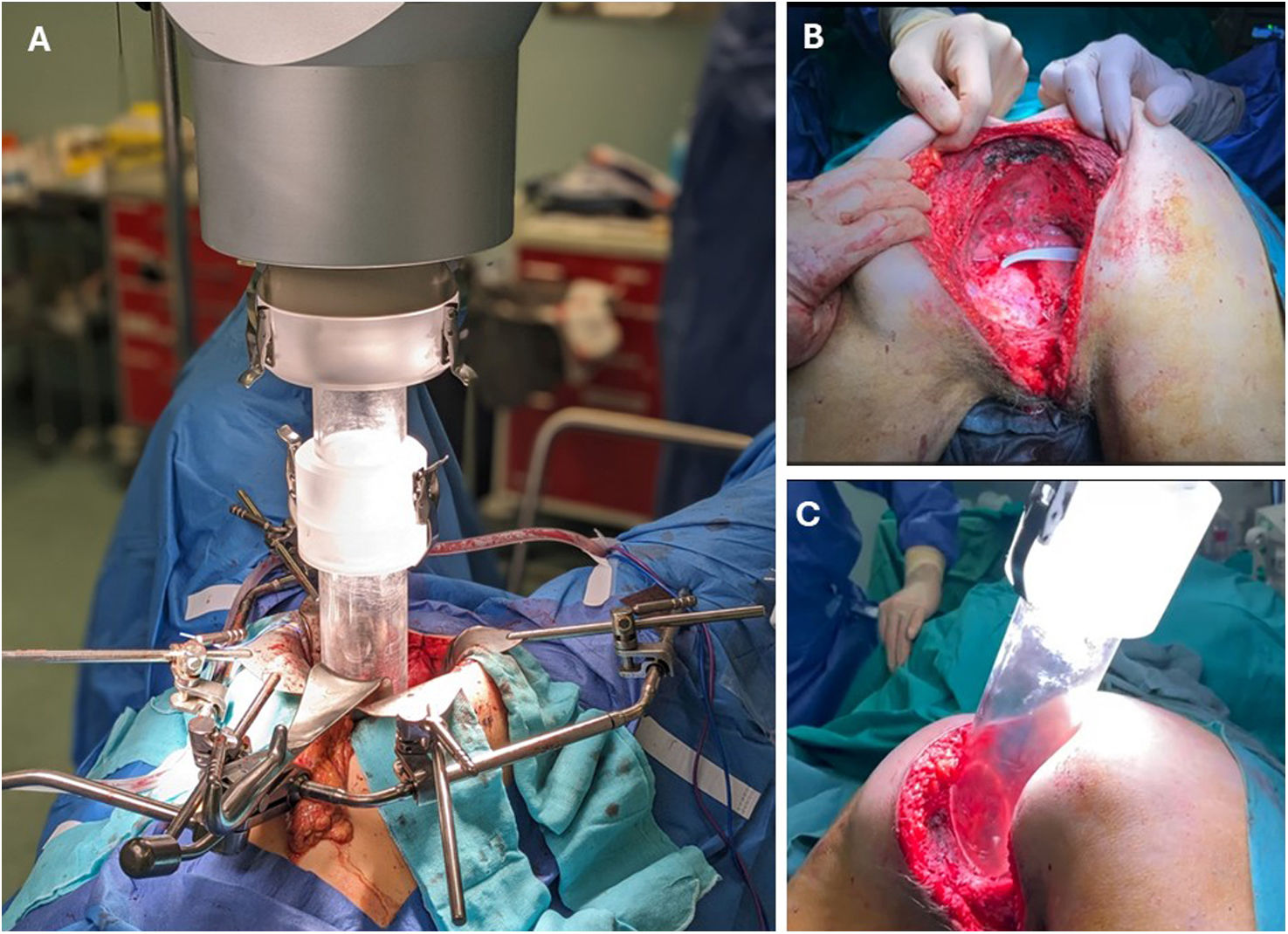
IORT, in its different modalities, allows for the administration of high doses (10–20 Gy) to the tumour bed after resection, minimising exposure to healthy tissue, unlike external beam radiation therapy (EBRT) (45–50 Gy). Furthermore, it has been shown to achieve excellent local control, improving disease-free survival and overall survival, without a significant increase in serious complications.77–80 IORT is especially useful in the context of pelvic exenteration (PE), although its routine use is not recommended, reserving it for areas with a high risk of local recurrence, especially in R1 resections or with compromised margins, either preoperatively or intraoperatively.63,77,79–83 Its application is more appropriate in patients who can assume a higher risk of complications in exchange for reducing recurrence.80 Its usefulness in macroscopic residual disease (R2) is limited.77 (Fig. 3).

Intraoperative radiotherapy (IORT) in patients undergoing pelvic exenteration. There are three types of IORT: intraoperative electron beam radiotherapy (IOERT) like imaging, high-dose-rate intraoperative brachytherapy (HDR-IORT), and low-kilovoltage IORT. IOERT has faster treatment times, simpler preparation, and greater tissue penetration. HDR-IORT, with more flexible applicators that better adapt to the pelvis, delivers more concentrated doses, but the treatment time is longer. Kilovoltage IORT has less penetration, requires less shielding, but its applicator is fixed and the treatment time is slower.80
(A) Case 1: IORT administration via laparotomy.
(B and C) Case 2: patient in the prone position, with IORT directed toward the lateral wall of the pelvis.
MIS, both laparoscopic and robotic84,85 has significantly advanced in pelvic procedures, showing similar mortality results compared to open approaches via laparotomy,86,87 which remains the most common approach. With increasing experience in MIS PE, a reduction in morbidity is expected.88 Studies consider it a safe and viable option in patients with favorable anatomy and tumours, offering lower blood loss and shorter hospital stays, albeit with longer surgical times.87,88
Genitourological reconstructionThere is no ideal urinary reconstruction, so it must be tailored to each patient. Urinary reconstruction options include proximal transection and reimplantation of the ureters, with or without a Boari flap, or ureterovesical reconstruction with an interposed ileal loop after partial cystectomy if the residual bladder volume is adequate to preserve bladder function. or a urinary diversion via an ileal (Bricker) conduit or colonic conduit in the case of radical cystectomy.18,30,89 More recent alternatives include the continent urostomy, which uses the transverse colon and ileum,39 and the increasing use of wet double-barrel colostomies in patients with short ureters or anatomical difficulties.90 Wet double-barrel colostomy simplifies management by unifying the diversions into a single stoma, reduces the risk of complications by avoiding intestinal anastomosis, separates faecal and urinary flows (compared to traditional wet colostomy), and promotes a faster recovery,91,92 As a last resort or temporary solution, cutaneous ureterostomy or nephrostomy may be used, although they carry considerable long-term morbidity due to the need for percutaneous drainage and frequent catheter changes.18 The use of intraoperative ureteral catheters facilitates their identification and protection.18 (Fig. 4).

Various techniques exist for the reconstruction of vaginal defects, ranging from the use of skin grafts or isolated segments of bowel to rectus abdominis myofascial flaps, deep inferior epigastric artery perforator (DIEP) flaps, bilateral flat gracilis flaps, or specialised gluteus maximus flaps.39,93
Bone Reconstruction/StabilisationThe functional benefits of bone reconstruction must be weighed against the risks of prolonged operative time, wound or prosthesis/allograft infections, and nonunion or bony nonunion. Prior radiation therapy limits prosthesis integration, so adequate soft tissue coverage is essential for any type of prosthesis. Biological reconstruction is preferable to prosthetic reconstruction in patients with high functional demands, as well as in those who cannot undergo frequent radiological and/or clinical follow-up.18 Although several fixation methods exist, there is no evidence to suggest the superiority of one over another.94
Vascular reconstructionVascular reconstruction is complex due to the presence of irradiated and contaminated tissue, which increases the risk of infection in synthetic grafts. The optimal method for vascular reconstruction has not yet been established. Autologous grafts, such as the great saphenous vein, are widely used due to their lower risk of thrombosis and infection, although they have limitations such as availability and differences in lumen size.68 For this reason, the use of vascular prostheses is also widespread. An alternative, although expensive, is the bovine biological graft. 81
To avoid ischaemia or compartment syndrome in the extremities, arterial reconstruction should be performed immediately.95 Recently, innovative approaches have been described that promote preventive grafting (crossed femoral-femoral95,96 or axilofemoral97 weeks before PE, reducing the risk of septic graft complications, total surgical time, and ischemia in the lower limbs.
Soft tissue reconstructionClosure options include primary repair (with or without the use of an omental pedicle), the use of biological or synthetic mesh to reconstruct the pelvic floor, autologous myocutaneous flaps, or the use of breast prostheses.81 However, evidence on the optimal technique remains limited.48
Reconstructive techniques vary depending on the affected region: for abdominal defects, rectus abdominis (VRAM)/ORAM flaps are used; for the gluteus, VY flaps or gluteal artery perforator flaps; for the thigh, anterolateral flaps with or without the vastus lateralis or bilateral gracilis; and for the gluteal/perineal fold, pudendal artery or perineal rotation flaps.l98 The VRAM/ORAM flap is one of the most common options due to its ease of harvest and robustness.48,81
Biological mesh, usually anchored at the level of the pubic symphysis and the sacral promontory, is a quick and safe option that prevents the small intestine from protruding into the pelvis and reduces the rate of hernias in the short term.99,100 (Fig. 5)

Perineal reconstructive techniques after pelvic exenteration. In both cases, Permacol® biological mesh is used, secured with interrupted, non-absorbable monofilament sutures in a crown-shaped pattern. Skin and subcutaneous tissue closure is performed by the Plastic Surgery team using a V-Y advancement flap (case A) and a double V-Y flap (case B).
An emerging technique is the use of 3D-printed, custom prostheses specifically tailored to each patient's defects.101
Palliative Pelvic Exenteration (PPE)PPE may be considered, exceptionally, without curative intent in patients with severe and intractable local symptoms—such as refractory pain, persistent bleeding, fistulisation into neighboring organs or the abdominal wall, and intestinal obstruction—when all conservative therapeutic alternatives have failed 32,102
In these selected cases, the patients should have a reasonable life expectancy and good functional status, who fully understand and accept the magnitude of the surgery and its sequelae, despite the lack of direct oncological benefit.
Although it can offer symptomatic relief in extreme scenarios, PPE remains a controversial procedure due to its high morbidity, its limited impact on survival, and the possible lack of recovery in quality of life, which tends to progressively deteriorate until death.32,33,39,81 Further studies are needed to more precisely define its indication and potential benefits.32
ResultsThe aetiological diversity, the multiple surgical technical options—including different types of pelvic exenteration (total, anterior, or posterior)—and the lack of standardisation in outcome measurement make comparative studies and meta-analyses difficult, resulting in highly variable figures in the literature.103 This technical variability significantly influences morbidity and survival, and interpreting results without considering these differences can be misleading. Coordinated international collaboration is needed to define key outcome sets, surgical classification criteria, and benchmarks that allow for more rigorous comparisons.103
Pelvic exenteration (PE) has a high overall morbidity, with rates ranging from 18% to 87%.7,10,31,104–106 These are particularly high in patients with local recurrence (LRRC: 82%) versus primary disease (LARC: 47%).104,105 The most common complications include infections, urinary or intestinal leaks, sepsis, and flap-related complications, with major complication rates ranging from 16.4% to 34.4%.107,112 Empty pelvis syndrome (EPS), responsible for up to 40% of postoperative complications, is associated with large resections and prior radiation therapy.48,99,109,115 The R0 resection rate varies widely by setting: up to 80% in LARC and 60% in LRRC.10–13,117,118
Despite its complexity, in-hospital mortality is low in specialised centres (1–5%),3,9,12,121 and 5-year survival rates reach up to 67% in some studies.9,106,122 Tumour recurrence, however, can affect up to 68% of patients, and may be local, distant, or combined.10 Quality of life after PE usually recovers progressively, especially in patients with R0 resection, and acceptable functional adaptation has been described in most cases,49,106,125,126 with variable impact on continence, urinary and sexual function, pain, and return to work.81
Table 3 presents a detailed summary of the results obtained after pelvic exenteration, along with key considerations that should be taken into account in the interpretation of these data.
Clinical results of pelvic exenteration.
| Category | Results and clinical commentas |
|---|---|
| Morbidity | Total: 18–87%7,10,31,104–106LARC: 47%, LRRC: 82%104,105Immediate postoperative: 40%9,107Risk factors: previous chemoradiotherapy, pelvic dead space, low tissue vascularisation, bacterial contamination92,108 |
| Hospìtal stay | Median: 9−29 days106 |
| Reinterventions | Long-term up to 17%106,109 |
| Common complications | Variable figures 3,92,106,110,111: sepsis, perineal wound infection, urinary complications (leaks, UTIs), gastrointestinal complications (anastomotic leaks), flap complications |
| Major complications (Clavien-Dindo III-IV) | 34.4% (n = 195)112; 16.4% (n = 604)107Intra-abdominal collections: up to 43.7%Woundinfection: 14%Sepsis: 3.7%–8.2%Iintestinal leak: 2.6–7.4%Urinary leak: 4.6%Postoperative bleeding: 4.4%–5.6%. |
| Pain | High prevalence113,114It is not usually measured or managed adequately42Better control of R0 margins vs. R1/R2 margins or after bone resection114 |
| Empty Pelvis Syndrome (EPS) | Up to 40% of postoperativecomplications48,115Causes: residual void after multivisceral resection, intestinal displacement into the pelvis, pelvic floor dysfunction, impaired healing due to chronic ischaemia99,109Aggravating factors: prior radiation, devascularisation of skin flaps, large tissue defects, bone exposure (high sacrectomy, pubic resection)19,30,99,108EPS-associated complications: pelvic collections, prolonged ileus, mechanical bowel obstruction, perineal dehiscence, perineal sinuses, enteroperineal fistulas99,115,116Prevention: reconstructive techniques, although they are also associated with morbidity48,98,99,109In cases of symptomatic obstruction or fistulisation, it is preferable to isolate the fistula or perform bypass surgery, avoiding more invasive resections109 |
| R0 resection rate | LARC: up to 80%; LRRC: up to 60%10–13,117,118Recent systematic revision106: 23.2%–98.4% (LARC), 14.9%–77.8% (LRRC)Urological: 84.6% (primary), 73.6% (recurrences)10Gynecological: 87% (endometrium), 76% (vagina), 73% (primary cervix), 79% (recurrent cervix), 64% (primary ovary), 52% (recurrent ovary)10 |
| Tumour recurrence | Global: 3.2%–68%. Local: 4.3%–68%. Distant: 11%–46%. Combined: 3.2–61%10Re-exenteration can be considered a curative option30,118–120 |
| Mortality | Inpatient: 3.11%121At 30 days: 0–9.1%106,121Specialised centres: 1%–5%3,9,12 |
| Survival | Variable figuresNo differentiation between open surgery and MIS 87Survival: 1 year: >80%; 3 years: 20%–75%; 5 years: 8%–67%9,106,122Disease-free survival: 5 years: 13%–89%; 10 years: 17.4%–50%9,106,122Critical factors: R0 resection, bone resection if necessary, specialised centres10,122,123; radiotherapy improves R0 rates but not survival105 |
| Quality of life (QoL) | Acceptable, with gradual recovery after the immediate postoperative period49,106,125R0 offers better long-term outcomes; most patients manage to adapt and maintain a stable functional status126Physical, functional, emotional, and social dimensions: urinary and sexual function, continence, pelvic pain, musculoskeletal function, and employability81 |
In recent years, the approach to PE has transcended morbidity and survival, incorporating quality of life and PROMS as key parameters for individualising treatment according to patient needs. These are standardised instruments that allow direct collection, through questionnaires, of patient perceptions of their physical, emotional, functional, and social health after surgery. In the case of PE, several studies have shown a significant impact on aspects such as urinary and sexual function, continence, pelvic pain, mobility, and return to work, especially in the first postoperative months. The outcome of these indicators may improve over time, depending on rehabilitation support and individual adaptation.23,24,109,124
Although annual volume is not the only indicator of quality, centres with more than 20 pelvic exenterations per year—considered high-volume—have shown better outcomes, supporting centralisation in specialised units and strengthening referral pathways to tertiary centres with established multidisciplinary teams.17
ConclusionsPelvic exenteration (PE) has become established as a curative therapeutic option for advanced or recurrent pelvic cancers, with R0 margins being the primary objective for improving survival. Advances in imaging, surgical techniques, and multidisciplinary management in specialised high-volume units have expanded the indications, improving oncological and functional outcomes with reduced morbidity. Furthermore, the integration of tools such as PROMS has reinforced the need for a comprehensive approach that considers not only tumour control but also patient quality of life.
CURRENT ADDRESS: Avinguda de Fernando Abril Martorell, 106, Quatre Carreres, 46026 València, Hospital Universitario y Politécnico La Fe, Torre G, planta 5, Coloproctology Unit. General and Digestive System Surgery Department.


