metricas
Cirugía Española (English Edition) Short-term results of unroofing and marsupialization compared to the Karydakis t...
Journal Information
Visits
282
Vol. 103. Issue 1.
Pages 25-33 (January 2025)
Original article
Full text access

Short-term results of unroofing and marsupialization compared to the Karydakis technique in the treatment of pilonidal sinus. A randomized prospective study

Resultados a corto plazo de la puesta a plano, curetaje y marsupialización frente a la técnica de Karydakis en el tratamiento del seno pilonidal. Estudio prospectivo aleatorizado
Visits
282
Pedro Antonio Parra Bañosa,
Corresponding author
pedroapb@yahoo.es

Corresponding author.
, Nuria Martínez Sanza, Francisco Miguel González Valverdea, Jorge Alejandro Benavides Bulejea, Miguel Ruiz Marína, Emilio Peña Rosa, Carmen Martínez Sanzb, Mari Fe Candel Arenasa
a Hospital General Universitario Reina Sofía, Avda. Intendente Jorge Palacios, 1, 30003, Murcia, Spain
b Centro de Atención Primaria San Andrés, Murcia C/ Escultor Sánchez Lozano, s/n, 30005, Murcia, Spain
This item has received
Article information
Abstract
Full Text
Bibliography
Download PDF
Statistics
Figures (4)
fig0005
fig0010
fig0015
fig0020
Show moreShow less
Tables (3)
Table 1. Characteristics of the patients and sinuses.
Tables
Table 2. Distribution of early postoperative complications (EPC).
Tables
Table 3. Values of the secondary objectives.
Tables
Show moreShow less
Abstract
Introduction

The treatment of pilonidal sinus (PS) is usually surgical, but no procedure is considered the gold standard. The Karydakis (K) technique is widely used, and unroofing and marsupialization (UM) is a simple surgery with good results.

Primary objective

To evaluate early postoperative complications (EPC) 30 days after UM surgery compared to the K technique.

Secondary objectives

To evaluate surgical time, postoperative pain, patient satisfaction, return to daily activity and early recurrence within 3 months.

Method

Prospective, single-center, randomized study in patients who underwent surgery for primary PS with no abscess between June 2016 and November 2017. They were randomized using a computer-generated block method. To analyze the main objective, a non-inferiority analysis was performed.
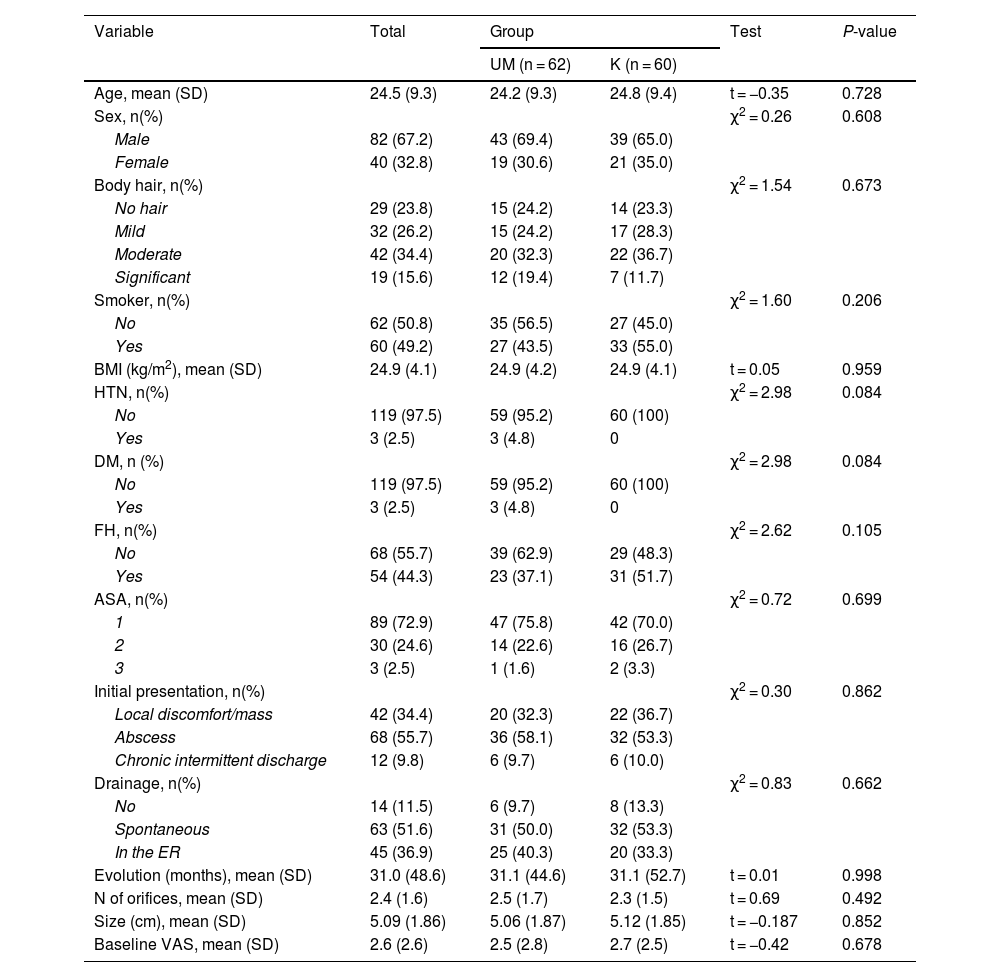
Results

122 patients with symptomatic primary PS were randomized: 60 in the K group and 62 in the UM group. Both groups were homogeneous.

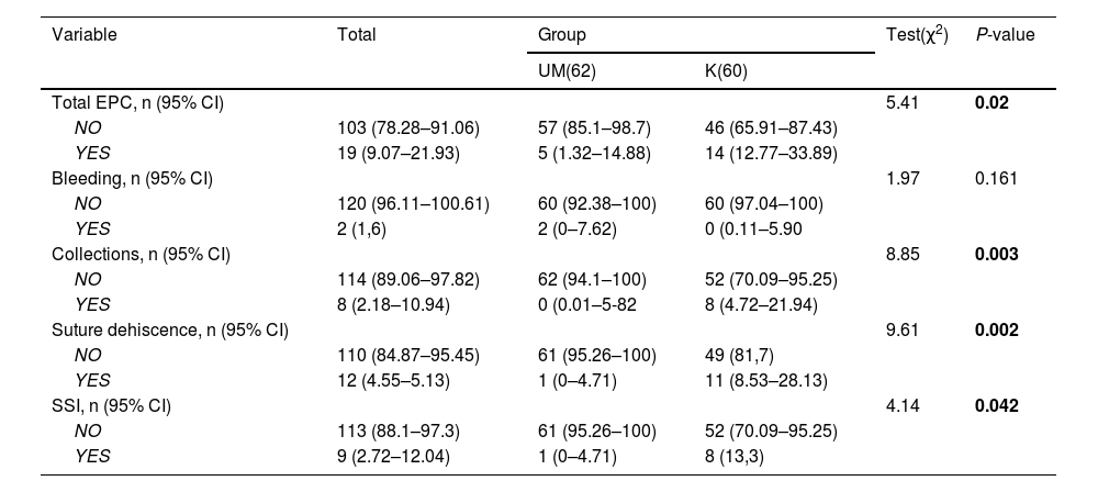
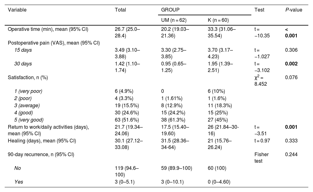
There were statistically significant differences between surgery and postoperative complications at 15 and 30 days in favor of UM. There were also differences in favor of UM in surgical time and return to daily activity. During the 90-day follow-up, there were 3 recurrences in the UM group and 0 in the K group.

Conclusions

UM is a simple, minimally invasive, easily reproducible technique that has a lower rate of early complications, with a shorter operative time and an earlier return to daily activity.

Keywords:
Pilonidal sinus
Sacrococcygeal region
Karydakis
Unroofing
Marsupialization
Surgical treatment
Postoperative complications
Resumen
Introducción

El tratamiento del sinus pilonidal (SP) es habitualmente quirúrgico, pero no existe un procedimiento considerado como “gold stándard”. La técnica de Karydakis (K) es ampliamente utilizada y la Puesta a Plano, Curetaje y Marsupialización de bordes (PPCYM) es una cirugía sencilla y con buenos resultados.

Objetivo primario

Evaluar las complicaciones postquirúrgicas precoces (CPP) a 30 días tras la cirugía de la PPCYM en comparación con la técnica K.

Objetivos secundarios

Evaluar el tiempo quirúrgico, dolor postoperatorio, satisfacción del paciente, reincorporación a las tareas habituales y recidiva precoz a los 3 meses.

Métodos

Estudio prospectivo, unicéntrico y aleatorizado en pacientes intervenidos de SP primario y sin absceso entre junio de 2016 y noviembre de 2017. Se aleatorizaron mediante un método de bloques balanceados generado por ordenador. Para analizar el objetivo principal se realizó un análisis de no inferioridad.

Resultados

122 pacientes aleatorizados con SP primario sintomático: 60 en el grupo K y 62 en el grupo PPCYM. Ambos grupos fueron homogéneos.

Hubo diferencias significativas en las complicaciones postoperatorias a los 15 y 30 días a favor de PPCYM. También hubo diferencias a favor de PPCYM en el tiempo quirúrgico y la reincorporación a las tareas habituales. Durante el seguimiento a 90 días hubo 3 recidivas en el grupo PPCYM y 0 en el grupo K.

Conclusiones

UM PPCYM es una técnica sencilla, mínimamente invasiva, fácilmente reproducible y que tiene una menor tasa de complicaciones precoces, con un menor tiempo quirúrgico y una reincorporación a las tareas habituales más precoz.

Palabras clave:
Seno pilonidal
Región sacrococcigea
Karydakis
Puesta a plano
Marsupialización
Tratamiento quirúrgico
Complicaciones postoperatorias
Graphical abstract
Full Text
Introduction

A pilonidal sinus (PS) is a pseudocyst containing hair follicles, located mainly in the sacrococcygeal region. Diagnosis is based on physical examination and clinical symptoms, which range from mild pain during sitting to an acute abscess with intense pain. Patients often report intermittent suppuration and pain alternating with asymptomatic periods.

Although there are publications that suggest inheritance is an important factor in the genesis of PS,1 the majority of the scientific community accepts that it is an acquired entity,2–4 in which hair penetration, favored by microtrauma, occurs in the subcutaneous cellular tissue, causing the formation of the pseudocyst as a result of a reaction to a foreign body. Multiple risk factors have been described that could predispose patients to the appearance of PS: age between 15 and 35 years,3 male sex,5 obesity,6 hirsutism,7 prolonged sitting,1 etc.

PS affects 26 out of every 100 000 inhabitants in the general population.5,8 However, there is a percentage of cases that remain asymptomatic, so it is difficult to establish the real prevalence of PS.9

Treatment of symptomatic chronic PS is usually surgical,10 and numerous techniques have been described that attempt to achieve the ideal operation that has low morbidity and recurrence, short hospital stay, good control of postoperative pain, and aesthetically acceptable results. The presentation, extension and severity are very heterogeneous, so there is no single surgical procedure that can be considered ideal or the gold standard.

The Karydakis (K) technique11,12 is a standardized procedure that is widely accepted and used by the scientific community.13–15 On the other hand, unroofing and marsupialization of margins is a technique described in the 1960s16 that has been gaining more acceptance in recent decades due to its simplicity and results.17,18

The primary objective of this study is to evaluate the early postoperative complications (EPC) within the first 30 days after unroofing and marsupialization surgery in the surgical treatment of PS compared to the Karydakis technique (K). As secondary objectives, we evaluated the surgical time, postoperative pain, patient satisfaction, return to work or daily activity, healing time and early recurrence within 3 months.

MethodsPatients and study design

A prospective, phase III, single-center, randomized, parallel-group, non-inferiority study was conducted in 122 patients undergoing primary PS surgery at the Reina Sofía Hospital in Murcia, Spain, from June 2016 to November 2017. The study was conducted in accordance with CONSORT guidelines for randomized studies18 (Addendum 1).

The study was approved by the hospital’s Research Ethics Committee, and all patients signed a consent form for inclusion in the study.

Inclusion and exclusion criteria

Our study included patients of legal age who had been diagnosed with symptomatic primary PS, with no abscess, no pathologies that contraindicated surgery or spinal anesthesia, and who had signed the consent form. Patients with recurrent PS, abscess or episode of inflammation in the previous 4 weeks, PS with bilateral secondary orifices or PS greater than 12 cm and immunosuppressed patients or those with previous radiotherapy on the sacrococcygeal region or who refused to be included were excluded from the study.

Surgical techniques

The surgeries were performed by the same 3 surgeons, under spinal anesthesia, and as part of a day surgery regimen. Patients received a prophylactic dose of IV antibiotic prior to the intervention (2 g of amoxicillin-clavulanic acid or 500 mg of metronidazole +240 mg of gentamicin in patients allergic to beta-lactams). In prone position and with the buttocks separated, patients in group K underwent sinus removal with a lateralized vertical elliptical excision. A thick flap was created by hollowing out the tissue under the medial side, which is advanced towards the midline to lateralize the entire suture line (Fig. 1).

Figure 1.

Diagram of the Karydakis technique: (A and B) lateralized incision; (C and D) preparation of the subcutaneous flap; (E) flap is advanced for suturing; (F) lateral closure at the midline. (Source: our own records).

In the patients of the UM group, the tracts were identified with a stylet, over which an incision was made through the skin, and the PS cavity and its tracts were opened longitudinally, allowing the entire lesion to be flattened out. The hair follicles were then removed, a curettage of the base was performed, and the cutaneous margins were then marsupialized, leaving the wound open to heal by secondary intention (Fig. 2).

Figure 2.

Diagram of the UM technique: (A) canalization of the tracts with a stylet; (B and C) unroofing and flattening; (D) curettage of the bed; (E) marsupialization of the edges, (F) final appearance. (Source: our own records).

Study variables

The study variables collected included sociodemographic data, medical-surgical history, presentation of the disease and evolution over time, treatments received, PS characteristics, and anthropometric data. Intraoperatively, surgical time was measured (from the start of the incision until the last stitch) and postoperatively, wound-related complications during the first month (surgical site infection, suture dehiscence, subcutaneous collections and postoperative bleeding), postoperative pain (using the Visual Analogue Scale), patient satisfaction (using a 5-point Likert scale, where 1 = very bad and 5 = very good), return to normal activities, healing time and recurrence (appearance of new holes accompanied by suppuration or pain) 3 months after surgery.

Sample size and randomization

The size of each treatment group was calculated based on the primary endpoint, which was EPC. The percentage of EPC is approximately 9% for the Karydakis technique according to studies by Karydakis19 and Bannura2 and 3% for UM in the study of Karakayali.20

To evaluate the primary objective of the study and declare the non-inferiority of UM compared to K, the lower limit of a two-sided 95% confidence interval for the difference in EPC rates between the experimental group and the reference group needed to be less than 5% (non-inferiority criterion δ).

For a power of 80% and a significance level of 5%, assuming that the proportion of EPC in the reference group (K) is 9% and in the experimental group (UM) 3%, that the proportion of experimental units in the reference group compared to the total is 50% and that the non-inferiority limit is 5%, with an expected dropout rate of 5%, it would be necessary to include 60 subjects in each group, requiring a sample size of 120 patients.

Patients were randomized using a computer-generated balanced block randomization method. The randomization codes were generated by an external computer system, remaining hidden and under the custody of a person outside the study, and only communicated to the surgeons by telephone just before starting the intervention.

Follow-up

After the intervention, the patients were reviewed by a surgeon not involved in the study after 15, 30 and 90 days.

Blinding

The patients, surgeons and outcome assessor could not be blinded to the technique that had been performed. However, the randomization sequence was concealed from the surgeons, and the data were analyzed by another collaborator who was unaware to which group each patient belonged.

Statistical analysis

For qualitative variables, the chi-squared or Fisher test was used. For quantitative variables, the Student’s t-test or Mann-Whitney U test was applied.

For the main objective of therapeutic safety, a non-inferiority analysis28 was performed, comparing the percentage of subjects in each treatment group with postoperative complications of the surgical wound during the first 30 days. The non-inferiority analysis for the primary endpoint was conducted using the Farrington-Manning Score, Miettinen-Nurminen Score, and Gart-Nam Score, establishing a significance level of 0.05 and a non-inferiority limit of 0.05.

The primary analysis of the study was based on an intention-to-treat (ITT) analysis of the population.

Data processing and the statistical analysis were performed using the SPSS 22.0 statistical program (IBM® SPSS® Statistics 22) for Windows.

Results

Out of the total of 122 patients, 60 were included in the K group and 62 in the UM group (Fig. 3). The characteristics of the population and PS are shown in Table 1. The groups were homogeneous.

Figure 3.

Flow diagram.

Table 1.

Characteristics of the patients and sinuses.

Variable  Total  GroupTest  P-value 
    UM (n = 62)  K (n = 60)     
Age, mean (SD)  24.5 (9.3)  24.2 (9.3)  24.8 (9.4)  t = −0.35  0.728 
Sex, n(%)        χ2 = 0.26  0.608 
Male  82 (67.2)  43 (69.4)  39 (65.0)     
Female  40 (32.8)  19 (30.6)  21 (35.0)     
Body hair, n(%)        χ2 = 1.54  0.673 
No hair  29 (23.8)  15 (24.2)  14 (23.3)     
Mild  32 (26.2)  15 (24.2)  17 (28.3)     
Moderate  42 (34.4)  20 (32.3)  22 (36.7)     
Significant  19 (15.6)  12 (19.4)  7 (11.7)     
Smoker, n(%)        χ2 = 1.60  0.206 
No  62 (50.8)  35 (56.5)  27 (45.0)     
Yes  60 (49.2)  27 (43.5)  33 (55.0)     
BMI (kg/m2), mean (SD)  24.9 (4.1)  24.9 (4.2)  24.9 (4.1)  t = 0.05  0.959 
HTN, n(%)        χ2 = 2.98  0.084 
No  119 (97.5)  59 (95.2)  60 (100)     
Yes  3 (2.5)  3 (4.8)     
DM, n (%)        χ2 = 2.98  0.084 
No  119 (97.5)  59 (95.2)  60 (100)     
Yes  3 (2.5)  3 (4.8)     
FH, n(%)        χ2 = 2.62  0.105 
No  68 (55.7)  39 (62.9)  29 (48.3)     
Yes  54 (44.3)  23 (37.1)  31 (51.7)     
ASA, n(%)        χ2 = 0.72  0.699 
1  89 (72.9)  47 (75.8)  42 (70.0)     
2  30 (24.6)  14 (22.6)  16 (26.7)     
3  3 (2.5)  1 (1.6)  2 (3.3)     
Initial presentation, n(%)        χ2 = 0.30  0.862 
Local discomfort/mass  42 (34.4)  20 (32.3)  22 (36.7)     
Abscess  68 (55.7)  36 (58.1)  32 (53.3)     
Chronic intermittent discharge  12 (9.8)  6 (9.7)  6 (10.0)     
Drainage, n(%)        χ2 = 0.83  0.662 
No  14 (11.5)  6 (9.7)  8 (13.3)     
Spontaneous  63 (51.6)  31 (50.0)  32 (53.3)     
In the ER  45 (36.9)  25 (40.3)  20 (33.3)     
Evolution (months), mean (SD)  31.0 (48.6)  31.1 (44.6)  31.1 (52.7)  t = 0.01  0.998 
N of orifices, mean (SD)  2.4 (1.6)  2.5 (1.7)  2.3 (1.5)  t = 0.69  0.492 
Size (cm), mean (SD)  5.09 (1.86)  5.06 (1.87)  5.12 (1.85)  t = −0.187  0.852 
Baseline VAS, mean (SD)  2.6 (2.6)  2.5 (2.8)  2.7 (2.5)  t = −0.42  0.678 

UM: unroofing and marsupialization; SD: standard deviation; HTN: hypertension; DM: diabetes mellitus; FH: family history; BMI: body mass index; ASA: American Society of Anesthesiologists. VAS: visual analogue scale.

Significant differences were found between surgery and postoperative complications within 15 days (33.3%, 95% CI: 21.4–45.2 in. K; and 8.06%, 95% CI: 0.12–14.6 in UM; P < .001) but at 30 days (23.3%, 95% CI:12.6–34.0 in K; and 8.06% 95% CI: 0.12–14.6 in UM; P = .02).

Complications in the first 30 postoperative days are shown in Table 2.

Table 2.

Distribution of early postoperative complications (EPC).

Variable  Total  GroupTest(χ2P-value 
    UM(62)  K(60)     
Total EPC, n (95% CI)        5.41  0.02 
NO  103 (78.28–91.06)  57 (85.1–98.7)  46 (65.91–87.43)     
YES  19 (9.07–21.93)  5 (1.32–14.88)  14 (12.77–33.89)     
Bleeding, n (95% CI)        1.97  0.161 
NO  120 (96.11–100.61)  60 (92.38–100)  60 (97.04–100)     
YES  2 (1,6)  2 (0–7.62)  0 (0.11–5.90     
Collections, n (95% CI)        8.85  0.003 
NO  114 (89.06–97.82)  62 (94.1–100)  52 (70.09–95.25)     
YES  8 (2.18–10.94)  0 (0.01–5-82  8 (4.72–21.94)     
Suture dehiscence, n (95% CI)        9.61  0.002 
NO  110 (84.87–95.45)  61 (95.26–100)  49 (81,7)     
YES  12 (4.55–5.13)  1 (0–4.71)  11 (8.53–28.13)     
SSI, n (95% CI)        4.14  0.042 
NO  113 (88.1–97.3)  61 (95.26–100)  52 (70.09–95.25)     
YES  9 (2.72–12.04)  1 (0–4.71)  8 (13,3)     

SSI: surgical site infection.

All tests performed in the non-inferiority analysis had a P-value <.05, and the value 0 was not included in any of the confidence interval limits of all tests. The results of the different variables of the secondary objectives are shown in Table 3.

Table 3.

Values of the secondary objectives.

Variable  Total  GROUPTest  P-value 
    UM (n = 62)  K (n = 60)     
Operative time (min), mean (95% CI)  26.7 (25.0–28.4)  20.2 (19.03–21.36)  33.3 (31.06–35.54)  t = −10.35  < 0.001 
Postoperative pain (VAS), mean (95% CI)           
15 days  3.49 (3.10–3.88)  3.30 (2.75–3.85)  3.70 (3.17–4.23)  t = −1.027  0.306 
30 days  1.42 (1.10–1.74)  0.95 (0.65–1.25)  1.95 (1.39–2.51)  t = −3.102  0.002 
Satisfaction, n (%)        χ2 = 8.452  0.076 
1 (very poor)  6 (4.9%)  6 (10%)     
2 (poor)  4 (3.3%)  1 (1.61%)  1 (1.6%)     
3 (average)  19 (15.5%)  8 (12.9%)  11 (18.3%)     
4 (good)  30 (24.6%)  15 (24.2%)  15 (25%)     
5 (very good)  63 (51.6%)  38 (61.3%)  27 (45%)     
Return to work/daily activities (days), mean (95% CI)  21.7 (19.34–24.06)  17.5 (15.40–19.60)  26 (21.84–30-16)  t = −3.51  0.001 
Healing (days), mean (95% CI)  30.1 (27.12–33.08)  31.5 (28.36–34-64)  21 (15.76–26.24)  t = 0.97  0.333 
90-day recurrence, n (95% CI)        Fisher test  0.244 
No  119 (94.6–100)  59 (89.9–100)  60 (100)     
Yes  3 (0–5.1)  3 (0–10.1)  0 (0–4.60)     

VAS: visual analogue scale. CI: confidence interval.

Operative time was significantly shorter in UM (20.2 min, 95% CI: 19.3–21.16 vs 33.3 min, 95% CI: 31.06–35.54; P < .001), while postoperative pain did not show differences between the 2 groups at 15 days but did at 30 days. Patient satisfaction with surgery was very good or good for both techniques, and there were no significant differences between the 2 groups. The mean time until returning to work or daily activities was 17.5 days (95% CI: 15.40–19.60) for UM and 26 days (95% CI: 21.84–30.16) for K, the difference being statistically significant (P = .01). Healing was later in UM, but no significant difference was found with K. Three recurrences had been observed within 90 days in the UM group and 0 in the K group.

Discussion

Randomization (controlled for selection bias) has allowed us to study 2 homogeneous groups. Furthermore, despite being limited to a population from a specific health area, the study population is diverse, and we believe that its characteristics can be extrapolated to the general population. UM is a simple technique that could be applied as a surgical option in the vast majority of chronic, symptomatic PS. On the other hand, the study lacks a long follow-up, and, although there have been no losses, it was not possible to evaluate long-term parameters like late complications or recurrences.

PS continues to be a prevalent pathology today with considerable morbidity. As PS is considered an acquired entity, surgical techniques for its treatment tend to be less aggressive and invasive.20,21 Some authors claim that radical removal techniques are an “overtreatment” of pilonidal disease22 and suggest techniques such as unroofing, curettage and marsupialization as appropriate therapy for this pathology.

Our overall complication rate with the K technique was 23.3%, which is lower than the rate reported by Alvandipour23 in 2019 (37 K patients) of 40.85% and higher than the rate reported by Caliskan24 in 2020 (53 K patients) with an early complication rate of 16.9%, but that does not take into account the 5.6% of wound dehiscences included among the late complications. In 2009, the Ersoy study13 (50 K patients) reported an infection rate of 26%, much higher than that of our study at 13.9%. In a 2016 RCT, Keshvari25 (161 K patients) reported an overall complication rate of 18.7%, which was somewhat higher but close to our results.

In the UM group, complications occurred in 8.8%, a higher percentage than that reported in most studies. In a 2005 RCT, Gencosmanoglu22 (73 UM patients) found a complication rate of 2.7%, similar to that found by Karakayali20 in a 2009 RCT (70 patients with UM) of 2.9%. In 2018, Mahran26 reported a complication rate of 6.6% for the UM group (although it only included 15 patients), while in 2013 Goyal27 reported an infection rate of 13% for a group of 15 patients. In our study, one patient in the UM group met the criteria for SSI28 (purulent exudate with + culture and perilesional cellulitis), and another presented dehiscence of the entire suture, thus losing the effect of marsupialization. However, there is a statistically significant difference in favor of the UM patient group.

In our search, we have found no studies of non-inferiority of the UM technique regarding EPC. When comparing this technique with K, the results of all the tests have a P-value <.05, so we can conclude that the UM technique is not inferior to the K technique in terms of EPC with a confidence level of 95%.

The operative time is also significantly shorter in UM at 20.2 min in our study, which is similar to the time reported by Mahran26 (22.6 min) and shorter than that reported by Yetisir29 in 2005 (31.5 min). Both techniques are fast, and a difference of 13 minutes does not seem very clinically relevant.

Postoperative pain the first 15 days after surgery did not present differences in the 2 groups, but at 30 days it did in favor of the UM group, although the clinical importance is small. Patients in the UM group were more satisfied with the surgery than those in the K group, although the differences were not significant. These results are similar to those reported by Popesku30,32 in 2020.

In our series, the return to work of patients with K was 26 ± 16 days, which is longer than the 11.59 ± 3.4 days reported by Alvandipour23 and the 14.5 ± 0.67 days of Keshvari,25 although it is closer to the reports by Abellatif31 and Tokac32 in 2015, which were 20 ± 6.1 days and 23.29 ± 6.42 days, respectively. In patients with UM, the return to daily activity was 17 ± 8 days after surgery, which was notably higher than the results of Gencosmanoglu22 at 3 (2–8) days or Mahran26 at 3.98 days, yet more similar to those reported by Karakayali20 (11.2 ± 5.8 days) and even lower than the results obtained by Abdeer33 (25.2 ± 5 days). Despite being an open wound that requires dressings, this earlier return to work could be influenced by less pain in patients with UM and the average size of PS in our series (5.06 ± 1.87 cm), which was not excessively large.

In our series, 3 (4.8%) recurrences occurred within 3 months of follow-up in the UM group, and none were registered in the K group. This figure is higher than in the Gencosmanoglu study22 (1.4% at 47 months) or the Mahran study26 (0 at 6 months) and lower than in the Abdeer study33 (7% over a 12-month follow-up). We think that these early recurrences are due to incomplete surgery in which a tract was left unroofed. However, this datum should be measured with a longer follow-up (more than 12 months) to be more reliable.

Conclusions

Unroofing and marsupialization of the margins is a simple, minimally invasive, easily reproducible technique that allows the patient to return to work shortly after surgery. It also has a lower rate of complications compared to the Karydakis technique, requiring less surgical time and causing less postoperative pain, with a similar degree of patient satisfaction. Finally, the population that is most affected is between 15 and 35 years old, hence the growing concern to find a surgery that is minimally invasive, can be performed on an outpatient basis and allows for faster return to work and daily activities, characteristics that can be attributed to UM. Nevertheless, studies with a larger patient cohort and longer follow-up would be needed to confirm these data and to study other important variables, such as late complications and recurrences.

CRediT authorship contribution statement

Study concept and design: Jorge Alejandro Benavides Buleje, Francisco Miguel. González Valverde and Emilio Peña Ros.

Data collection: Nuria Martínez Sanz.

Data analysis and interpretation: All authors.

Composition and review of the manuscript: Pedro Antonio Parra Baños.

Approval of the final version of the manuscript: All authors.

Agreement to be responsible for all aspects of the study: All authors.

Ethics approval and consent for participation

The study protocol was established in accordance with the ethical guidelines of the Declaration of Helsinki and was approved by the Research Ethics Committee of the Hospital General Universitario Reina Sofía in Murcia, Spain. Informed, written consent was obtained from all participants.

Funding

The authors declare that no funding was received for the research, authorship and/or publication of this article.

Acknowledgements

The authors would like to thank all the participating patients and researchers involved in this study.

References
[1]
T. Yildiz, B. Elmas, A. Ycak, H.T. Turgut, Z. Ilce.
Risk factors for pilonidal sinus disease in teenagers.
Indian J Pediatric, 84 (2017), pp. 134-138
[2]
G. Bannura.
Enfermedad pilonidal sacro-coccígea: factores de riesgo y tratamiento quirúrgico.
Rev Chil Cir, 63 (2011), pp. 527-533
[3]
X. Barth, E. Tissot, O. Monneuse.
Traitement chirurgical de la maladie pilonidale.
Elsevier Masson SAS. Techniques chirurgicales - Appareil digestif, EMC, (2010), pp. 1-7
[4]
B. Kanat, S. Sözen.
Disease that should remember. Sacrococcygeal pilonidal sinus disease and short history.
World J Clin Cases, 3 (2015), pp. 876-879
[5]
TL Hull, Wu James.
Pilonidial disease.
Surg Clin North Am, 82 (2002), pp. 1169-1185
[6]
S. Bolandparvaz, P.M. Dizaj, R. Salahi, S. Paydar, M. Bananzadeh, H.R. Abbasi, et al.
Evaluation of the risk factors of pilonidal sinus: a single center experience.
Turk J Gastroenterol, 23 (2012), pp. 535-537
[7]
A. Harlak, O. Mentes, S. Kilic, K. Coskun, K. Duman, F. Yilmaz.
Sacrococcygeal pilonidal disease: analysis of previously proposed risk factors.
Clinics, 5 (2010), pp. 125-131
[8]
I. Mc Callum, M. King Peter, J. Bruce.
Healing by primary closure versus open healing after surgery for pilonidal sinus: systematic review and meta-analysis.
[9]
E. Aysan, M. Ilhan, H. Bektas, E.A. Kaya, B. Sam, N. Buyukpinarbasili, et al.
Prevalence of sacrococcygeal pilonidal sinus as a silent disease.
Surg Today, 43 (2013), pp. 1286-1289
[10]
F. Blanco, N. Silva.
Sinus pilonidal.
Manual de la AEC de proctología para atención primaria, 1st ed., pp. 66-75
[11]
G. Karydakis.
New approach to the problem of pilonidal sinus.
[12]
PR Kitchen.
Pilonidal sinus: experience with the Karydakis flap.
Br J Surg, 83 (1996), pp. 1452-1455
[13]
E. Ersoy, A.O. Devay, R. Aktimur, B. Doganay, M. Özdogan, R.H. Gündogdu.
Comparison of the short-term results after Limberg and Karydakis procedures for pilonidal disease: randomized prospective analysis of 100 patients.
Colorectal Dis, 11 (2009), pp. 705-710
[14]
M.F. Can, M.M. Sevinc, O. Hancerliogullari, M. Yilmaz, G. Yagci.
Multicenter prospective randomized trial comparing modified Limberg flap transposition and Karydakis flap reconstruction in patients with sacrococcygeal pilonidal disease.
Am J Surg, 200 (2010), pp. 318-327
[15]
SS Bessa.
Comparison of Short-term results between the modified Karydakis flap and the modified Limberg flap in the management of pilonidal sinus disease: a randomized controlled study.
Dis Colon Rectum, 56 (2013), pp. 491-498
[16]
DJ Abramson.
A simple marsupialization technic for treatment of pilonidal sinus: long-term follow up.
[17]
P.J. Lee, S. Raniga, D.K. Biyani, A.J.M. Watson, I.G. Garagher, F.A. Frizelle.
Sacrococcygeal pilonidal disease.
Colorectal Dis, 10 (2008), pp. 639-652
[18]
K.F. Schulz, D.G. Altman, D Moher, the CONSORT Group.
CONSORT 2010 Statement: updated guidelines for reporting parallel group randomised trials. Schulz et al..
[19]
G. Karydakis.
Easy and successful treatment of pilonidal sinus after explanation of its causative process.
Aust N Z J Surg, 62 (1992), pp. 385-389
[20]
F. Karakayali, E. Karagulle, Z. Karabulut, E. Oksuz, G. Moray, M. Haberal.
Unroofing and Marsupialization vs. Rhomboid Excision and Limberg flap in pilonidal disease: a prospective, randomized, clinical trial.
Dis Colon Rectum, 52 (2009), pp. 496-502
[21]
J. Bascom.
Repeat pilonidal operations.
Am J Surg, 154 (1987), pp. 118-122
[22]
R. Gencosmanoglu, R. Inceoglu.
Modified lay-open (incision, curettage, partial lateral wall excision and marsupialization) versus total excision with primary closure in the treatment of chronic sacrococcygeal pilonidal sinus. A prospective, randomized clinical trial with a complete two-year follow-up.
Int J Colorectal Dis, 20 (2005), pp. 415-422
[23]
M. Alvandipour, M.S. Zamani, M. Ghorbani, J.Y. Charati, M.Y. Karami.
Comparison of Limberg Flap and Karydakis flap surgery for the treatment of patients with pilonidal sinus disease: a single-blinded parallel randomized study.
Ann Coloproctol, 35 (2019), pp. 313-318
[24]
M. Caliskan, K. Kosmaz, I.E. Subasi, A. Acar, I. Evren, G. Bas, et al.
Comparison of common surgical procedures in non-complicated pilonidal sinus disease, a 7-year follow-up trial.
World J Surg, 44 (2020), pp. 1091-1098
[25]
A. Keshvari, M.R. Keramati, M.S. Fazeli, A. Kazemeini, M.K. Nouritaromlou.
Risk factors for complications and recurrence after the Karydakis flap.
J Surg Res, 204 (2016), pp. 55-60
[26]
M.M. Mahran, M.S. Shehata, A.M.M. Abd-Elwahab.
A comparative study between sinotomy with or without marsupialization versus sinotomy and primary closure with or without drain in the management of pilonidal sinus.
Egypt J Hosp Med, 73 (2018), pp. 7133-7140
[27]
S.K. Goyal, S. Garg, G. Singhal.
Assessment of various surgical methods in the management of Pilonidal disease.
J Adv Med Dent Sci, 1 (2013), pp. 167-170
[28]
E Nve Obiang, JM Badía Pérez.
Infección del sitio quirúrgico: definición, clasificación y factores de riesgo.
Infecciones Quirúrgicas. Guías Clínicas de la Asociación Española de Cirujanos, pp. 99-120
[29]
F. Yetişir, O. Kaya, I. Baran.
Comparación del método de marsupialización y colgajo de Limberg en el tratamiento del seno pilonidal.
Rev Turca Cir, 21 (2005), pp. 184-190
[30]
S.G. Popeskou, B. Pravini, S. Panteleimonitis, A.F. Di Tor Vajana, A. Vanoni, M. Schmalzbauer, et al.
Conservative Sinusectomy vs. excision and primary off-midline closure for pilonidal disease: a randomized controlled trial.
Int J Colorec Dis, 35 (2020), pp. 1193-1199
[31]
M.E. Abellatif, N. Noaman, A. Negm, A. Abass, A. El-Hadidy, G. El-Morsy, et al.
A prospective randomised trial comparing rhomboid excision and limber flap closure versus karydakis flap reconstruction for treatment of sacrococcygeal pilonidal disease.
Benha MJ, 28 (2011), pp. 9-23
[32]
M. Tokac, E.G. Dumlu, M.S. Aydin, A. Yalcın, M. Kilic.
Comparison of modified Limberg flap and Karydakis flap operations in pilonidal sinus surgery: prospective randomized study.
Int Surg, 100 (2015), pp. 870-877
[33]
A.F. Abdeer, A.M. Elhefny, W.B. Gerges, J.S.M. Sawires.
Modified Sinotomy with marsuplization versus excision with lay open in treatment of pilonidal sinus disease.
N Y Sci J, 12 (2019), pp. 35-40
Download PDF