
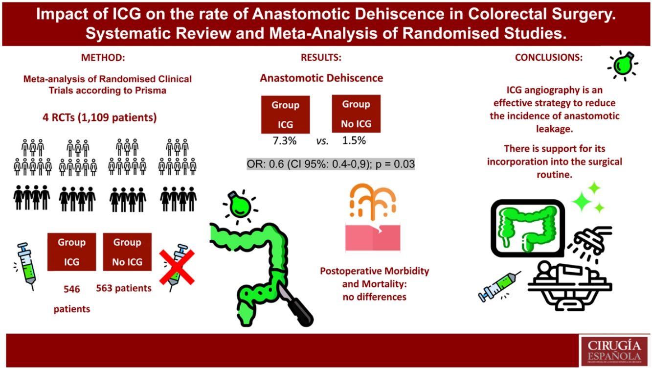
Anastomotic dehiscence, a feared complication in colorectal surgery, motivates the search for effective strategies to mitigate its risk. This meta-analysis encompasses all published randomized trials investigating and comparing the impact of indocyanine green (ICG) angiography on this complication. With four studies and 1,109 patients, the intraoperative ICG angiography group demonstrated a significant reduction in the overall rate of anastomotic dehiscence compared to the non-angiography group (7.3% vs. 11.5%; OR: 0.6; 95% CI: 0.4−0.9; p = 0.03). These differences were maintained in rectal surgery subgroup, with no prolongation of surgical time or increase in morbidity and mortality. There were no differences in the left colon surgery group. The evidence provided by this meta-analysis would support the effectiveness of ICG angiography in reducing the incidence of anastomotic leakage in colorectal surgery, advocating for its integration into routine surgical practice.
La dehiscencia anastomótica, temida en cirugía colorrectal, motiva la búsqueda de estrategias eficaces para mitigar su riesgo. El presente metaanálisis incluye todos los ensayos aleatorizados publicados que investigan y comparan el impacto de la angiografía con verde de indocianina (ICG) en esta complicación. Con cuatro estudios y 1.109 pacientes, el grupo con angiografía intraoperatoria ICG mostró una significativa reducción en la tasa de dehiscencia anastomótica en comparación con el grupo sin angiografía (7,3% vs 11,5%; OR:0,6; IC95%:0,4−0,9; p = 0,03). Estas diferencias sólo se mantuvieron en el subgrupo de cirugía rectal, sin prolongación del tiempo quirúrgico, ni aumento en morbimortalidad. No hubo diferencias en el grupo de cirugía de colon izquierdo. La evidencia proporcionada por este metaanálisis respaldaría la efectividad de la angiografía ICG para reducir la incidencia de fuga anastomótica en cirugía colorrectal, abogando por su integración en la práctica quirúrgica rutinaria.
Anastomotic dehiscence represents one of the most worrisome complications in colorectal surgery, with notable associated morbidity and mortality.1 Although the aetiology of anastomotic leak is multifactorial, it is postulated that infusion insufficiency plays a substantial role in its pathogenesis.2 The importance of ensuring adequate perfusion is critical, as insufficient blood flow is the main cause. This fact has motivated surgeons to run checks, both before and after the procedure. Traditionally, surgeons have relied on several clinical signs, such as intestinal serous colour, palpable pulsation, peristaltic motion, and active marginal arterial bleeding to assess intestinal perfusion. However, this subjective assessment of perfusion quality may vary depending on the individual interpretation of the surgeon and manifests a certain degree of inconsistency. Therefore, for a more objective and reproducible assessment of colonic viability, intraoperative angiography with indocyanine green is proposed as the ideal test.3
ICG is a fluorescent dye metabolised exclusively by the liver, excreted through bile, and its half-life (usually between 3 and 4 min) is linked to liver function. Developed by Kodak in the 1950s, it received FDA approval for use in humans in 1959.4 Since then, it has been used in various medical applications and in various specialties.5
The application of ICG in colorectal surgery has proven to be a promising tool. Notable applications in this area include intraoperative visualisation of the ureter, identification of the sentinel node, observation of lymphatic drainage, and assessment of perfusión.6 The last of these makes it possible to intraoperatively assess arterial blood flow in the anastomotic ends, making changes possible in the extension of the resection. However, the results of recent randomised clinical trials that have evaluated ICG’s impact on anastomotic dehiscence have been contradictory, and previously published meta-analyses include observational data sets that increase the risk of bias and affect their validity.7–14
The main objective of this meta-analysis was to analyse the impact of the application of indocyanine green angiography on the reduction of the rate of anastomotic dehiscence in colorectal surgery, as well as its associated complications.
MethodsSearch strategy and study selection criteriaA systematic review and meta-analysis were undertaken following the PRISMA methodology (Preferred Reporting Items for Systematic reviews and Meta-Analysis)15 and details of the protocol used were extracted from the latest edition of the Cochrane Handbook.16 To this end, an exhaustive literature review was undertaken through a search of various electronic databases, which included MEDLINE (PubMed), the Cochrane Library database, OVID and ClinicalTrials.gov, with a time limit set until June 2023. No language restrictions were applied to any of these searches. PubMed's "related articles" feature was used to broaden the scope of the search process. In addition, we explored other potentially relevant studies in the reference lists of relevant literature reviews and meta-analyses.
Search terms (Medical Subject Headings-MeSH) covered the following keywords: "indocyanine", "indocyanine green", "ICG", "colorectal" and "surgery". The search was run independently by two authors (ZB and IB). Accessory Table 1 summarises the search strategy to enable adequate reproduction of this study.The study was registered with PROSPERO under the number CRD42023416857.
Characteristics of the studies included in the meta-analysis, in alphabetical order.
| Author & Year | Country of Origin | No. patients | Agea | Sex (M/F) | BMIa | DM (%) | ASA Rating | Malignant Neopl. (%) | Surgery Durationa (min) | Hosp. staya (days) | Ileus postop No. | Mortality No. | Anastomotic dehiscence | ||||||||||
|---|---|---|---|---|---|---|---|---|---|---|---|---|---|---|---|---|---|---|---|---|---|---|---|
| Total | ICG | N | I (%) | II (%) | III (%) | RHC | LHC | ARR | Grade A | Grade B | Grade C | TOTAL | |||||||||||
| Alekseev 202019 | Russia | 377 | ICG | 187 | 63 | 92/95 | N.E. | 15 (8) | 18 (9,6) | 145 (77,6 | 24 (12,8) | 183 (97,9) | 176 ± 53,3 | 8 ± 4,6 | 7 | 0 | 1/76 | 16/111 | 7 | 6 | 4 | 17/187 | |
| No ICG | 190 | 63 | 92/98 | N.E. | 17 (8,9) | 22 (11,6) | 143 (75,3) | 25 (13,1) | 184 (96,8) | 185 ± 47,5 | 8 ± 4,6 | 5 | 0 | – | 4/86 | 27/104 | 21 | 7 | 3 | 31/190 | |||
| From Nardi 201920 | Italy | 240 | ICG | 118 | 66,1 | 60/58 | 25,2 | 17 (14,4) | 10 (8,5) | 82 (69,5) | 26 (22) | 86 (72,9) | 192,5 ± 54,5 | 6 ± 8 | 4 | 0 | – | 2/62 | 4/56 | 0 | 2 | 4 | 06/118 |
| No ICG | 122 | 65,1 | 66/56 | 25,6 | 19 (15,6) | 7 (5,7) | 92 (75,4) | 23 (18,9) | 85 (69,7) | 187,5 ± 49,7 | 7 ± 3,3 | 1 | 1 | – | 4/69 | 7/53 | 1 | 3 | 7 | 11/122 | |||
| Jafari 202121 | USA | 347 | ICG | 178 | 57,2 ±11,4 | 109/69 | 27,8 ± 5,6 | 22 (12,4) | – | – | – | 178 (100) | – | – | 6 | 0 | – | – | 16/178 | 4 | 12 | 0 | 16/178 |
| No ICG | 169 | 57 ±11,4 | 99/70 | 28,2 ± 5,9 | 23 (13,6) | – | – | – | 169 (100) | – | – | 13 | 1 | – | – | 16/169 | 2 | 14 | 0 | 16/169 | |||
| Ren 201822 | China | 145 | ICG | 63 | 61 | 36/27 | – | – | – | – | – | 63 (100) | 158,3 ± 43,6 | – | – | 0 | 0/5 | 0/20 | 1/23 | – | – | – | 1/63 |
| No ICG | 82 | 62 | 40/42 | – | – | – | – | – | 82 (100) | 147,6 ± 52,6 | – | – | 1 | 1/9 | 2/16 | 4/32 | – | – | – | 7/82 | |||
ICG, Indocyanine Green; M, Male; F, Female; BMI, Body Mass Index; DM, Diabetes Mellitus; RH, Right Hemicolectomy; LH, Left/Sigma Hemicolectomy; ARR, Anterior Rectal Resection.
In this study randomised clinical trials that met the following criteria were included: (1) patients undergoing colorectal surgery (right, left, sigmoidectomy, and anterior rectal resection) with anastomosis, (2) comparative analysis of the rate of postoperative anastomotic dehiscence, (3) intraoperative ICG angiography in one group (ICG Group), (4) absence of ICG angiography in the other group (Control group) and (5) clear presentation of the variables assessed in this review.
The PICO strategy was as follows: P (Patient): patients undergoing colorectal surgery with anastomosis; I (Intervention): intraoperative ICG angiography; C (Comparison): absence of intraoperative ICG angiography; O (Outcomes): rate of anastomotic dehiscence.
Exclusion criteriaWe excluded articles that did not provide results that were of interest, or where calculating the outcomes from the original data was impossible. We also excluded those that were irrelevant, editorials, letters to the editor, reviews, single-arm cohort studies, case reports, conference abstracts, editorials, and expert opinions. In the case of patient series included in multiple studies, only publications with the most complete study information were considered.
Variables analysedThe primary endpoint of the study was the rate of anastomotic dehiscence within 30 days of colorectal surgery: anastomotic dehiscence, such as the presence of a failure in the integrity of the intestinal wall in the colorectal or coloanal anastomosis, resulting in communication between the intraluminal and extraluminal compartments, as defined by Rahbari.17 It is classified into three groups according to its clinical management: Group A, "radiological leak", when the leak is detected by radiological examinations, it is not associated with clinical symptoms or alteration of analytical parameters; and does not require active treatment; Group B, when it involves active therapeutic intervention without reoperation (antibiotic therapy and/or placement of percutaneous drainage); and Group C, if reoperation is required, most patients undergo anastomosis resection and terminal colostomy, including those previously treated with a diverting stoma. However, some may be treated with bypass ileostomy if they did not have this previously or with anastomosis reinforcement or reconstruction if they already had a bypass ileostomy.
Study selection and data extractionTwo independent review authors (ZB and IB) compiled the information from the studies selected and completed an electronic database that included details such as the name of the first author and the year of publication, country of origin, study design, number of patients included, mean age of the patients, tumour location and all information related to the variables defined. Any discrepancies between the reviewers were resolved through the intervention of a third author.
Assessment of the quality of the studies includedFor the assessment of the quality of randomised clinical trials (RCTs), we applied the Cochrane Risk of Bias (RoB2.0) tool, which is based on six criteria: (1) bias derived from the randomisation process, (2) deviations from planned interventions, (3) lack of outcome data, (4) outcome measurement, (5) selection of reported outcome, and (6) overall bias. Each of these aspects was assessed in terms of low, moderate, high, or critical risk of bias.18
The methodological quality and risk of bias of each study included were assessed jointly by two review authors (ZB and IB), resolving any disagreement in interpretation by consensus.
Statistical analysisStatistical analysis was run using Review Manager (RevMan) Version 5.3 Copenhagen software. A meta-analysis was undertaken for the analysed variable, pooling the data and calculating the odds ratio (OR) with 95% confidence intervals (CI 95%) for dichotomous variables and mean difference (MD) for continuous variables. A random-effects model was chosen for data analysis, considering the inherent clinical and statistical heterogeneity between studies, especially in the field of surgical research. Forest plots represented the combined results of the studies: each individual study is represented by a square, the size of which reflects the statistical weight it had in the estimate of the overall effect, therefore larger studies would show a greater contribution to the overall result of the meta-analysis. The diamond in the centre of the forest plot represents the estimate of the overall effect of the meta-analysis, and its size indicates the accuracy of the estimate. A larger diamond indicates greater accuracy, while a smaller diamond indicates lower accuracy. The vertical line through the diamond shows the 95% confidence interval for the estimation of the overall effect
The statistical heterogeneity of the studies included was assessed using Cochrane's Q test and inconsistency statistics (I2). Their interpretation was based on the most recent Cochrane guidelines, classifying these as follows: 0–40% as mild, 30–60% as moderate, 50–90% as substantial, and 75–100% as considerable. In the presence of substantial statistical heterogeneity (I2 > 50%), the calculations were repeated by removing each study included individually, as part of a sensitivity analysis, in order to assess the influence of each study on the overall outcome.
To examine publication bias, a visual inspection of funnel charts was undertaken, supplemented with Egger's test. Publication bias was considered to exist if the P-value of Egger's test was <0.05 and the funnel plot was skewed. Otherwise, we concluded that the study did not exhibit publication bias. None of these procedures were performed if the review included fewer than 10 studies.
ResultsSelection of studies includedThe electronic search identified a total of 1651 studies (894 in OVID, 666 in PubMed, 77 in Cochrane and 14 in ClinicalTrials.gov). After removing 666 duplicate articles, we reviewed 985 studies based on the title and abstract, of which 668 were excluded. Following a full-text review, an additional 313 articles were rejected. Finally, a total of 4 articles were included in the meta-analysis (Fig. 1).
Characteristics of the studies includedThe characteristics of the studies included are summarised in Table 1. A total of 4 randomised clinical trials were incorporated,19–22 involving 1,109 patients.
Risk of bias of the studies includedTwo studies were assessed to be at a slight risk of bias, while two studies were judged to be at an unclear risk, according to the Cochrane Risk of Bias assessment tool (RoB 2.0) (Supplementary material).
Meta-analysis of anastomotic dehiscenceA total of 4 studies and 1,109 patients assessed the rate of global anastomotic dehiscence (Fig. 2A). The leakage rate in the ICG group was 7.9% (40/546 patients) versus 11.5% in the non-ICG group (65/563 patients), with statistically significant differences and slight heterogeneity between studies (OR: 0.6; 95%; CI:0.4−0.9; p = 0.03; I2 = 3%). This meta-analysis indicates that the odds of experiencing dehiscence were approximately 0.6-fold lower in the group of patients who underwent intraoperative angiography with ICG, compared to the group of patients who did not undergo ICG.
When patients were divided according to the type of colorectal resection, patients who underwent anterior rectal resection and angiography with ICG had a dehiscence rate of 10.1% compared to 15.1% in patients without ICG angiography, with significant differences and no heterogeneity in results (OR: 0.6; 95% CI: 0.4–1.0; p = 0.04; I2 = 0%). However, the rate of dehiscence in left colon surgery did not show statistically significant differences in the group with ICG angiography and no heterogeneity was observed in the results (1.9% vs 5.8%; OR: 0.4; 95% CI: 0.1–1.2; p = 0.1; I2 = 0%. The rate of specific dehiscence in patients undergoing right hemicolectomy could not be analysed due to lack of available studies (Fig. 2B).
In the subgroup analysis according to the degree of anastomotic dehiscence, the overall rate of grade C leakage (requiring repeat surgery) was 1.6% in the ICG group, compared with 2% in the group without ICG, with no statistically significant differences or heterogeneity being shown (OR: 0.8; 95% CI: 0.3–2.1; p = 0.69; I2 = 0%). In relation to grade B of dehiscence, the overall rate was 4.1% in the ICG group and 5% in the non-ICG group, with no significant differences or heterogeneity (OR: 0.8; 95% CI: 0.4–1.5; p = 0.49; I2 = 0%). Finally, in group A, no significant differences in the rate of dehiscence were observed, although there was moderate heterogeneity in the results (2.3% in the ICG group vs. 5% in the group without ICG; OR: 0.5; 95% CI: 0.16–1.9; p = 0.37; I2 = 42%) (Fig. 2C).
Four studies examined patients in the ICG group who did not show adequate perfusion on angiography, requiring extended resection and a change in the planned level of anastomosis, with a range of 11–19% of cases. After making this adjustment, none of the patients experienced anastomotic dehiscence (Table 2).
Review of the impact of changes in the planned level of anastomosis according to ICG angiography without adequate perfusion on the rate of anastomotic dehiscence.
| Study | ICG group (No.) | ICG Dosage | Route of administration | System fluorescence | ICG Usage | ICG Time | Dehiscence (No.) | No. Change (%) |
|---|---|---|---|---|---|---|---|---|
| Alekseev19 | 187 | 0.2 mg/kg | iv | Karl Storz | Beforea | 2−3 min | 17 | 36 (19,2%) |
| DeNardi20 | 118 | 0.3 mg/kg | iv | Karl Storz | Before & Aftera | 1 min | 6 | 13 (11%) |
| Jafari21 | 178 | 3 ml (2.5 mg/mL) | iv | Pinpoint | Before & Aftera | – | 16 | – |
| Ren22 | 63 | 2.5 mg/mL | iv | Olympus | Beforea | – | 1 | – |
Three studies examined the duration of surgery, with a total of 762 patients, and no statistically significant differences were observed between the two groups (MD:1; 95% CI: -11−13; p = 0.8; I2 = 62%) (Fig. 3A). To assess the moderate heterogeneity of the results, a sensitivity analysis was run. By excluding Alekseev's article,11 heterogeneity disappeared and the lack of differences between the two groups was maintained (MD:7,4; 95% CI:−2.7–17.5; p = 0.15; I2 = 0%).
Meta-analysis of length of hospital stayTwo articles, including 617 patients, evaluated the length of hospitalisation in the patients who underwent surgery, and no statistically significant differences were observed in the different groups (MD:-0.3; 95% CI: −1.2−0.6; p = 0.5; I2 = 14%) (Fig. 3B).
Meta-analysis of postoperative morbidity and mortalityThree articles, with a total of 964 patients, analysed postoperative complications, such as postoperative ileus or surgical wound infection, with no statistically significant differences between the two groups (OR: 0.9; 95% CI: 0.3–3.2; p = 0.9; I2 = 58% and OR: 1.37; 95% CI: 0.3–5.5; p = 0.6; I2 = 30%, respectively). A meta-analysis of the rate of stenosis in anastomosis was not possible due to the lack of available studies.
Four articles, covering 1,109 patients, investigated the impact of ICG angiography on the postoperative mortality rate. No deaths were recorded in the group treated with ICG, while the three deaths in the group without ICG were attributed to the unfavourable evolution of anastomotic dehiscence, without any significant differences (Fig. 4).
DiscussionThis study investigates the efficacy of intraoperative indocyanine green angiography (ICG) for a more accurate and objective assessment of intestinal perfusion in patients undergoing resection of the right, left, or rectal colon. In addition, the impact of this approach on reducing the rate of anastomotic dehiscence was explored, with the aim of providing high-quality evidence in this clinical setting. Meta-analysis of randomised trials has conclusively revealed that ICG angiography significantly reduces the rate of anastomotic dehiscence, with an OR of 0.6 (95% CI: 0.4−0.9) and mild heterogeneity. That is, patients who underwent intraoperative angiography with ICG showed an approximately 40% reduction in the odds of experiencing anastomotic dehiscence compared to those in whom this technique was not used. It should be noted that the results of three clinical trials included in this study, individually, did not show significant differences. However, these differences were manifested when pooling the data in this meta-analysis.19–21
At the same time, a more detailed evaluation was carried out through subgroup analysis, focussing specifically on the type of surgery performed. This strategy made it possible to remove the slight heterogeneity of the results and demonstrated that intraoperative angiography in rectal surgery significantly reduced the risk of dehiscence (OR: 0.6; 95% CI: 0.4–1), although no differences were identified in left colon surgery. The low incidence of leakage observed in studies of left colon surgery could explain the absence of statistically significant differences in the results obtained, without neglecting the positive trend towards the benefit of angiography on the forest plot. Evaluation of the benefit of ICG angiography in right colon surgery was limited by the scarcity of available studies, highlighting the need for further research to corroborate its usefulness. Although the results of our meta-analysis highlight the clinical importance of ICG angiography in rectal surgery, the relevance of its use in other types of surgery still requires a more precise assessment. Nevertheless, given the current available evidence, its application seems justified in colorectal surgery.
On the other hand, when performing the subgroup analysis according to the degree of anastomotic dehiscence, no significant benefit of ICG was observed in any category, including reoperation rates (grade C). Unlike previous research, such as the meta-analysis conducted by Lin,23 which suggested that the use of ICG could reduce the rate of reoperation, our study failed to conclusively demonstrate this benefit. This discrepancy could be attributed to limitations in our sample size, as only three studies included provided data on reoperation rates.
ICG angiography revealed poor perfusion at the proximal resection margin, despite appearing normal on visual inspection and assessment of active bleeding, even in up to 20% of patients. This implies a modification in the surgical strategy, such as enlarging the level of resection of the proximal bowel and reconsidering the level of anastomosis. These findings are consistent with the figures reported in recent systematic reviews.11,24–26
It is important to note that, in our review, no patient who experienced a change in the level of transection due to poor perfusion presented anastomotic dehiscence, which reinforces the validity of the test to evaluate such perfusion. On the contrary, previous studies, such as Shen's meta-analysis, indicate that patients with modifications in the surgical plan showed higher rates of anastomotic leakage, even higher than those with adequate perfusion (19% vs. 5%),25 which could be attributed to an increase in suture tension. However, it is important to note that patients with good perfusion on angiography had a significantly lower rate of anastomotic leakage compared to the control group (7.3% vs. 11.5%), thus reducing their susceptibility to anastomotic ischemia. This phenomenon could be attributed to the fact that patients initially identified with poor perfusion had other risk factors, such as the presence of arteriosclerosis, affecting systemic tissue perfusion. Consequently, it is suggested that ICG angiography is not only beneficial in preventing anastomotic leakage but also demonstrates its usefulness in identifying patients with high-risk factors and anticipating their likelihood of dehiscence. These results suggest that, in the face of inadequate perfusion on angiography, the possibility of a more proximal extended bowel resection should be considered to alter the transection site or even consider performing an intestinal bypass instead of proceeding to an anastomosis.
Regarding the doses of ICG used during surgery, there is no standard consensus for their evaluation. The dosages adopted for the amount of ICG dye in the studies included were 0.2−0.3 mg/kg or a single dose of 3 mL (2.5 mg/mL). On the other hand, the visualisation time of intestinal perfusion, since its intravenous administration, was also different in the different studies. Alekseev suggested that 1 min after intravenous administration was sufficient to determine adequate perfusión,19 while DeNardi defined insufficient perfusion when the ICG was not visible at 180 seconds.20 Similarly, there is also disagreement as to the optimal time to perform angiography; all performed it after dissection of the mesentery at the planned level, prior to anastomosis, while two articles performed it both before and after completion of the anastomosis.20,21 These discrepancies could therefore lead to heterogeneity in the results.
In relation to surgery duration, in our meta-analysis no differences were observed between the two groups. Most studies described an extra 10−20 min of intraoperative angiography; however, this time tends to decrease with frequent repetition of the technique.
No influence was observed over postoperative stay or morbidity. No studies reported adverse effects due to the use of ICG and, as a precaution, this treatment was not given in patients with prior allergies or hypersensitivity to iodine or indocyanine green. There were also no cases of death in the group that received ICG and, although all three deaths occurred in the non-ICG group due to the unfavourable evolution of anastomotic dehiscence, no significant differences were reached. Therefore, the results of the present meta-analysis failed to demonstrate that ICG angiography can reduce postoperative mortality, probably in relation to the low incidence of perioperative mortality.
This study has several strengths: firstly, to mitigate potential bias, we conducted a comprehensive search of multiple electronic databases with no language restrictions. Secondly, we included only recent randomised clinical trials, thus strengthening the robustness of the present evidence. Thirdly, all studies included used the same definition of anastomotic dehiscence. Fourthly, advanced statistical methods, such as sensitivity analysis and subgroup analysis, were used to reaffirm the robustness of the results.
Nevertheless, there are several limitations to our meta-analysis. Firstly, several different fluorescence imaging systems were used, lacking indicators to quantify fluorescence and perfusion parameters. The incorporation of specific software could favour greater standardisation in the interpretation of the signal and could enable the identification of non-arterial perfusion, such as venous return. Second, differences were observed in the dose of ICG administered intravenously, with determining the optimal dose as a potential goal for future research. And thirdly, the limitation in the number of high-quality studies, with the absence of data in some of these, could limit certain results in the present meta-analysis. It should be noted that the final result is attributable to the impact of the study by Alekseev,19 where the rate of dehiscence in low anastomoses in the group without ICG was 25.7%, a figure that contrasts with the recently published data and exceeds the quality standards that are considered required in a colorectal unit."
To conclude, it is important to note that this study is the first meta-analysis of randomised clinical trials on intraoperative angiography in colorectal surgery that includes lower pelvic anastomosis. It also stands out for considering a sufficient number of patients to ensure adequate statistical power.
The evidence provided by this meta-analysis of randomised trials suggests that ICG fluorescence angiography may be a safe, useful, and effective strategy for reducing the incidence of postoperative anastomotic leakage, particularly in rectal surgery, supporting its inclusion in routine surgical practice.
FundingThis study was funded by the Spanish Association of Surgeons through the Meta-analysis Grant, awarded in 2023.
Declaration of competing interestNo conflict of interest.