


Controversy exists in the literature about the best treatment for type III acromioclavicular dislocations. The aim of this study is to compare functional results between surgical and conservative treatment in type III acromioclavicular joint dislocations.
Material and methodWe retrospectively evaluated the records of 30 patients from our area with acute type III acromioclavicular dislocations that were treated from January 1st, 2016 to December 31st, 2020. Fifteen patients were treated surgically and 15 conservatively. Follow-up mean time was 37.93 months in operative group and 35.73 months in non-operative group. Results obtained on the Constant score was the main variable analysed and results obtained on the Oxford score and the Visual Analogue Scale for pain were the secondary variables. Epidemiological variables were analysed, as well as range of mobility in injured shoulder and subjective and radiological variables (distance between the superior border of the acromion and the superior border of the clavicle's distal end and presence of osteoarthritis in the acromioclavicular joint).
ResultsFunctional evaluation scores did not show differences between the two groups (Constant: operative 82/non-operative 86.38, p 0.412; Oxford: operative 42/non-operative 44.80, p 0.126) nor did Visual Analogue Scale (operative 1/non-operative 0.20, p 0.345). Subjective evaluation of the injured shoulder was excellent or good in 80% of the patients in both groups. Measurement of the distance between the superior border of the acromion and the superior border of the clavicle's distal end were significantly higher in non-operative group (operative 8.95/non-operative 14.21, p 0.008).
ConclusionsAlthough radiographic results were better in the surgical treatment group, functional evaluation scores did not show significant differences between the two groups. These results do not support the routine use of surgical treatment for grade III acromioclavicular dislocations.
Existe controversia en la literatura sobre el tratamiento más adecuado para las luxaciones acromioclaviculares (LAC) tipo III. El objetivo principal de este estudio es comparar el resultado funcional a medio plazo de los pacientes con esta patología manejados de forma conservadora y mediante tratamiento quirúrgico.
Material y métodoSe evaluaron de forma retrospectiva los datos de 30 pacientes con LAC tipo III desde el 1 de enero del 2016 hasta el 31 de diciembre del 2020. Se trató de forma quirúrgica a 15 pacientes y otros 15 se abordaron de manera conservadora. El tiempo de seguimiento medio fue de 37,93 meses en el grupo de tratamiento quirúrgico y de 35,73 meses en el grupo de tratamiento conservador. La variable principal estudiada fue el resultado obtenido en la escala Constant; los resultados de la escala de Oxford y la escala visual analógica (EVA) para el dolor fueron las variables secundarias. Se analizaron variables epidemiológicas, rango de movimiento del hombro, variables subjetivas y radiológicas (distancia entre el borde superior del acromion y el borde superior del extremo distal de la clavícula y presencia de cambios degenerativos en la articulación acromioclavicular).
ResultadosNo se encontraron diferencias significativas entre ambos grupos en las escalas de evaluación funcional (Constant: quirúrgico 82/no quirúrgico 86,38, p=0,412; Oxford: quirúrgico 42/no quirúrgico 44,80, p=0,126) ni en la escala EVA para el dolor (quirúrgico 1/no quirúrgico 0,20, p=0,345). En ambos grupos, la evaluación subjetiva del resultado fue buena o excelente en 80% de los casos. La distancia entre el borde superior del acromion y el borde superior del extremo distal de la clavícula fue significativamente mayor en el grupo de tratamiento conservador (quirúrgico 8,95/no quirúrgico 14,21, p=0,008).
ConclusionesA pesar de que los resultados radiográficos fueron mejores en el grupo de tratamiento quirúrgico, las escalas de evaluación funcional no mostraron diferencias significativas entre ambos grupos. Estos resultados no apoyan el tratamiento quirúrgico de forma rutinaria para el tratamiento de las LAC tipo III.
Acromioclavicular dislocation (ACL) is a common injury, especially in athletes, and represents 12% of shoulder injuries.1 It is due to a rupture of the acromioclavicular (anterior, posterior and superior) and coracoclavicular (conoid and trapezoid) ligaments, the latter being the most important for acromioclavicular stability.2 Tossy et al.3 described three types (I–III), later Rockwood et al.4 added three more subgroups (IV–VI), and thus the classification that is used today emerged. Injuries classified as grade III are characterised by superior displacement of the distal end of the clavicle equal to or greater than 25% of the diameter of the clavicle on anteroposterior (AP) radiograph. In this type of injury, the acromioclavicular and coracoclavicular ligaments are ruptured with loss of horizontal and vertical stability, resulting in a complete dislocation.5
There is controversy in the literature regarding the appropriate treatment for ACL. Classically, grades I and II have been treated non-surgically and grades IV–VI surgically, with the treatment of grade III injuries being uncertain.6–8 There are studies that seem to show advantages in favour of surgical treatment,9 but favourable data have also been published in patients with conservative management,10 as well as studies that do not demonstrate differences between the two.11
Surgery has been advocated to restore the anatomy of the acromioclavicular joint, but this carries a significant risk of complications: migration of the devices used, bone erosion by the fixation systems, failure of the implants, recurrences of the deformity, painful or non-aesthetic scar, osteoarthritis or pain in the acromioclavicular joint and the need for revision surgery to remove the implants.4 On the other hand, conservative treatment, even if it does not restore the anatomy, allows patients a faster recovery12 and does not require a hospitalization; however it may fail due to the appearance of pain, instability or limitation of shoulder mobility, including scapular dyskinesia.13 Although in recent years the number of publications on the surgical procedure is increasing, there is still no evidence on what is the gold standard for the treatment of grade III ACL.14
The main objective of this study is to analyse the mid-term functional outcome of patients undergoing surgery for grade III ACL and compare it with the results of those treated conservatively.
Material and methodA retrospective observational cohort study was conducted between January 1st, 2016 and December 31st, 2020. The study population was patients from our healthcare area diagnosed with grade III ACL over 18 years of age and treated (orthopedically or surgically) by doctors specialising in Orthopaedic Surgery and Traumatology belonging to the Upper Limb Unit. Patients who had a new ACL dislocation or who presented associated ipsilateral injuries (glenohumeral dislocation, fracture of the clavicle or proximal humerus), those who did not give their consent to participate in the study, and subjects unable to follow the indicated guidelines were excluded.
The diagnosis of ACL type III was established at the time of the patient's emergency attendance based on the definition of the Rockwood classification: superior displacement of the distal end of the clavicle equal to or greater than 25% of the diameter of the clavicle with respect to the superior border of the acromion on the AP shoulder radiograph.
The treatment decision in each case was agreed upon with the patient based on their functional demands and expectations. Risks and benefits of both approaches were explained and definitive treatment was decided jointly.
The main objective of this study was to analyse the mid-term functional outcome of patients undergoing surgery for grade III ACL and compare it with the findings of subjects treated conservatively. For this purpose, the score obtained in the validated Spanish version of the specific questionnaire for shoulder pathology Constant Shoulder Score15 was established as the main variable. Secondary variables included the scores obtained on the validated version of the specific questionnaire for shoulder pathology Oxford Shoulder Score16 and on the Visual Analogue Scale (VAS) for pain.
Two secondary objectives were also established. On the one hand, analyse the result subjectively perceived by patients who underwent ACL grade III surgery and compare it with the results obtained in those with conservative treatment and, on the other hand, analyse the radiological variables (distance between the upper edge of the acromion and the superior border of the distal end of the clavicle and presence of degenerative changes).
Once located, the patients were contacted by telephone to follow-up on their pathology through an in-person review in Traumatology outpatient clinics and the taking of an AP X-ray of the injured shoulder. The functional evaluation was carried out by determining the range of mobility of the affected arm (abduction and antepulsion), the Constant scale, the Oxford scale and the VAS for the evaluation of pain. To measure force (kg), the patient was asked, with the elbow extended and the forearm pronated, to raise the shoulder laterally holding adjustable dumbbells with progressive increases in weight according to the intervals defined on the Constant scale. This measurement was carried out twice consecutively and subsequently the arithmetic mean of both results was taken for analysis.
For the subjective evaluation of the result obtained after the treatment received, the patient was asked to define the current functional situation of the injured shoulder in one of the following categories: excellent, good, average or failure. They were also asked about returning to previous activity (work, sports and/or recreation). Radiographic evaluation was performed using simple AP radiography of the affected shoulder in standing position. No complementary imaging test was performed to diagnose the lesion or to decide treatment. In the AP shoulder radiograph, the distance between the upper edge of the acromion and the upper edge of the distal end of the clavicle was measured and the presence of degenerative changes in the acromioclavicular joint was assessed, defined as the presence of subchondral sclerosis, osteophytes, narrowing of the joint space or joint deformity.
The functional and radiological evaluation of the patients was carried out by three traumatologists (in training and area specialist) and, subsequently, the data were analysed by an independent traumatologist (area specialist), none of whom were involved in the treatment (surgical or conservative) of the subjects.
Surgical treatmentSurgical treatment consisted in all cases, except one, of fixation with a double-button coracoclavicular cortical suspension band-type system (Tight Rope, Arthrex, Naples, FL, USA; ENDOBUTTON TwinBridge, Smith&Nephew, Andover, MA, USA). In this study, this technique was performed openly and with fluoroscopic control. In one case, aplasty of the acromioclavicular ligament and fixation with Kirschner wires was performed as a complementary reinforcement method to maintain the reduction and promote healing of the plasty. Postoperatively, sling immobilisation was maintained for three weeks, combined with the performance of isometric scapular exercises with increasing intensity started as soon as the patient tolerated them. From the third week, passive and assisted mobilisation exercises were carried out, allowing abduction and antepulsion above 90° after the sixth postoperative week, from which active mobilisation began. Resistance training activities were postponed until the twelfth week.
Conservative treatmentWhen it was decided to opt for conservative treatment, no attempt was made to reduce the dislocation and patients were instructed to use a simple sling (without an anti-rotation control device) for the shortest time necessary to control pain, up to a maximum of three weeks. Depending on the degree of pain of each individual during this period, isometric shoulder exercises were started with the aim of improving scapular stability. From the third week, passive and assisted exercises were carried out.
Statistical analysisA descriptive analysis was carried out, presenting the qualitative variables with their absolute frequency and percentage, and the quantitative variables with their mean (m) and standard deviation (SD) or median and percentiles if they did not fit a normal distribution.
A univariate analysis was initially carried out to determine which variables had an independent effect on functional results (Constant scale, Oxford scale and VAS for pain). To compare quantitative variables between the two groups, evaluate if there were differences and if they were significant, the normality of the data distribution in each of the cohorts was analysed and the parametric Student's t test or the non-parametric Mann–Whitney U test was applied. To compare the qualitative variables between the two cohorts, the χ2 test was used.
SPSS 22.0 software was used for data analysis and .05 was considered the accepted significance level α for all hypothesis contrasts.
To calculate the sample size, the study published by Kukkonen et al.,17 was taken as a reference. This study reported the minimum clinically important difference (MCID) for the Constant scale score, accepting an alpha risk of .05 and a beta risk of less than .2 in a bilateral contrast. Fifteen subjects are needed in the first group and 15 in the second to detect a difference equal to or greater than 10.4 units. The common SD is considered to be 10. A loss to follow-up rate of 10% was estimated.
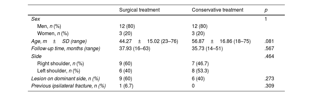
ResultsThe total number of subjects included in the study was 30. Fifteen patients were included in the conservative treatment group and 15 in the surgical group. Each was made up of 12 (80%) men and 3 (20%) women. The mean age in the surgical treatment group was 44.27 years (23–76) and 56.87 years (18–75) in the conservative group. The mean follow-up time was 37.93 months in the surgical group with a minimum of 16 months and a maximum of 63. Patients in the conservative treatment group had a mean follow-up of 35.73 months with a minimum of 14 months and a maximum of 51. The rest of the epidemiological characteristics of the patients in the sample are shown in Table 1. There were no statistically significant differences, so both groups were comparable.
Epidemiological results.
| Surgical treatment | Conservative treatment | p | |
|---|---|---|---|
| Sex | 1 | ||
| Men, n (%) | 12 (80) | 12 (80) | |
| Women, n (%) | 3 (20) | 3 (20) | |
| Age, m±SD (range) | 44.27±15.02 (23–76) | 56.87±16.86 (18–75) | .081 |
| Follow-up time, months (range) | 37.93 (16–63) | 35.73 (14–51) | .567 |
| Side | .464 | ||
| Right shoulder, n (%) | 9 (60) | 7 (46.7) | |
| Left shoulder, n (%) | 6 (40) | 8 (53.3) | |
| Lesion on dominant side, n (%) | 9 (60) | 6 (40) | .273 |
| Previous ipsilateral fracture, n (%) | 1 (6.7) | 0 | .309 |
m: mean; SD: standard deviation.
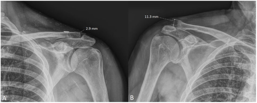
The results of the variables studied are shown in Tables 2 and 3. In addition to these data, a measurement in millimetres (mm) of the distance from the upper edge of the acromion to the upper edge of the distal end of the clavicle was also made (Fig. 1). In the conservative treatment group this distance was on average 14.21 (SD=4.74, range 8.7–23.8) and in the surgical treatment group it was 8.95 (SD=5.52, range 2–20), resulting in this difference being statistically significant with a p value of .008. In relation to the subjective results, in the conservative group eight (53.3%) patients defined the result as excellent, four (26.7%) as good and three (20%) as average. In the surgical group, six (40%) subjects defined the result as excellent, six (40%) as good, one (6.7%) as average and two (13.3%) as failure. This variable did not show statistical significance with a p of .297.
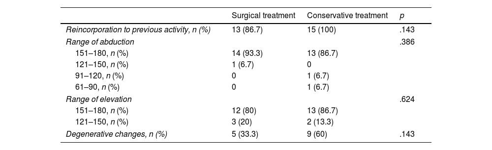
Variables results.
| Surgical treatment | Conservative treatment | p | |
|---|---|---|---|
| Reincorporation to previous activity, n (%) | 13 (86.7) | 15 (100) | .143 |
| Range of abduction | .386 | ||
| 151–180, n (%) | 14 (93.3) | 13 (86.7) | |
| 121–150, n (%) | 1 (6.7) | 0 | |
| 91–120, n (%) | 0 | 1 (6.7) | |
| 61–90, n (%) | 0 | 1 (6.7) | |
| Range of elevation | .624 | ||
| 151–180, n (%) | 12 (80) | 13 (86.7) | |
| 121–150, n (%) | 3 (20) | 2 (13.3) | |
| Degenerative changes, n (%) | 5 (33.3) | 9 (60) | .143 |
 of the distance from the upper edge of the acromion to the upper edge of the distal end of the clavicle. (A) Patient undergoing surgical treatment (2.9mm). (B) Patient managed conservatively (11.3mm).")
In recent years, articles have been published comparing the results obtained after treatment, both conservative and surgical, of type III ACL. Despite this, the best option has not yet been clearly established.18 Furthermore, in the current literature there are more than 150 surgical techniques identified for its treatment,19 so the gold standard for the surgical approach has also yet to be defined. The device used in the majority of patients undergoing surgical treatment in this study was a system that replicates the stability of the acromioclavicular joint, allowing for a more physiological stabilisation.20
In many of the articles published to date, surgical treatment is recommended in athletes and young patients who have to lift weights overhead in their work activity.9,21 However, several studies have already presented good results after conservative treatment in all patient groups,12 so the recommendations regarding the surgical approach in the literature should be reconsidered.
It must also be taken into account that when comparing conservative treatment versus surgical treatment, it may include various techniques that have different advantages, disadvantages and results. In cases of acute unstable injuries, the acromioclavicular joint can be stabilised through repair or reconstruction techniques. Contrasting different techniques, it is accepted that non-anatomical reconstructions are biomechanically inferior to anatomical techniques. However, the latter present a greater risk of fracture in the clavicle and, potentially, in the coracoid, which must be taken into account if this treatment is considered in contact athletes.22 In our study, coracoclavicular cortical suspension band-type systems were used in the majority of patients undergoing the surgical approach. These systems, used in the acute phase of the injury, act as internal support and maintain the reduction of the joint, allowing the ligaments to heal. Another possible technique is the locked hook plate, which allows for more biomechanically resistant reconstruction,23 but requires revision surgery for its removal. Standard techniques, such as fixation with Kirschnner wires and suturing of the ligaments, are no longer used, since they do not achieve adequate stability. However, there are no clear differences in results to recommend one technique over another in grade III ACL.19
In our study, the main objective was to analyse the mid-term clinical and functional outcome evaluated using the Constant and Oxford scales and the VAS for pain. In these categories, both groups presented good results and there were no statistically significant differences. This contrasts with that which was published by Gstettner et al.,9 who report better results in the group of patients who underwent surgical intervention using a hook plate compared to those treated conservatively, with the first group obtaining a score on the Constant scale of 90.4 versus 80.7 from the second group. With these results, the authors recommend surgical treatment in young, active patients who need good mobility and strength.
Regarding the assessment of pain, in our study 14 patients in the conservative group and 11 in the surgical group expressed a result of 0 on the VAS for pain without statistically significant differences, which represents a good result in both groups. Joukainen et al.,24 conducted a randomised clinical trial with a follow-up of between 18 and 20 years on nine patients treated conservatively and 16 surgically, and found that no subject in the non-surgical group presented pain, while two individuals who underwent surgery had pain in the acromioclavicular joint on palpation and on the crossed arm test. The presence of greater pain in patients undergoing surgery may be related to the development of calcifications in the coracoclavicular or acromioclavicular ligaments.18,24 However, in the present study no patient required the regular use of analgesics or anti-inflammatory drugs to control pain and those with more pain did not present worse functional results.
Based on the AP shoulder radiographs taken during follow-up, the surgical treatment improved the position of the joint in the majority of the patients in the present study, these results being better than those obtained by Calvo et al.,25 who used the Phemister technique and only achieved an anatomical reduction in half of the subjects and a higher incidence of osteoarthritis and ossification of the coracoclavicular ligament in those who underwent surgery.
In relation to the above, the distance between the upper tip of the acromion and the upper tip of the distal end of the clavicle, that is, the degree of subluxation of the acromioclavicular joint, presented statistically significant differences in both groups (p=.008). However, the persistence of subluxation of the acromioclavicular joint did not influence the functional results, which matches that published by Fremerey et al.26 and Calvo et al.25
The AP shoulder radiograph also assessed the presence of degenerative changes in the acromioclavicular joint. Five patients in the surgical treatment group and nine in the conservative group presented with osteoarthritis, this difference not being statistically significant (p=.143), as has been previously reported in other studies.12 In this same study, after one year of follow-up, almost all patients had returned to work, regardless of the treatment performed. These data concur with those obtained in the present study, since all subjects in the conservative treatment group returned to their previous activity and only two in the surgical group had to adapt their work or sports life after the intervention.
Korsten et al.8 and De Carli et al.27 published better subjective and aesthetic results in young and active patients undergoing surgical treatment compared to the conservative approach. However, when asked about perceived satisfaction, 80% of patients in both groups defined their result as excellent or good and none reported concern about the aesthetic result, neither in relation to the shoulder deformity nor the surgical scar, thus demonstrating that both treatment options may be appropriate.
Beitzel et al.19 analysed 14 studies comparing 706 patients with ACL type III who had undergone surgical or conservative treatment. They published favourable results in 88% of patients undergoing surgical intervention and 86% in those managed conservatively. The authors found that the subjects in the conservative group had a faster recovery, so they were able to return to their work or sports activities sooner than those treated surgically. We have not compared the time of return to previous activity, but all patients in the conservative treatment group returned to their previous activity and only two in the surgical group had to adapt their work or sports life after the intervention.
LimitationsOur study has several limitations. Firstly, it is a retrospective study with the biases inherent to this type of design, the most important being the absence of treatment randomisation. Furthermore, the sample size may be small, but according to the literature, it should be sufficient to find a significant difference on the Constant scale. Finally, other limitations that could influence the final results are the lack of evaluation of associated complications, such as scapular dyskinesia, and the lack of information on the moment of incorporation into the previous activity.
ConclusionsExcellent results can be achieved in type III ACL with both surgical and conservative treatment. Although the radiographic results are better in the surgical group (less distance between the upper edge of the acromion and the upper edge of the distal end of the clavicle and anatomical reconstruction of the joint), the functional evaluation scales used (Constant, Oxford and VAS for pain) did not show significant differences between both groups. These results do not support the routine use of surgical treatment for grade III ACLs.
Level of evidenceLevel of evidence III.
FundingThis study did not receive any type of funding.
Conflict of interestsThe authors have no conflict of interests to declare.
