metricas
covid
Buscar en
Radiología (English Edition)
Toda la web
Inicio Radiología (English Edition) Pulmonary radiofrequency ablation (Part 2): Procedure and follow-up
Journal Information
Vol. 57. Issue 4.
Pages 287-302 (July - August 2015)
Share
Share
Download PDF
More article options
Visits
2031
Vol. 57. Issue 4.
Pages 287-302 (July - August 2015)
Update in Radiology
Pulmonary radiofrequency ablation (Part 2): Procedure and follow-up
Radiofrecuencia pulmonar (Parte 2): procedimiento y seguimiento
Visits
2031
J.M. Plasencia Martínez
Servicio de Radiología, Hospital General Universitario Morales Meseguer, Murcia, Spain
This item has received
Article information
Abstract
Full Text
Bibliography
Download PDF
Statistics
Figures (14)
Show moreShow less
Additional material (2)
Abstract

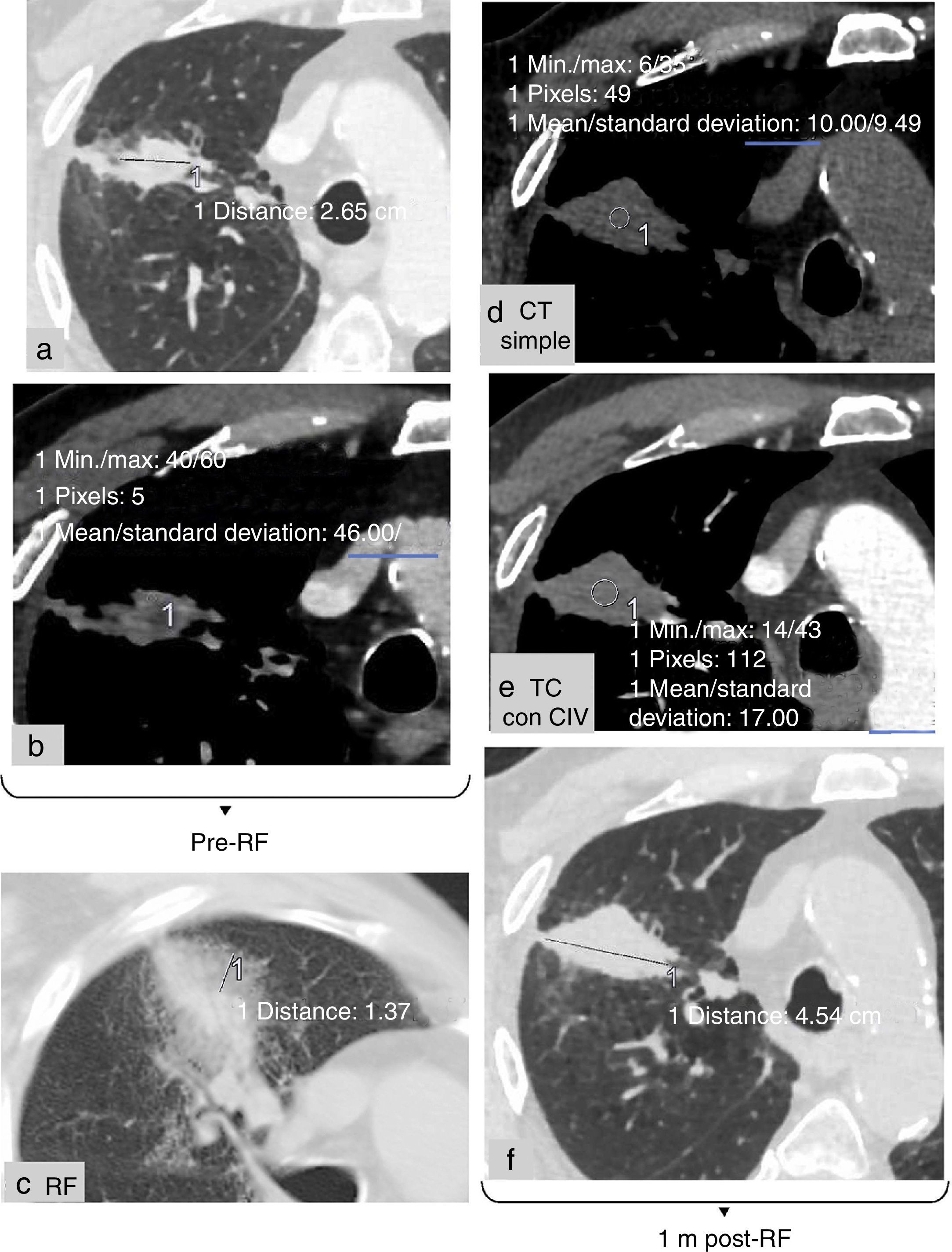
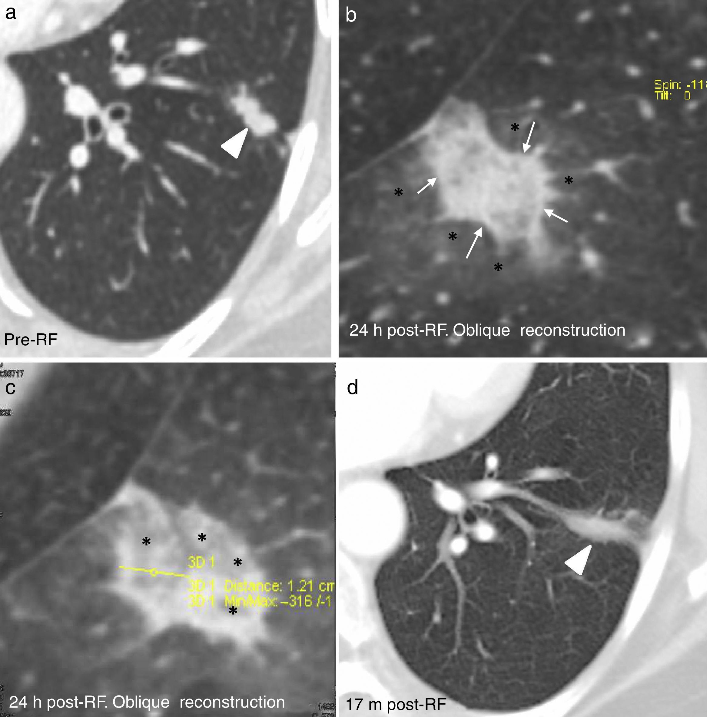
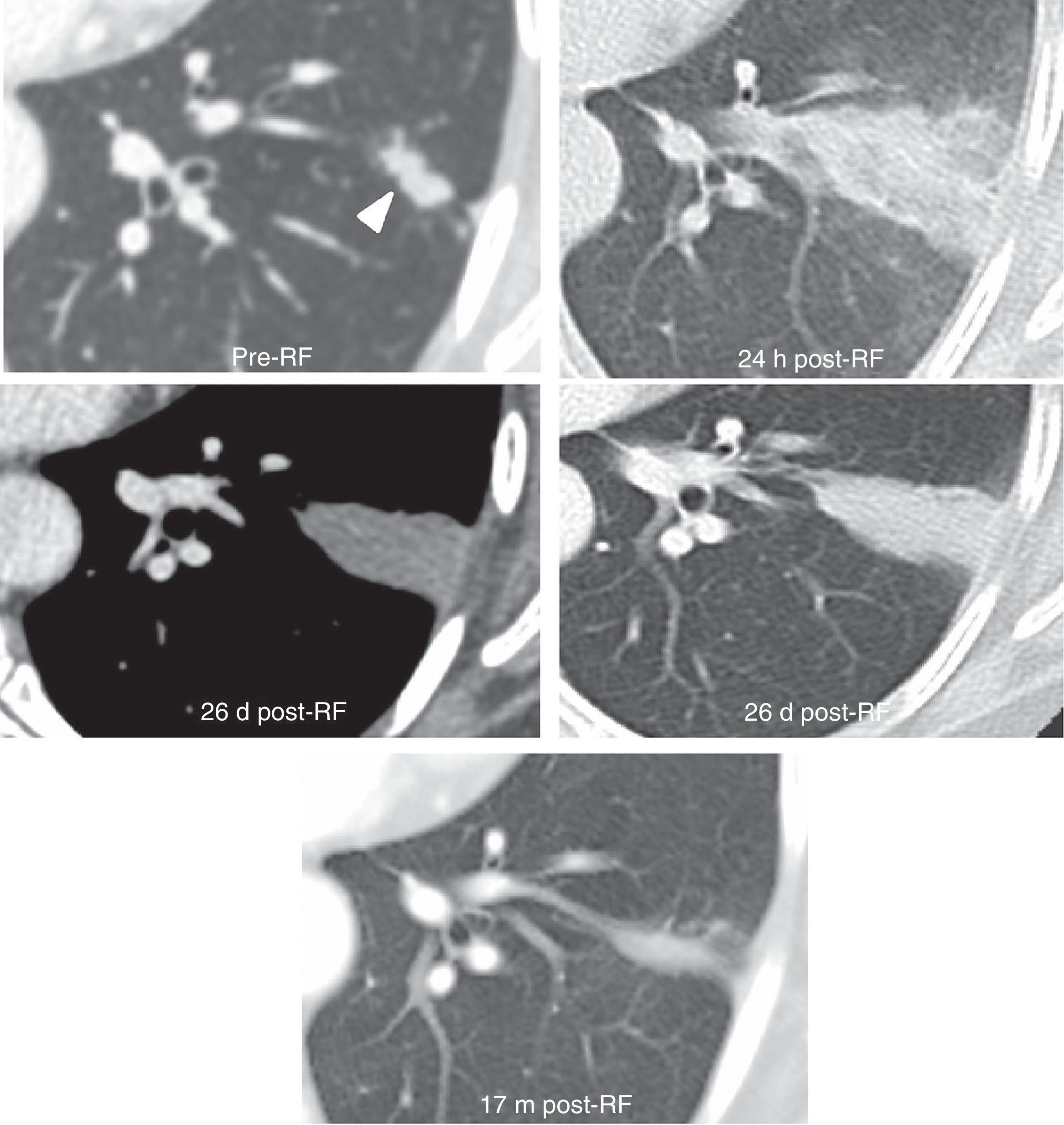
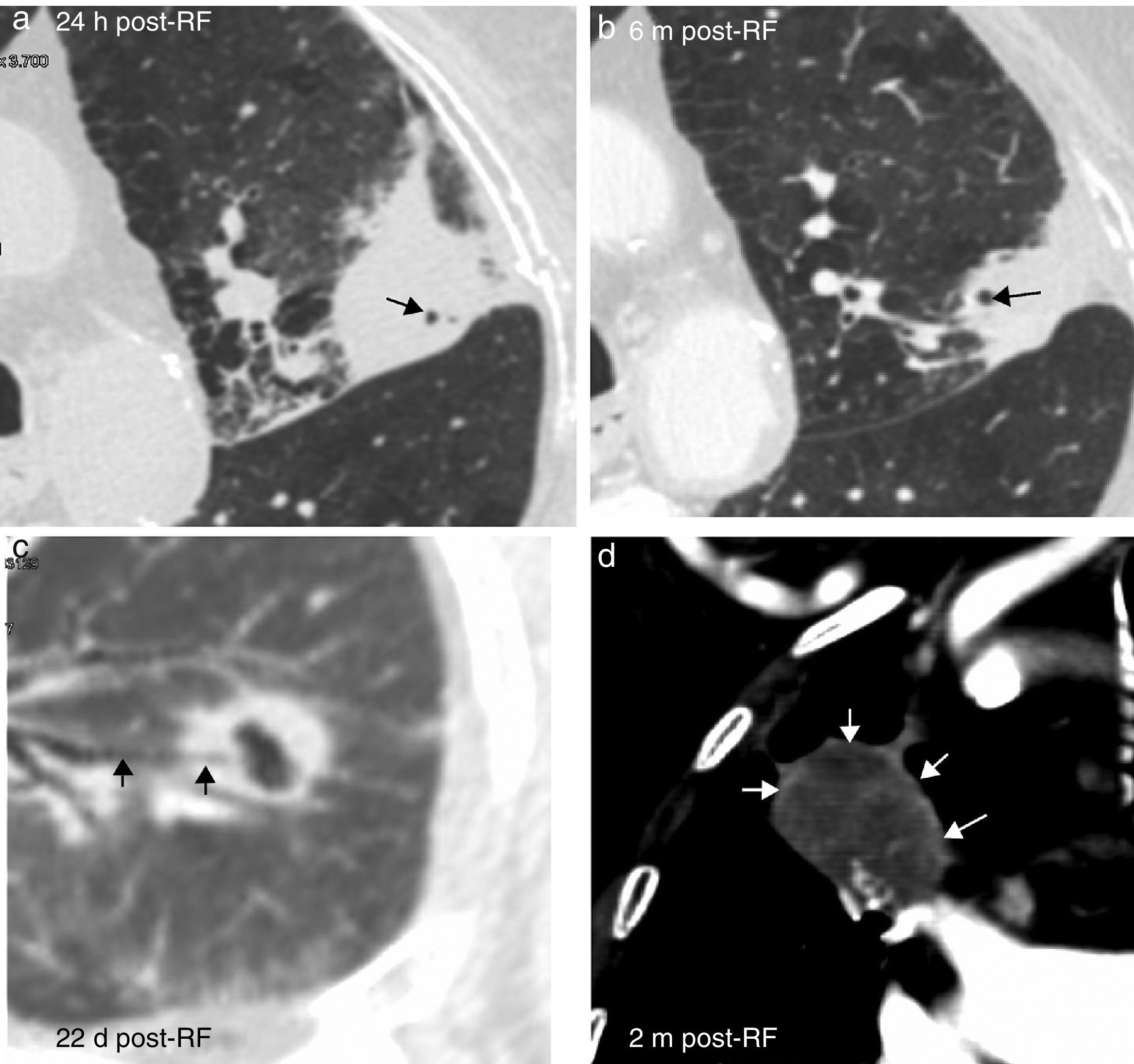
Pulmonary radiofrequency ablation requires more than just interventional radiology skills. Patients must be selected carefully, and the acts that need to be done before, during, and after the procedure must be coordinated. To guarantee patient safety, radiologists need to know the variants of the technique, the precautions that must be taken, the complications that can occur, and the risks involved. Early differentiation between tumor tissue and normal changes secondary to treatment on imaging tests will make it possible to repeat the treatment without delays, and this will increase survival. This article describes how to coordinate and carry out pulmonary radiofrequency ablation, the complications of the technique, and the current evidence in follow-up.

Keywords:
Radiofrequency ablation
Lung tumors
Image-guided procedures
Iatrogenesis
Follow-up
Resumen

Para la ablación pulmonar con radiofrecuencia no solo son necesarias habilidades intervencionistas. Tras seleccionar adecuadamente al paciente, hay que coordinar las actuaciones previas, durante y posteriores al procedimiento. Conocer las variantes de la técnica, las precauciones, las complicaciones, los riesgos y las recomendaciones para el seguimiento garantizará la seguridad del paciente. Diferenciar precozmente en las pruebas de imagen el tejido tumoral de los cambios normales secundarios al tratamiento permitirá volver a tratar pronto al paciente, lo que aumentará su supervivencia. El objetivo de este trabajo es describir cómo coordinar y realizar la ablación pulmonar con radiofrecuencia, sus complicaciones y la evidencia actual en el seguimiento.

Palabras clave:
Ablación con radiofrecuencia
Tumores pulmonares
Guía de imagen
Iatrogenia
Seguimiento

Article

These are the options to access the full texts of the publication Radiología (English Edition)
Subscriber
Subscriber

If you already have your login data, please click here .

If you have forgotten your password you can you can recover it by clicking here and selecting the option “I have forgotten my password”
Purchase
Purchase article

Purchasing article the PDF version will be downloaded

Price 19.34 €

Purchase now
Contact
Phone for subscriptions and reporting of errors
From Monday to Friday from 9 a.m. to 6 p.m. (GMT + 1) except for the months of July and August which will be from 9 a.m. to 3 p.m.
Calls from Spain
932 415 960
Calls from outside Spain
+34 932 415 960
E-mail
Article options
es en pt

¿Es usted profesional sanitario apto para prescribir o dispensar medicamentos?

Are you a health professional able to prescribe or dispense drugs?

Você é um profissional de saúde habilitado a prescrever ou dispensar medicamentos