


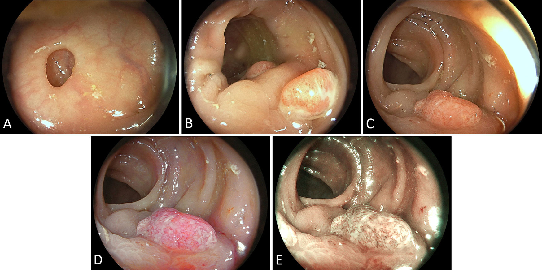
A 70-year-old male patient was referred for outpatient ileocolonoscopy. Medical history was significant for vague left lower abdominal pain for about three days duration six weeks before, potentially suggesting a minor episode of diverticulitis. Indeed, the recent lower GI endoscopy indicated some unremarkable diverticula in the sigmoid (Fig. 1A). By contrast, there was a short segment at 30cm from the dentate line with edematous changes and minor putrid discharge from adjacent diverticula, from which two polypoid lesions estimated at 10mm arose (Fig. 1B). Further characterization including image-enhanced endoscopy revealed a dense network of irregular vessels, however, no abnormal pit pattern to suggest a neoplastic lesion (Fig. 1C–E). Given an optical diagnosis of most likely granulation polyps, complicating subacute diverticulitis, we abstained from endoscopic resection and instead biopsied the lesions. Histopathology likewise confirmed the presence of granulation polyps with exuberant leukocytic infiltration and lack of intact surface epithelium. The patient was informed about the diagnosis and a wait-and-see approach was consented.
 An unremarkable diverticulum in the sigmoid. (B) Short segmental edema with residual putrid discharge from an adjacent diverticulum as well as two polypoid lesions of an estimated 10mm size. (C) Detailed endoscopic characterization including (D) linked colour imaging (LCI) and (E) blue laser imaging (BLI) indicated a dense network of irregular vessels, however, no pit pattern suggestive of neoplasia.")
(A) An unremarkable diverticulum in the sigmoid. (B) Short segmental edema with residual putrid discharge from an adjacent diverticulum as well as two polypoid lesions of an estimated 10mm size. (C) Detailed endoscopic characterization including (D) linked colour imaging (LCI) and (E) blue laser imaging (BLI) indicated a dense network of irregular vessels, however, no pit pattern suggestive of neoplasia.
Albeit granulation polyp formation is a common reaction of the colorectal epithelium to variable forms of tissue injury, there are, to the best of our knowledge, only three other reports in a setting of diverticulitis.1 Correct optical diagnosis may be instrumental in avoidance of considerations of overly aggressive procedures, such as (with a view to adjacent diverticula) technically demanding endoscopic or even surgical resection.
Conflict of interestNothing to declare.
