


31-year-old patient with no history of interest. Consults due to the appearance of a tumour on his back for a week, dry cough, weight loss of 9 kg and sweating for the past 3 weeks.
Physical examination reveals a painful right-sided tumour of 10 × 9 cm, soft, covered by healthy skin and with a slight increase in local temperature (Fig. 1). Upon oral inspection, oral sepsis is observed.

A chest X-ray is performed (Fig. 2). The patient remembers that his cousin and his aunt had tuberculosis 4 years earlier, but he was not screened for latent tuberculosis infection nor did he receive chemoprophylaxis. The lab tests show 19.77 thousand/mcl leukocytes and CRP of 32.7 mg/dl.

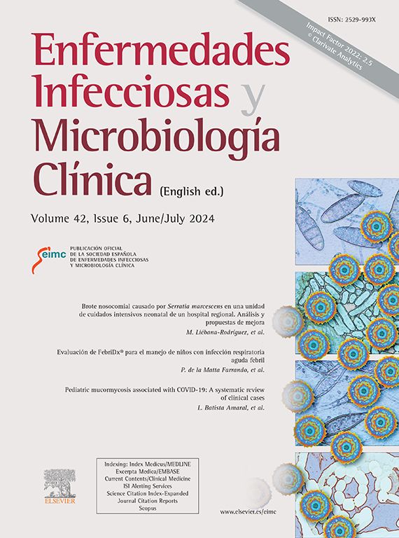
A puncture is made obtaining purulent material (Fig. 3).
Outcomes
3 sputum samples are collected for Ziehl-Neelsen staining that are negative, as well as the tuberculin test and Gram and Ziehl-Neelsen stains of the content of the abscess. A chest CT is performed that shows a liver abscess of 7.3 × 5.8 cm with extension to the chest wall forming a collection in communication with the pleural cavity (Fig. 4). A drain is placed, and treatment with ceftriaxone is started 2 g/24 hours and metronidazole 500 mg/8 hours iv.

On day 7° of the culture, growth of Fusobacterium nucleatum in the pus is reported. Subsequently, the result of the 16S PCR is received, in which Fusobacterium nucleatumis also identified. Pathological examination reveals positive branching filamentous structures for thePAS and Gomori-Grocott stains, that did not stain on the Gram stain.
He is treated by percutaneous drainage and amoxicillin-clavulanic acid for a total of 75 days (33 days intravenously during hospitalisation and 6 more weeks orally after drain removal). At 6 months in the ultrasound follow-up there are no signs of recurrence.
CommentsThe finding of a chest wall mass at a first approximation implies a differential diagnosis between entities such as tuberculous cold abscess, pyogenic abscess and neoplastic involvement of the chest wall. The tuberculous cold abscess of the chest wall is a rare entity, but it should never be overlooked in the differential diagnosis of any chest wall mass, especially in a patient with an epidemiological history and compatible radiological findings. In our case it was the first clinical suspicion, a possibility that was ruled out when the result of the abdominal CT was available which revealed the hepatic origin of the abscess.
Actinomycosis is caused by bacteria of the genus Actinomyces, a branched gram-positive bacillus, facultative anaerobe. The disease is characterised by abscess and fistulous tract formation along with the discharge of purulent material containing yellow sulphur granules. There are 3 main forms of involvement: cervicofacial (65%), abdominal (20%) and thoracic (15%)1. The presentation of this liver abscess with chest wall fistula is suggestive of actinomycosis and in the cytological examination this diagnostic possibility was suggested. For this reason, a Gram stain was conducted on the cytology sample and it was verified that the filamentous structures observed were not stained. The negativity of this staining and the absence of growth in culture after 2 weeks of incubation, together with the PCR identification of the Fusobacterium, made us doubt the presence of Actinomyces. However, liver abscesses are often polymicrobial (between 11 and 40% depending on the series)2 and we could not completely rule out in our case the coexistence of Actinomyces and Fusobacterium.
F. nucleatum is a small anaerobic spindle-shaped gram-negative bacillus, non-mobile and non-spore forming, which is frequently part of normal oral flora3. In a review of 48 cases of liver abscess due to fusobacteria, of the 22 caused by F. nucleatum, 9 were attributed to periodontal disease, 4 gastrointestinal disease, one to Lemierre syndrome and 8 were considered cryptogenic. Median duration of treatment was 6 weeks and recovery was complete in all cases4. Most occurred, just like in our case, in immunocompetent young people with periodontal disease or recent pharyngitis as the only risk factor for haematogenous spread of spherobacteria.
Please cite this article as: Van den Eynde E, Capilla S, Parra T. Absceso de pared torácica. Enferm Infecc Microbiol Clin. 2020;38:446–447.
