


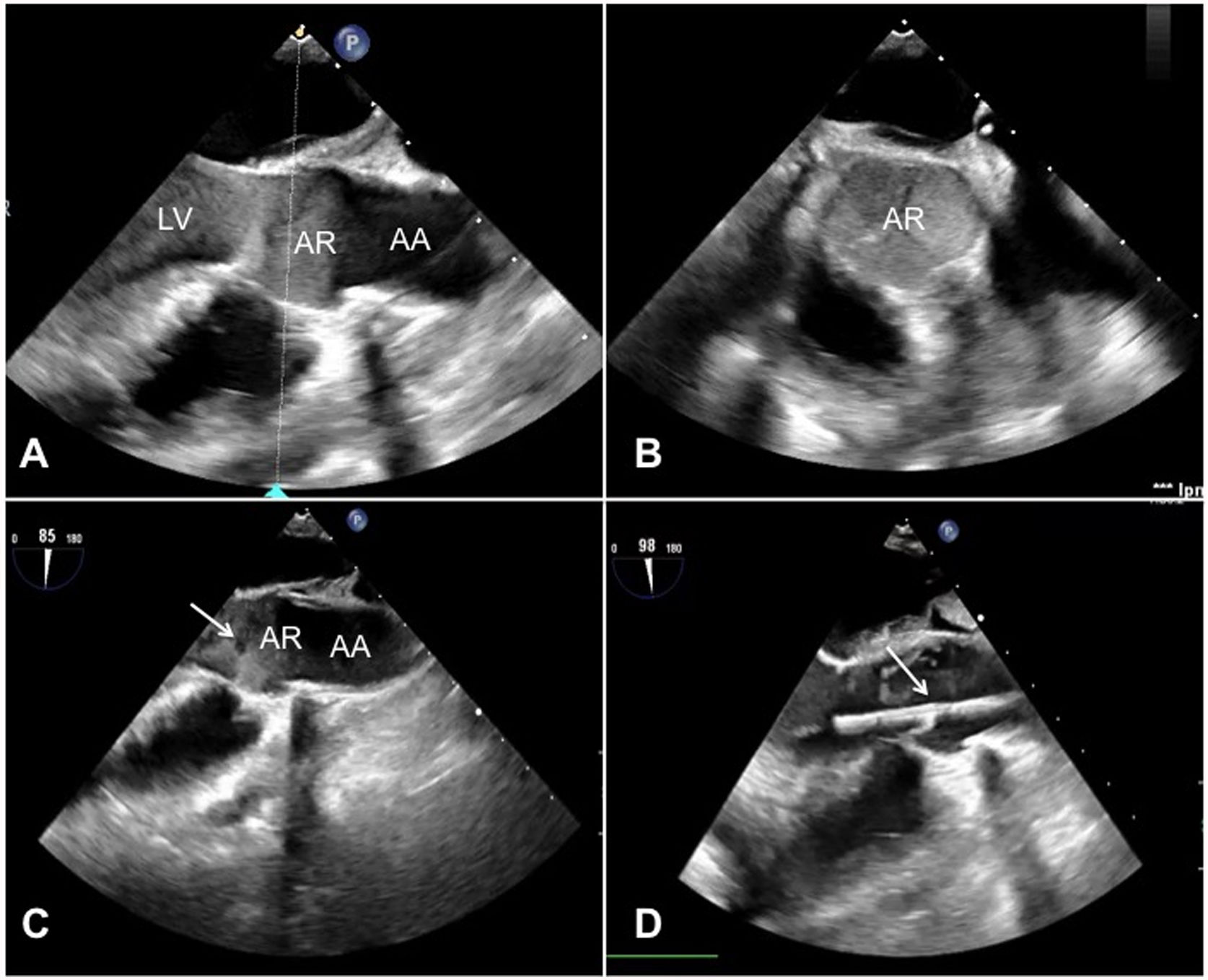
A 62-year-old patient developed cardiogenic shock after acute myocardial infarction and was given venoarterial ECMO support with the extraction cannula placed in the left atrium (LAVA-ECMO). During ECMO he lost pulsatility. A transoesophageal echocardiogram was performed, showing no contractility, closure of the aortic valve, and spontaneous contrast in the left ventricle and aortic root (Fig. 1A and B, Video 1). Valve opening was achieved with chest compressions (Fig. 1C, Video 2). A left intraventricular catheter was connected to the extraction cannula for definitive management (Fig. 1D, Video 3). Chest compressions can be a rescue measure to prevent left chamber thrombosis during left ventricular distension syndrome in which an active left ventricular drainage mechanism is established.
. A) 3-chamber view showing spontaneous echo contrast in the left ventricle and aortic root. B) Mid-oesophageal aortic valve short axis cross-section showing spontaneous echo contrast in the aortic root. C) 3-chamber view showing aortic valve opening (white arrow). D) Real-time TOE showing opening of the aortic valve (white arrow) after start of drainage (3-chamber view). AA: Ascending aorta; AR: Aortic root; LV: Left ventricle.")
Transoesophageal echocardiography (TOE). A) 3-chamber view showing spontaneous echo contrast in the left ventricle and aortic root. B) Mid-oesophageal aortic valve short axis cross-section showing spontaneous echo contrast in the aortic root. C) 3-chamber view showing aortic valve opening (white arrow). D) Real-time TOE showing opening of the aortic valve (white arrow) after start of drainage (3-chamber view).
AA: Ascending aorta; AR: Aortic root; LV: Left ventricle.
No funding.
Conflict of interest disclosureThe authors declare that they have no competing interests.
Patient consent statementWritten informed consent for patient information and images to be published were provided by the patient or a legally authorized representative.
The following are Supplementary data to this article:
